Introduction
Pneumocystis jirovecii (PJ), an opportunistic
fungal pathogen, is a major cause of Pneumocystis pneumonia (PCP)
in immunocompromised individuals, such as those with HIV/AIDS,
organ transplant recipients, and patients receiving chemotherapy or
long-term immunosuppressive therapy (1-4).
PCP is characterized by severe respiratory symptoms, and if not
diagnosed and treated promptly, it can lead to respiratory failure
and death (3). Therefore, early
and accurate diagnosis of PJ infection is crucial for improving
patient outcomes.
Currently, the diagnostic methods commonly used for
PJ detection include microscopic examination of bronchoalveolar
lavage fluid (BALF) smears, PCR-based assays and targeted
next-generation sequencing (tNGS) (5-9).
Microscopy and conventional PCR are the traditional ‘gold
standards’ for PJ detection. Microscopy exhibits low sensitivity,
particularly in cases with low PJ burdens (10,11).
PCR has higher sensitivity than microscopy, but it requires
expensive thermal cyclers and trained personnel, and its detection
process is relatively time-consuming (12-15).
tNGS has the advantage of high-throughput detection but is costly
and not widely available in clinical settings, especially in
resource-limited areas (7,16-19),
and entails a relatively long detection time (20). Thus, there is an urgent need for a
rapid, simple, sensitive and cost-effective diagnostic method for
PJ detection.
Nucleic acid detection technologies have
revolutionized the field of clinical diagnosis, pathogen
surveillance and genetic analysis, with continuous advancements
aiming to address the limitations of traditional methods such as
microscopy and PCR-based assays (21). Among the emerging alternatives,
isothermal amplification techniques have garnered considerable
attention due to their inherent advantages of simplicity, rapidity
and independence from expensive thermal cyclers, making them
particularly promising for applications in resource-limited
settings (22,23). For instance, loop-mediated
isothermal amplification (LAMP) has been extensively explored for
the detection of a broad spectrum of targets, including
Plasmodium knowlesi, severe acute respiratory syndrome
coronavirus (SARS-CoV)-2 and various bacteria, fungi and sexually
transmitted infection-related pathogens (24-28).
Advances in molecular diagnostics have also promoted the
application of isothermal amplification techniques in specialized
fields such as tuberculosis diagnosis, where they serve as valuable
alternatives for the detection of Mycobacterium tuberculosis
(29). Beyond pathogen detection,
LAMP has also found utility in crop breeding, disease diagnosis and
genetic screening - for instance, in identifying single-nucleotide
polymorphisms and gene translocations associated with cancer,
highlighting its potential as a versatile tool for mutation
screening (25,30). Another notable isothermal
amplification technique is recombinase polymerase amplification
(RPA), which has been successfully applied in the detection of
bacteria, fungi, viruses, parasites, drug-resistant genes,
genetically modified foods and SARS-CoV-2 (31-33).
Recombinase-aided amplification (RAA) is a novel
isothermal nucleic acid amplification technology that has emerged
in recent years (31,34,35).
It utilizes recombinase, single-stranded DNA-binding protein and
DNA polymerase to achieve rapid amplification of target nucleic
acids at a constant temperature (37-42˚C) (36-38).
RAA utilizes recombinase sourced from bacteria or fungi, while RPA
employs recombinase primarily derived from the T4 bacteriophage
(39). RAA has the advantages of
rapid amplification (40-42)
(usually completed within 30 min), simple operation, no need for
complex thermal cycling equipment, and high sensitivity and
specificity (38). It has been
successfully applied for the detection of various pathogens, such
as bacteria, viruses and parasites (37,43-47).
Its application scope further extends to the agricultural and
livestock sectors, enabling effective detection of viruses that
cause infections in livestock (42,48,49).
The RAA-amplified products can be detected by agarose gel
electrophoresis or a real-time fluorescence detection platform
(46,50,51).
Its compatibility with point-of-care testing (POCT) (52) scenarios is further enhanced by
integration with CRISPR-Cas technology (53), a combination that enables more
convenient and intuitive interpretation of detection results while
significantly expanding the application potential of RAA in
pathogen detection (35,54,55).
However, no reports have documented the application of RAA for PJ
detection.
In the present study, specific primers and probes
targeting PJ were designed, the RAA reaction conditions were
optimized and the analytical performance (sensitivity, specificity,
repeatability) of the assay was evaluated. Additionally, the
clinical performance of the RAA assay was validated using clinical
BALF samples, aiming to provide a rapid and reliable diagnostic
tool for PJ infection.
Materials and methods
Ethical approval and consent
This study was approved by the Ethics Committee of
Weifang Second People's Hospital (Weifang, China; approval no.
KY2024-023-01; approval date: March 15, 2024) and conducted in
strict accordance with the principles of the Declaration of
Helsinki. Written informed consent was obtained from all patients
prior to BALF sample collection and study inclusion; for patients
unable to provide consent due to critical illness (e.g., severe
respiratory failure requiring mechanical ventilation or altered
consciousness), consent was obtained from their legal
representatives. All participants were informed about the purpose
of the study, the procedures involved, potential risks and
benefits, and their right to withdraw at any time without affecting
their clinical care. Patient data were anonymized and de-identified
prior to analysis to ensure confidentiality.
Case definition
PJ infection was defined based on the following
composite criteria: i) Clinical manifestations consistent with PCP,
including progressive dyspnea, nonproductive cough, fever and
hypoxemia; ii) characteristic chest imaging findings of diffuse
bilateral ground-glass opacities, interstitial infiltrates or
cystic lesions on computed tomography (CT); and iii) laboratory
confirmation of PJ nucleic acid detection by tNGS from BALF
samples. Patients meeting all three criteria were classified as
confirmed PJ infection cases. Patients with negative tNGS results
and no clinical or radiological evidence of PCP were classified as
non-PJ controls.
Inclusion and exclusion criteria
Inclusion criteria were as follows: i) Patients aged
≥18 years who underwent bronchoscopy and BALF collection for
suspected lower respiratory tract infection at Weifang Second
People's Hospital between March 18, 2024 and December 31, 2024; ii)
BALF samples with sufficient volume (≥3 ml) and quality for nucleic
acid extraction and molecular testing; iii) complete clinical,
laboratory and imaging data available for review; iv) for
PJ-positive cases, confirmation by tNGS detection of PJ with
consistent clinical and radiological findings. Exclusion criteria
were as follows: i) BALF samples with insufficient volume (<3
ml) or visible blood contamination exceeding 50% of the sample
volume; ii) patients with incomplete medical records or missing
tNGS results; iii) samples stored for >72 h at 4˚C or subjected
to repeated freeze-thaw cycles (>2 cycles) prior to testing; iv)
patients receiving empirical anti-PCP therapy
(trimethoprim-sulfamethoxazole, pentamidine or atovaquone) for
>72 h before BALF collection; and v) concurrent diagnosis of
other fungal infections (e.g., invasive aspergillosis,
mucormycosis) that could confound the clinical presentation. The
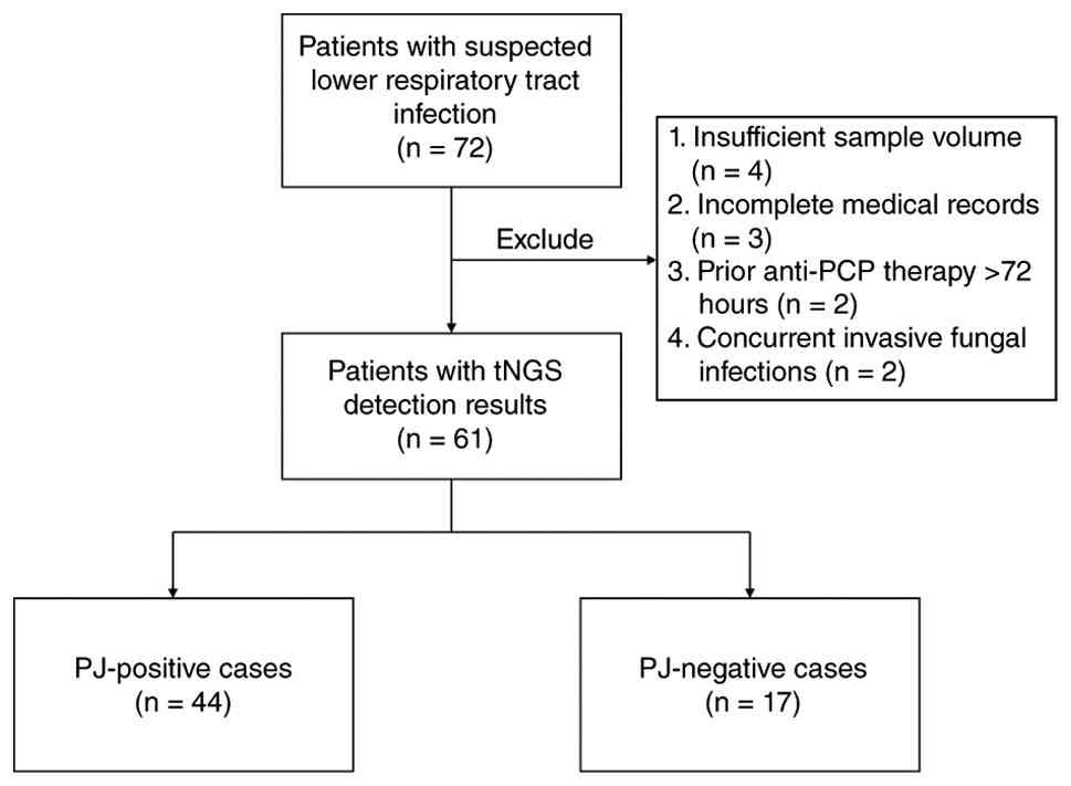
participant selection process is summarized in Fig. 1.
Experimental materials. Clinical
sample collection
A total of 44 BALF samples positive for PJ by tNGS
and 17 BALF samples negative for PJ by tNGS were collected. All
samples were processed within 2 h of collection.
Pathogen controls. Genomic DNA of common
clinically relevant pathogens, including Acinetobacter
baumannii, Klebsiella pneumoniae, Pseudomonas
aeruginosa, Proteus mirabilis, Stenotrophomonas
maltophilia, Enterobacter aerogenes, Enterobacter
cloacae, Serratia marcescens, Candida albicans
and Candida krusei, was obtained from the Clinical
Microbiology Laboratory of Weifang No. 2 People's Hospital
(Weifang, China), having been isolated from the patient's clinical
specimen during diagnostic examination.
Kits and instruments. The RAA Nucleic Acid
Amplification kit (Conventional Method; cat. no. B00000A-96; lot
no. BH7OA0) and RAA Nucleic Acid Amplification kit (Fluorescent
Method; cat. no. F00001A-96; lot no. FH9KD0) were purchased from QT
Biotech Co., Ltd. Thermo Scientific™ PCR Premix (2X) (cat. no.
K0172) was purchased from Thermo Fisher Scientific, Inc. The
MGIEasy Nucleic Acid Extraction Kit (lot no. 111942507003; cat. no.
RM0766) was purchased from BGI Genomics Co., Ltd. The Equalbit 1X
dsDNA HS Assay Kit (cat. no. EQ121-02; lot no. 7E1352F5) was
purchased from Vazyme Biotech Co., Ltd. DL1500 Ladder (100 bp to
1,500 bp; cat. no. MD1019) was purchased from Simgen Biotechnology
Co., Ltd. Instruments included a Qubit 4 Fluorometer (Thermo Fisher
Scientific, Inc.), CFX96™ Real-Time PCR System (Bio-Rad
Laboratories, Inc.), T100™ Thermal Cycler (Bio-Rad Laboratories,
Inc.) and ChemiDoc™ Imaging System (Bio-Rad Laboratories,
Inc.).
Nucleic acid extraction. Nucleic acid
extraction was performed using the MGIEasy Nucleic Acid Extraction
Kit, with steps as follows: i) Reagent and sample preparation:
Lysis buffer: Antifoaming agent was added to lysis buffer; Wash
Buffer A/B: Anhydrous ethanol was added to Wash Buffer A/B,
respectively. For BALF samples: Transferred 350 µl of BALF to a
clean 1.5 ml centrifuge tube. ii) Preparation and addition of
extraction reaction mix for each sample. iii) Magnetic bead
extraction. Viscous residues were homogenized and the mixture was
then incubated at 56˚C for 10 min. Isopropanol and magnetic beads
were added and after vortexing, the mixture was left to stand at
room temperature for 10 min (invert every 3 min). Samples were
centrifuged, placed on a magnetic rack and the supernatant was
discarded. Beads were washed with Wash Buffer A/B sequentially and
then air-dried at room temperature, followed by addition of
nuclease-free water and elution of nucleic acid, which was stored
at -20˚C. iv) Quality control. The DNA concentration was measured
with the Equalbit 1X dsDNA HS Assay Kit (Vazyme Biotech Co., Ltd.)
and Qubit 4 Fluorometer (Thermo Fisher Scientific, Inc.).
Samples were considered to meet the requirements for
subsequent library preparation if their DNA concentration, as
determined by NanoDrop DS-11, was ≥1 ng/µl (total amount ≥10 ng),
and if the A260/A280 ratio and A260/A230 ratio, as measured by
spectrophotometry (NanoDrop DS-11; Thermo Fisher Scientific, Inc.),
were 1.8-2.0 and ≥1.8, respectively.
Construction of recombinant plasmid standard.
A 149 bp conserved region of the PJ major surface glycoprotein gene
(GenBank ID: KP168716.1) was synthesized and cloned into the pUC57
vector by Sangon Biotech, generating a recombinant plasmid. The
plasmid was serially diluted to concentrations ranging from
104 to 10 copies/µl with nuclease-free water. The number
of copies/µl was calculated using the following formula:
Primer and probe design. The 149 bp conserved
region of the PJ major surface glycoprotein gene was selected as
the target for RAA development. Specific primers and an exo probe
for RAA were designed using Oligo 7 software (Molecular Biology
Insights, Inc.). The probe was labeled with a FAM fluorophore at
the 5' end and a BHQ-1 quencher at the 3' end, and a
tetrahydrofuran spacer was included to facilitate cleavage by the
exonuclease. All oligonucleotides were synthesized by Sangon
Biotech (Shanghai, China). Sequences are listed in Table I.
 | Table ISequences of primers and probe for PJ
RAA. |
Table I
Sequences of primers and probe for PJ
RAA.
| Primer name | Sequence
(5'-3') | Target region | Length, bp |
|---|
| PJ-RAA-F |
CGACTCATACCAGCACATCCACAACCACAT | 2858-2887 | 30 |
| PJ-RAA-R |
ACTCATCCTCAACCCTTCACTCGGCTTCAC | 2977-3006 | 30 |
| PJ-RAA-Probe |
TCAAGAATAACACTCACTTCGACAAGAAGA
[i6FAMdT][idsp][iBHQ1dt]AAACCGACCAAGTGT [C3Spser] | 2899-2947 | 49 |
Specificity of primers and RAA assay. To
evaluate the specificity of PJ-RAA-forward(F)/reverse (R) primers
and confirm the amplification capability of the fluorescent RAA
assay for PJ, conventional PCR was performed. Conventional PCR used
genomic DNA templates including 8 common respiratory bacteria, 2
opportunistic fungi and a PJ-positive BALF sample. Nuclease-free
water served as the negative control (NC) for both assays.
Workflow of tNGS. tNGS was performed using
the PTseq Respiratory Pathogen Targeted High-Throughput Genetic
Detection Kit (lot no. 513392407006; cat. no. RM1261; MGI Tech Co.,
Ltd.) on the MGI DNBSEQ-G99 platform according to the
manufacturer's instructions. The tNGS workflow encompasses sample
collection, nucleic acid extraction, multiplex targeted enrichment,
library construction and bioinformatics analysis, covering a total
of 274 common respiratory pathogens, including 72 bacteria, 95
fungi, 50 viruses and 15 other pathogens. Bioinformatics analysis
was performed using the MGI platform to align reads against the
reference genome. A positive result was defined as detection of 13
or more unique reads with >95% coverage of the target
region.
Evaluation of clinical diagnostic
efficacy of RAA for PJ. PCR amplification system
The total volume of the PCR reaction system was 25
µl. Each reaction contained 12.5 µl of Thermo Scientific™ PCR
Premix (2X) (containing Taq DNA polymerase, dNTPs and reaction
buffer), 2 µl of 10 µM PJ-F primer, 2 µl of 10 µM PJ-R primer, 2 µl
of template DNA (genomic DNA extracted from PJ-positive samples,
non-PJ pathogens or NC) and 6.5 µl of nuclease-free water to bring
the volume to 25 µl.
PCR amplification program. Thermal cycling
was carried out on a PCR instrument with the following parameters:
First, pre-denaturation was performed at 94˚C for 10 min to fully
denature genomic DNA. This was followed by 30 amplification cycles,
each consisting of denaturation at 94˚C for 30 sec (to separate
double-stranded DNA), annealing at 60˚C for 30 sec (to enable
specific binding of primers to target sequences) and extension at
72˚C for 30 sec (to synthesize new DNA strands via Taq polymerase).
After the cycling process, a final extension step was conducted at
72˚C for 6 min to ensure complete extension of all amplified
products, and the reaction was then held at 4˚C indefinitely to
preserve the amplified products.
Agarose gel electrophoresis. PCR products
were analyzed by agarose gel electrophoresis to confirm
amplification specificity. A 1% agarose gel was prepared using 1X
Tris-acetate-EDTA (TAE) buffer, with ethidium bromide added for
subsequent nucleic acid visualization. Electrophoresis was run at a
constant voltage of 90 V; the running time was adjusted based on
gel separation efficiency, typically 20-30 min, until DNA bands
were clearly distinguishable. The PCR products were mixed with 6X
DNA loading buffer. For sample loading, 1.3 µl of DNA Marker
(GeneRuler 100 bp DNA Ladder; cat. no. SM0241; lot no. 3498729;
Thermo Fisher Scientific, Inc.) and 4 µl of PCR product from each
reaction were loaded into separate wells of the gel. After
electrophoresis, gel images were captured using the ChemiDoc™
Imaging System (cat. no. 12003153; Bio-Rad Laboratories, Inc.), and
the presence or absence of target bands was used to evaluate the
specificity of the PJ primers.
Establishment and optimization of RAA
for PJ. RAA reaction system
The RAA assay was performed in a 50 µl reaction
volume using an RAA Nucleic Acid Amplification kit according to the
manufacturer's instructions. For fluorescent RAA amplification, the
system included 25 µl of RAA reaction buffer, 2.6 µl of the forward
primer PJ-RAA-F (10 µM) and 2.6 µl of the reverse primer PJ-RAA-R
(10 µM), 0.8 µl of PJ-RAA-Probe (10 µM), 12 µl of nuclease-free
water, 5 µl magnesium acetate and 2 µl of template DNA (25 ng/µl).
For conventional (electrophoresis-based) RAA amplification, the
system excluded the 0.8 µl of PJ-RAA-Probe (10 µM), and the volume
of nuclease-free water was adjusted to 12.8 µl to maintain a total
reaction volume of 50 µl. Fluorescent reactions were incubated at
40˚C for 20 min on the CFX96™ Real-Time PCR System, with
fluorescence signals recorded every 20 sec (corresponding to 1
cycle per 20 sec).
Optimization of reaction conditions. Key
parameters were optimized using DNA extracted from clinical samples
as the template, with tests performed across a series of
conditions: Temperatures of 37, 38, 39, 40, 41 and 42˚C (each with
a 20 min incubation). For fluorescent RAA, samples that gave rise
to an exponential amplification curve above the NC threshold within
20 min were judged as positive. Optimal reaction conditions were
defined as the combination that yielded the earliest amplification
initiation and the fastest plateau phase arrival.
Evaluation of clinical diagnostic
efficacy of RAA for PJ. Sensitivity
For sensitivity evaluation, the recombinant pUC57-PJ
plasmid was serially diluted to generate a concentration gradient
ranging from 104 to 101 copies/µl
(104, 10³, 10², 10) for fluorescent RAA assays.
Fluorescent RAA was performed under the optimized reaction
conditions. All dilutions were tested in triplicate to ensure
reproducibility.
Clinical validation. The optimized
fluorescent RAA assay was used to detect 44 PJ-positive and 17
PJ-negative BALF samples. Diagnostic performance indicators,
including sensitivity, specificity, positive predictive value,
negative predictive value, total agreement and κ value (κ:
0.61-0.80=substantial agreement; 0.41-0.60=moderate agreement) were
calculated using SPSS 26.0 (IBM Corp.).
Statistical analysis
Data were analyzed using ImageJ (version 1.52a;
National Institutes of Health) and SPSS 26.0. The clinical
diagnostic performance of the RAA assays was evaluated, with tNGS
used as the reference standard. Analyses were based on a 2x2
contingency matrix using the chi-square test. A two-tailed
P<0.05 was considered to indicate statistical significance.
Results
Primer/probe validation and plasmid
construction
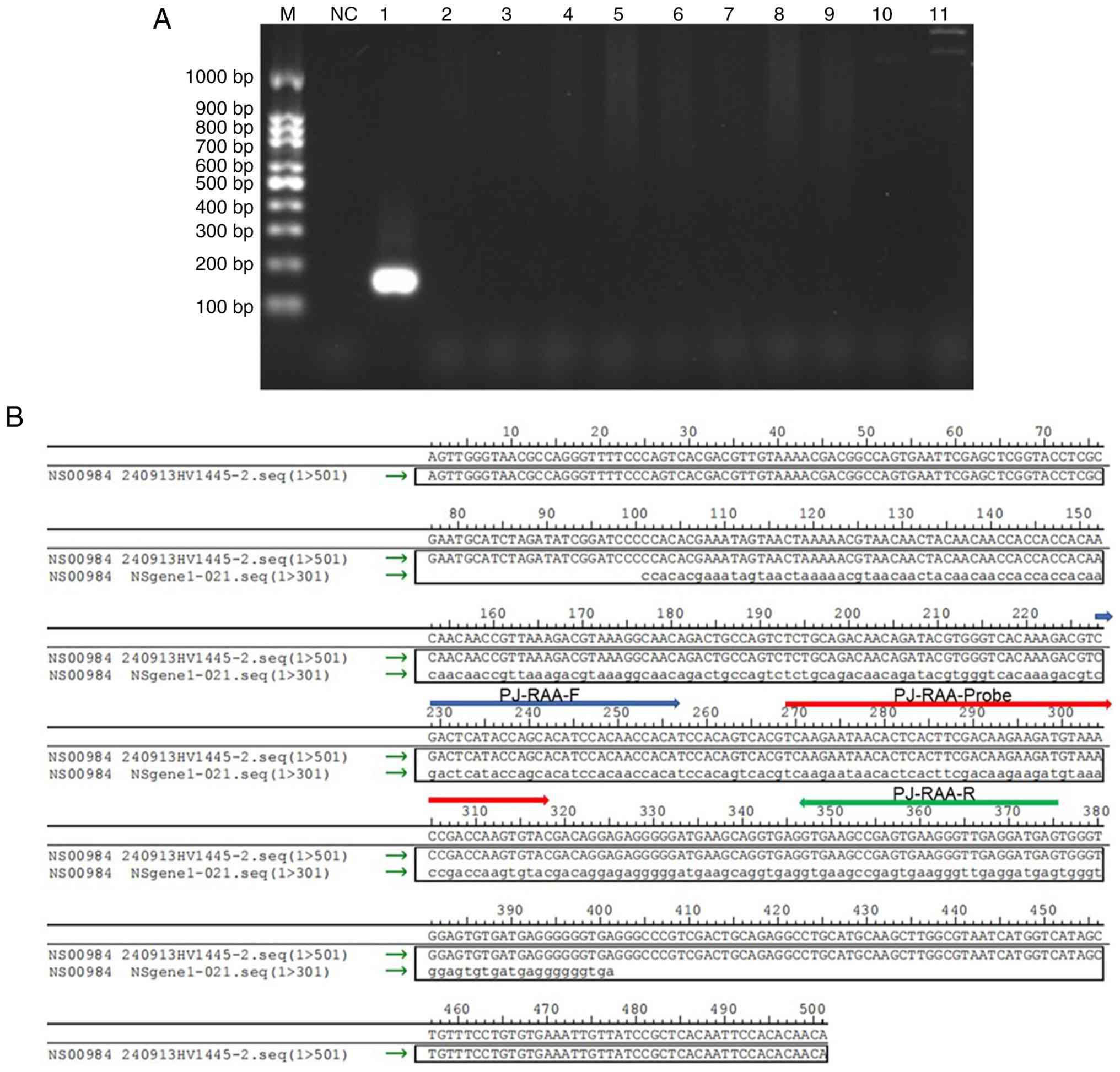
Conventional PCR using PJ-RAA-F/R primers amplified
a 149 bp fragment from PJ-positive BALF DNA (Fig. 2A), confirming the validity of the
target region within the PJ major surface glycoprotein gene. A
specific amplification band was only visible in the PJ-positive
sample, while no bands were detected in the NC and all non-PJ
pathogens. This result confirmed the high specificity of the
primers for PJ detection. NGS of pUC57-PJ showed 100% homology with
the reference PJ major surface glycoprotein sequence gene (GenBank
ID: KP168716.1), confirming successful plasmid construction
(Fig. 2B).
Optimization of RAA reaction
conditions
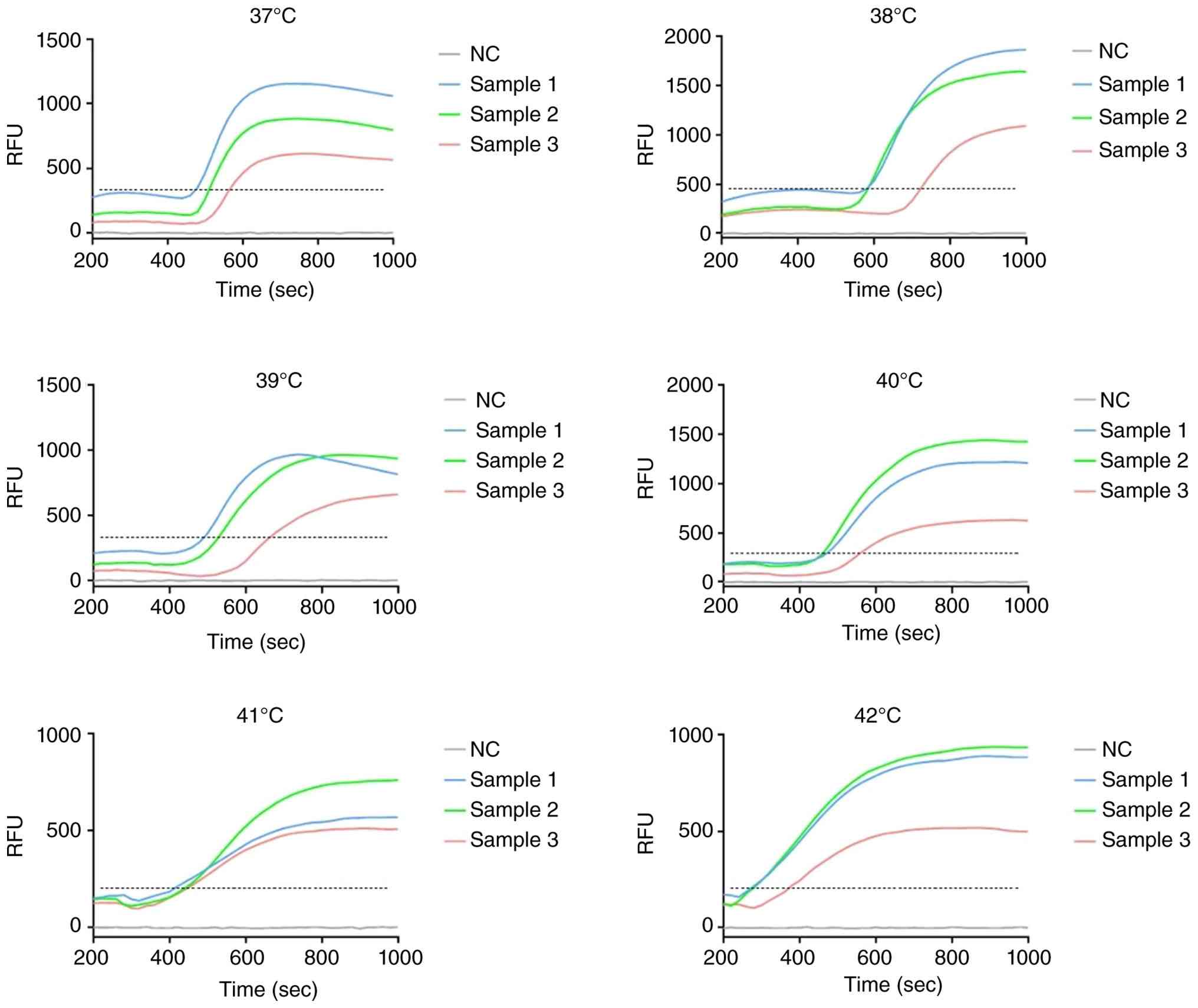
Fluorescence amplification was successfully detected
across all tested temperatures (37-42˚C) in the detection of 3
PJ-positive BALF samples. At temperatures <40˚C (37, 38 and
39˚C), amplification initiation was relatively delayed, and the
arrival time to the plateau phase was prolonged. By contrast, the
system exhibited the earliest amplification initiation (mean: 20
cycles) and the fastest arrival at the plateau phase at 40˚C
(Fig. 3), demonstrating the most
robust and efficient amplification kinetics. This superior
performance could be attributed to the highest and most stable
recombinase activity at 40˚C in the fluorescent RAA system, which
ensured consistent and rapid nucleic acid amplification. When the
temperature exceeded 40˚C (41 and 42˚C), the amplification curves
showed suboptimal morphological characteristics, with slower
overall kinetics compared to those at 40˚C. Collectively, these
results indicated that 40˚C was the optimal amplification
temperature, as it balanced both the speed of amplification
initiation and the efficiency of plateau phase arrival.
Analytical performance of RAA assay.
Sensitivity
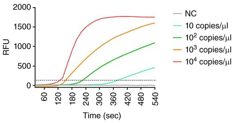
To evaluate the sensitivity of the fluorescent RAA
assay for PJ detection, serial dilutions of the PJ-positive plasmid
(104, 10³, 10² and 10 copies/µl) were used as templates
for amplification under the optimized reaction conditions, and the
corresponding curves are shown in Fig.
4. The amplification curves exhibited a concentration-dependent
trend: The template with the highest concentration (104
copies/µl) initiated amplification earliest (within 5-10 cycles)
and produced the strongest final relative fluorescence unit (RFU)
signal. As the plasmid concentration decreased (10³, 10² and 10
copies/µl), the initiation cycle of amplification was gradually
delayed, and the final RFU signal was sequentially reduced.
Notably, even the lowest concentration template (10 copies/µl)
still generated a detectable amplification signal (crossing the
baseline within 25 cycles), indicating that the fluorescent RAA
assay has good sensitivity and can effectively amplify PJ target
sequences at low copy numbers.
Clinical validation. To evaluate the
diagnostic performance of fluorescent RAA, 44 BALF samples from
patients suspected of PCP were tested with tNGS as the reference
standard. Among 44 PJ-positive samples confirmed by tNGS, 36 were
detected by fluorescent RAA, and no false-positive results were
observed in 17 PJ-negative samples (100% specificity for all
assays). Diagnostic efficacy metrics (Table II) showed that fluorescent RAA
achieved 81.82% sensitivity, 86.89% overall agreement and a κ value
of 0.715 (P<0.001). The fluorescent RAA assay exhibited good
rule-out diagnostic value, coupled with excellent clinical
diagnostic performance. Among the 61 patients, 48 (78.7%) were male
and 13 (21.3%) were female, with a mean age of 70.2±9.8 years. In
the PJ-positive group, 36 (81.8%) were male and 8 (18.2%) were
female, with a mean age of 70.4±10.8 years; in the PJ-negative
group, 12 (70.6%) were male and 5 (29.4%) were female, with a mean
age of 69.5±6.9 years. The characteristics of the cohort are
summarized in Table III. There
were no significant differences between the two groups in terms of
gender or age (P>0.05).
| Table IIDiagnostic performance of fluorescent
RAA in clinical bronchoalveolar lavage fluid samples. |
Table II
Diagnostic performance of fluorescent
RAA in clinical bronchoalveolar lavage fluid samples.
| Parameter | N fluorescence on
RAA | P fluorescence on
RAA | Sensitivity, % | Specificity, % | PPV, % | NPV, % | Overall agreement,
% | κ value | P-value |
|---|
| tNGS P/tNGS N | 36/8 | 0/17 | 81.82 | 100 | 100 | 68 | 86.89 | 0.715 | 0.001 |
| Table IIICharacteristics of patients of the
two groups at baseline. |
Table III
Characteristics of patients of the
two groups at baseline.
| Characteristic | All patients
(n=61) | PJ-positive
(n=44) | PJ-negative
(n=17) | P-value |
|---|
| Sex, n (%) | | | | 0.486 |
|
Male | 48 (78.7) | 36 (81.8) | 12 (70.6) | |
|
Female | 13 (21.3) | 8 (18.2) | 5 (29.4) | |
| Age, mean ± SD | 70.2±9.8 | 70.4±10.8 | 69.5±6.9 | 0.444 |
Discussion
In this study, a novel RAA assay for the rapid and
sensitive detection of PJ was successfully developed and optimized.
The establishment of a robust molecular diagnostic tool for PCP is
of paramount clinical importance, particularly for
immunocompromised patients, where timely diagnosis can
significantly impact outcomes.
The RAA assay demonstrated several key advantages
over microscopy and PCR. It exhibited superior analytical
sensitivity, detecting down to 10 copies/µl of the target plasmid.
The RAA sensitivity was comparable to, or even exceeded, the
sensitivity of numerous previously reported molecular assays for PJ
(56-60).
The ability to detect low pathogen loads is critical for PCP
diagnosis, as patients can present with subclinical or early-stage
infections.
The isothermal nature of the RAA technology is a
major strength. The optimized reaction at 40˚C eliminates the need
for sophisticated and expensive thermal cyclers, which are required
for qPCR. This feature, combined with the rapid turnaround time
(results within 20-30 min depending on the template concentration),
makes the RAA assay a promising candidate for POCT and use in
resource-limited settings (61-63).
The entire process, from DNA extraction to result interpretation,
can be streamlined and potentially performed in decentralized
laboratories.
In terms of cost-effectiveness, the RAA assay offers
significant economic advantages over tNGS. The estimated reagent
cost per RAA test is ~50-80 RMB (~7-12 USD), which is substantially
lower than the tNGS cost of 400-600 RMB (~59-88 USD) per sample on
the MGI DNBSEQ-G99 platform. Furthermore, the RAA assay does not
require expensive thermal cyclers or high-throughput sequencing
platforms, reducing both capital investment and maintenance costs.
The total turnaround time from sample processing to result
reporting is ~40-50 min for RAA, compared with 24-48 h for tNGS,
which may translate into reduced hospitalization costs and earlier
initiation of targeted therapy. These features make RAA
particularly suitable for resource-limited clinical settings where
budget constraints limit access to advanced molecular
diagnostics.
The clinical performance of the RAA assay was highly
encouraging. When tested on 44 tNGS-positive BALF samples, the RAA
assay detected PJ in 36 samples, demonstrating a high sensitivity
that supports its utility as a rapid screening tool. Notably, the
assay achieved perfect specificity, with no false-positive results,
which is clinically valuable for avoiding unnecessary antifungal
therapy and reducing patient risk. The substantial agreement
between the RAA assay and the tNGS reference standard further
validates its reliability, while the observed false-negative cases
highlight the need for complementary testing in high-risk settings.
Overall, these findings position the RAA assay as a promising
diagnostic option for PJ, particularly in resource-constrained
environments where timely detection is critical.
The systematic optimization process was crucial for
maximizing the assay's performance. Of note, 40˚C was identified as
the optimal temperature, which provided a balance between
amplification efficiency and enzyme stability.
Despite these promising results, the present study
has certain limitations. The sample size was relatively small.
Future studies should include a larger, more diverse cohort,
including clinically suspected samples, to comprehensively evaluate
the clinical sensitivity and specificity. Additionally, formal
specificity testing against a broader panel of respiratory
pathogens is warranted to conclusively demonstrate the assay's
exclusivity for PJ.
In addition to these study limitations, several
practical challenges should be considered when implementing the RAA
assay in clinical practice. The high analytical sensitivity of RAA,
while enabling detection of low pathogen loads, concomitantly
increases the risk of false-positive results due to
cross-contamination and nonspecific amplification arising from
short target amplicons or primer dimers (35). Furthermore, on-site nucleic acid
extraction remains a practical bottleneck for point-of-care
deployment, and achieving multiplexed target detection under
single-tube closed-tube conditions represents an unresolved
technical challenge that may limit the assay's broader application
(35). In the present clinical
evaluation, the observed sensitivity of 81.82% indicates that a
subset of PJ-positive cases may be missed, necessitating
complementary testing with tNGS or qPCR in high-risk patients with
strong clinical suspicion. Additionally, the assay has been
validated only with BALF samples, and its performance with less
invasive specimens (such as expectorated sputum, induced sputum,
nasopharyngeal swabs and oropharyngeal swabs), along with the
long-term stability of lyophilized reagents under decentralized
storage conditions, requires further investigation. Addressing
these operational and technical hurdles is essential to maximize
the clinical utility of RAA in resource-limited settings.
In conclusion, in the present study, a rapid,
sensitive and specific RAA assay for the detection of PJ was
developed. Its isothermal nature, speed and compatibility with
portable fluorescence detectors position it as a valuable tool for
the rapid diagnosis of PCP, potentially facilitating earlier
treatment and improved patient management.
The current study established and optimized a RAA
assay for the detection of PJ. The assay demonstrates high
sensitivity, with a detection limit of 10 copies/µl, and can
deliver results within 20 min under an isothermal incubation at
40˚C. The clinical evaluation revealed excellent concordance with
tNGS results, underscoring the superior diagnostic performance of
the RAA assay for PJ detection. With its simplicity, rapidity and
potential for point-of-care application, the RAA assay represents a
significant advancement in the molecular diagnosis of PCP.
Acknowledgements
Not applicable.
Funding
Funding: This work was supported by Weifang Municipal Health
Commission Scientific Research Program (grant nos. WFWSJK-2024-025
and WFZYY2025-4-008).
Availability of data and materials
The data generated in the present study may be
requested from the corresponding author.
Authors' contributions
HZ and BY performed the conceptual design, designed
the primers and probes, and drafted the original manuscript. BY was
responsible for methodology and performed the RAA experiments. KC
collected the clinical BALF samples and clinical data, participated
in the nucleic acid extraction experiments and critically revised
the manuscript. FX performed the analytical performance evaluation
of the RAA assay, analyzed the clinical validation data and
contributed to writing the original manuscript. MQ quantified and
analyzed the data, prepared the figures and tables, and critically
revised the manuscript. YT conceived and designed the study,
supervised the project, was responsible for project administration
and funding acquisition, and reviewed and edited the manuscript. MQ
and YT checked and confirmed the authenticity of all the raw data.
All authors have read and approved the final manuscript.
Ethics approval and consent to
participate
The present study was approved by the Ethics
Committee of the Weifang No. 2 People's Hospital (approval no.
KY2024-023-01; Weifang, China) and was performed in accordance with
the Declaration of Helsinki.
Patient consent for publication
Not applicable.
Competing interests
The authors declare that they have no competing
interests.
Use of artificial intelligence tools
During the preparation of this work, artificial
intelligence tools were used to improve the readability and
language of the manuscript, and subsequently, the authors revised
and edited the content produced by the artificial intelligence
tools as necessary, taking full responsibility for the ultimate
content of the present manuscript.
References
|
1
|
Arsić Arsenijevic V, Vyzantiadis TA, Mares
M, Otasevic S, Tragiannidis A and Janic D: Diagnosis of
pneumocystis jirovecii pneumonia in pediatric patients in serbia,
greece, and romania. Current status and challenges for
collaboration. J Fungi (Basel). 6(49)2020.PubMed/NCBI View Article : Google Scholar
|
|
2
|
Lemiale V, Resche-Rigon M, Zerbib Y,
Mokart D, De Prost N, Wallet F, Perez P, Kouatchet A, Argaud L,
Decavèle M, et al: Adjunctive corticosteroids in non-AIDS patients
with severe Pneumocystis jirovecii pneumonia (PIC): A multicentre,
double-blind, randomised controlled trial. Lancet Respir Med.
13:800–808. 2025.PubMed/NCBI View Article : Google Scholar
|
|
3
|
Lécuyer R, Issa N, Camou F, Lavergne RA,
Gabriel F, Morio F, Canet E, Raffi F, Boutoille D, Cady A, et al:
Characteristics and prognosis factors of pneumocystis jirovecii
pneumonia according to underlying disease: A retrospective
multicenter study. Chest. 165:1319–1329. 2024.PubMed/NCBI View Article : Google Scholar
|
|
4
|
Chen YH, Fang XY, Li YT, Liu YL, Hang YP,
Xiao YP, Cao XW, Zhong QS and Hu LH: Characterization of
Pneumocystis jirovecii pneumonia at three tertiary comprehensive
hospitals in southern China. Braz J Microbiol. 51:1061–1069.
2020.PubMed/NCBI View Article : Google Scholar
|
|
5
|
Alshahrani MY, Alfaifi M, Ahmad I,
Alkhathami AG, Hakami AR, Ahmad H, Alshehri OM and Dhakad MS:
Pneumocystis Jirovecii detection and comparison of multiple
diagnostic methods with quantitative real-time PCR in patients with
respiratory symptoms. Saudi J Biol Sci. 27:1423–1427.
2020.PubMed/NCBI View Article : Google Scholar
|
|
6
|
Bateman M, Oladele R and Kolls JK:
Diagnosing Pneumocystis jirovecii pneumonia: A review of current
methods and novel approaches. Med Mycol. 58:1015–1028.
2020.PubMed/NCBI View Article : Google Scholar
|
|
7
|
Yin Y, Zhu P, Guo Y, Li Y, Chen H, Liu J,
Sun L, Ma S, Hu C and Wang H: Enhancing lower respiratory tract
infection diagnosis: implementation and clinical assessment of
multiplex PCR-based and hybrid capture-based targeted
next-generation sequencing. EBioMedicine.
107(105307)2024.PubMed/NCBI View Article : Google Scholar
|
|
8
|
Almeida-Siva F, Almeida-Paes R,
Serra-Damasceno L, Motta-Santos E, Ferreira LC, Pereira-Quintella
L, De Faria Ferreira M, De Medeiros-Muniz M and Zancopé-Oliveira
RM: The conventional diagnosis challenge: Real-time PCR and nested
PCR correlation with the scoring system for individuals at
high-risk of Pneumocystis jirovecii pneumonia. Biomedica. 43(Sp
1):255–266. 2023.PubMed/NCBI View Article : Google Scholar : (In English,
Spanish).
|
|
9
|
Lagrou K, Chen S, Masur H, Viscoli C,
Decker CF, Pagano L and Groll AH: Pneumocystis jirovecii disease:
Basis for the revised EORTC/MSGERC invasive fungal disease
definitions in individuals without human immunodeficiency virus.
Clin Infect Dis. 72 (Suppl 2):S114–S120. 2021.PubMed/NCBI View Article : Google Scholar
|
|
10
|
White PL, Backx M and Barnes RA: Diagnosis
and management of Pneumocystis jirovecii infection. Expert Rev Anti
Infect Ther. 15:435–447. 2017.PubMed/NCBI View Article : Google Scholar
|
|
11
|
Wills NK, Lawrence DS, Botsile E, Tenforde
MW and Jarvis JN: The prevalence of laboratory-confirmed
Pneumocystis jirovecii in HIV-infected adults in Africa: A
systematic review and meta-analysis. Med Mycol. 59:802–812.
2021.PubMed/NCBI View Article : Google Scholar
|
|
12
|
Ruiz-Ruiz S, Ponce CA, Pesantes N,
Bustamante R, Gatti G, San Martin V, Gutierrez M, Bórquez P, Vargas
SL, Magne F, et al: A real-time PCR assay for detection of low
pneumocystis jirovecii levels. Front Microbiol.
12(787554)2022.PubMed/NCBI View Article : Google Scholar
|
|
13
|
Liu W, Li M, Xu Y, Wang F, Wang J, Wang H,
Xu X, Wang Y and Sun H: Evaluation of the performance of a
multiplex real-time PCR assay for the identification of
aspergillus, cryptococcus neoformans, and pneumocystis jirovecii
simultaneously from sputum in multicenter. Infect Drug Resist.
15:6009–6017. 2022.PubMed/NCBI View Article : Google Scholar
|
|
14
|
Belanger CR, Locher K, Velapatino B,
Dufresne PJ, Eckbo E and Charles M: Quick versus quantitative:
Evaluation of two commercial real-time PCR assays for the detection
of pneumocystis jirovecii from bronchoalveolar lavage fluids.
Microbiol Spectr. 11(e0102123)2023.PubMed/NCBI View Article : Google Scholar
|
|
15
|
Alsayed AR, Talib W, Al-Dulaimi A, Daoud S
and Al Maqbali M: The first detection of Pneumocystis jirovecii in
asthmatic patients post-COVID-19 in Jordan. Bosn J Basic Med Sci.
22:784–790. 2022.PubMed/NCBI View Article : Google Scholar
|
|
16
|
Shrestha S, Addae A, Miller C, Ismail N
and Zwerling A: Cost-effectiveness of targeted next-generation
sequencing (tNGS) for detection of tuberculosis drug resistance in
India, South Africa and Georgia: A modeling analysis.
EClinicalMedicine. 79(103003)2024.PubMed/NCBI View Article : Google Scholar
|
|
17
|
Liu Y, Wu W, Xiao Y, Zou H, Hao S and
Jiang Y: Application of metagenomic next-generation sequencing and
targeted metagenomic next-generation sequencing in diagnosing
pulmonary infections in immunocompetent and immunocompromised
patients. Front Cell Infect Microbiol. 14(1439472)2024.PubMed/NCBI View Article : Google Scholar
|
|
18
|
Chen Z, Liu X, Tan L, Lyu X, Long Q, Wu W,
Guo Z, Liu Z, Li Z and Hu M: Comparative of metagenomic and
targeted next-generation sequencing in lower respiratory tract
fungal infections. Front Cell Infect Microbiol.
15(1534519)2025.PubMed/NCBI View Article : Google Scholar
|
|
19
|
Wang H, Chen X, Wu X, Cao Q, Wu Y, Wang F,
Wang Y, Zhou Y, Tang Y, Ren T and Wang M: Combined multiplex
polymerase chain reaction-based targeted next-generation sequencing
and serum 1, 3-β-D-glucan for differential diagnosis of
Pneumocystis pneumonia and Pneumocystis colonization. Front Cell
Infect Microbiol. 15(1611391)2025.PubMed/NCBI View Article : Google Scholar
|
|
20
|
Chen Q, Yi J, Liu Y, Yang C, Sun Y, Du J,
Liu Y, Gu D, Liu H, Xu Y and Chen Y: Clinical diagnostic value of
targeted next-generation sequencing for infectious diseases
(Review). Mol Med Rep. 30(153)2024.PubMed/NCBI View Article : Google Scholar
|
|
21
|
Zhu H, Zhang H, Xu Y, Laššáková S,
Korabečná M and Neužil P: PCR past, present and future.
Biotechniques. 69:317–325. 2020.PubMed/NCBI View Article : Google Scholar
|
|
22
|
Soroka M, Wasowicz B and Rymaszewska A:
Loop-mediated isothermal amplification (LAMP): The better sibling
of PCR? Cells. 10(1931)2021.PubMed/NCBI View Article : Google Scholar
|
|
23
|
Ngoc LTN and Lee YC: Current trends in RNA
virus detection via nucleic acid isothermal amplification-based
platforms. Biosensors (Basel). 14(97)2024.PubMed/NCBI View Article : Google Scholar
|
|
24
|
Lai MY, Abdul Hamid MH, Jelip J, Mudin RN
and Lau YL: Recombinase-aided loop-mediated isothermal
amplification on human plasmodium knowlesi. Am J Trop Med Hyg.
110:648–652. 2024.PubMed/NCBI View Article : Google Scholar
|
|
25
|
Park JW: Principles and applications of
loop-mediated isothermal amplification to point-of-care tests.
Biosensors (Basel). 12(857)2022.PubMed/NCBI View Article : Google Scholar
|
|
26
|
Ahmadi Y, Yu Y, Cui Z, Huang WE and
Andersson MI: Loop-mediated isothermal amplification (LAMP) for the
diagnosis of sexually transmitted infections: A review. Microb
Biotechnol. 18(e70153)2025.PubMed/NCBI View Article : Google Scholar
|
|
27
|
Owoicho O, Olwal CO, Tettevi EJ, Atu BO
and Durugbo EU: Loop-mediated isothermal amplification for Candida
species surveillance in under-resourced setting: A review of
evidence. Expert Rev Mol Diagn. 22:643–653. 2022.PubMed/NCBI View Article : Google Scholar
|
|
28
|
Gieroń M, Żarnowiec P, Zegadło K, Gmiter
D, Czerwonka G, Kaca W and Kręcisz B: Loop-mediated isothermal
amplification of DNA (LAMP) as an alternative method for
determining bacteria in wound infections. Int J Mol Sci.
25(411)2023.PubMed/NCBI View Article : Google Scholar
|
|
29
|
Acharya B, Acharya A, Gautam S, Ghimire
SP, Mishra G, Parajuli N and Sapkota B: Advances in diagnosis of
Tuberculosis: An update into molecular diagnosis of Mycobacterium
tuberculosis. Mol Biol Rep. 47:4065–4075. 2020.PubMed/NCBI View Article : Google Scholar
|
|
30
|
Srividya A, Maiti B, Chakraborty A and
Chakraborty G: Loop mediated isothermal amplification: A promising
tool for screening genetic mutations. Mol Diagn Ther. 23:723–733.
2019.PubMed/NCBI View Article : Google Scholar
|
|
31
|
Munawar MA: Critical insight into
recombinase polymerase amplification technology. Expert Rev Mol
Diagn. 22:725–737. 2022.PubMed/NCBI View Article : Google Scholar
|
|
32
|
Tan M, Liao C, Liang L, Yi X, Zhou Z and
Wei G: Recent advances in recombinase polymerase amplification:
Principle, advantages, disadvantages and applications. Front Cell
Infect Microbiol. 12(1019071)2022.PubMed/NCBI View Article : Google Scholar
|
|
33
|
Feng X, Liu Y, Zhao Y, Sun Z, Xu N, Zhao C
and Xia W: Recombinase polymerase amplification-based biosensors
for rapid zoonoses screening. Int J Nanomedicine. 18:6311–6331.
2023.PubMed/NCBI View Article : Google Scholar
|
|
34
|
Lu X, Cao Y, Zhang P, Chen X, Irwin DM and
Shen Y: Visual recombinase aided amplification technology for
detecting feline coronavirus. Vet J. 312(106356)2025.PubMed/NCBI View Article : Google Scholar
|
|
35
|
Li X, Zhu S, Zhang X, Ren Y, He J, Zhou J,
Yin L, Wang G, Zhong T, Wang L, et al: Advances in the application
of recombinase-aided amplification combined with CRISPR-Cas
technology in quick detection of pathogenic microbes. Front Bioeng
Biotechnol. 11(1215466)2023.PubMed/NCBI View Article : Google Scholar
|
|
36
|
Trinh TND and Nam NN: Isothermal
amplification-based microfluidic devices for detecting foodborne
pathogens: A review. Anal Methods. 16:1150–1157. 2024.PubMed/NCBI View Article : Google Scholar
|
|
37
|
Yan C, Zhou Y, Du S, Du B, Zhao H, Feng Y,
Xue G, Cui J, Gan L, Feng J, et al: Recombinase-aided amplification
assay for rapid detection of hypervirulent klebsiella pneumoniae
(hvKp) and Characterization of the hvKp pathotype. Microbiol
Spectr. 11(e0398422)2023.PubMed/NCBI View Article : Google Scholar
|
|
38
|
Duan X, Ma W, Jiao Z, Tian Y, Ismail RG,
Zhou T and Fan Z: Reverse transcription-recombinase-aided
amplification and CRISPR/Cas12a-based visual detection of maize
chlorotic mottle virus. Phytopathol Res. 4(23)2022.PubMed/NCBI View Article : Google Scholar
|
|
39
|
Pang F and Long Q: Recent advances in
diagnostic approaches for orf virus. Appl Microbiol Biotechnol.
107:1515–1523. 2023.PubMed/NCBI View Article : Google Scholar
|
|
40
|
Zhang X, Guo L, Ma R, Cong L, Wu Z, Wei Y,
Xue S, Zheng W and Tang S: Rapid detection of salmonella with
recombinase aided amplification. J Microbiol Methods. 139:202–204.
2017.PubMed/NCBI View Article : Google Scholar
|
|
41
|
Xiao X, Yang X, Xu K, Huang F, Zhang Y,
Jiang Y, Shi Y, Zhou Q, Wang L, Lu J, et al: DNA fragment fusion
and nucleic acid detection by fusion recombinase-aided
amplification. Anal Chem. 97:6538–6547. 2025.PubMed/NCBI View Article : Google Scholar
|
|
42
|
He Y, Chen W, Fan J, Fan S, Ding H, Chen J
and Yi L: Recombinase-aided amplification coupled with lateral flow
dipstick for efficient and accurate detection of porcine
parvovirus. Life (Basel). 11(762)2021.PubMed/NCBI View Article : Google Scholar
|
|
43
|
Wang S, Zhuang Q, Jiang N, Zhang F, Chen
Q, Zhao R, Li Y, Yu X, Li J, Hou G, et al: Reverse transcription
recombinase-aided amplification assay for avian influenza virus.
Virus Genes. 59:410–416. 2023.PubMed/NCBI View Article : Google Scholar
|
|
44
|
Ceruti A, Faye M, Diagne MM, Kobialka RM,
Makiala-Mandanda S, Faye O, Faye O, El Wahed AA and Weidmann M:
Rapid detection of Ebolavirus using isothermal recombinase-aided
amplification. J Med Virol. 96(e29744)2024.PubMed/NCBI View Article : Google Scholar
|
|
45
|
Wang K, Huang R, Zhang L, Liu D and Diao
Y: Recombinase-aided amplification combined with lateral flow
(LF-RAA) assay for Rapid AAV genome detection. ACS Omega.
7:47832–47839. 2022.PubMed/NCBI View Article : Google Scholar
|
|
46
|
Lin H, Zhao S, Ye Y, Shao L, Jiang N and
Yang K: A fluorescent recombinase aided amplification assay for
detection of babesia microti. Korean J Parasitol. 60:201–205.
2022.PubMed/NCBI View Article : Google Scholar
|
|
47
|
Liu N, Ning X, Zhang X, Zhou Z, Fu M, Wang
Y and Wu T: Gradient galectin-1 coating technology: Bionic
multichannel nerve guidance conduits promote neural cell migration.
Adv Technol. Neurosci. 1:276–289. 2024.
|
|
48
|
Wang S, Li Y, Zhang F, Jiang N, Zhuang Q,
Hou G, Jiang L, Yu J, Yu X, Liu H, et al: Reverse transcription
recombinase-aided amplification assay for H5 subtype avian
influenza virus. Virol J. 19(129)2022.PubMed/NCBI View Article : Google Scholar
|
|
49
|
Liu J, Qian W, Wang J, Bai Y, Gui Y, Xia
L, Gong G, Ge F, Shen H, Chang X and Zhao H: A recombinase-aided
amplification assay for the detection of chlamydia felis. Pol J
Microbiol. 72:339–343. 2023.PubMed/NCBI View Article : Google Scholar
|
|
50
|
Wang ZH, Zhang W, Zhang XZ, Yao XR, Huang
W, Jia H, Liu XL, Hou SH and Wang XJ: Development of a real-time
recombinase-aided amplification (RT-RAA) molecular diagnosis assay
for sensitive and rapid detection of Toxoplasma gondii. Vet
Parasitol. 298(109489)2021.PubMed/NCBI View Article : Google Scholar
|
|
51
|
Li Q, Wang N, Pang M, Miao H, Dai X, Li B,
Yang X, Li C and Liu Y: Rapid and highly sensitive detection of
mycobacterium tuberculosis utilizing the recombinase aided
amplification-based CRISPR-Cas13a system. Microorganisms.
12(1507)2024.PubMed/NCBI View Article : Google Scholar
|
|
52
|
Samuel L: Point-of-care testing in
microbiology. Clin Lab Med. 40:483–494. 2020.PubMed/NCBI View Article : Google Scholar
|
|
53
|
Wang Y, Du P, Shao Y, Wang W, Liu Y, Ma Y,
Hu P, Cao J, Wang X and Abd El-Aty AM: An innovative and efficient
fluorescent detection technique for salmonella in animal-derived
foods Using the CRISPR/Cas12a-HCR system combined with PCR/RAA. J
Agric Food Chem. 72:8831–8839. 2024.PubMed/NCBI View Article : Google Scholar
|
|
54
|
Zhang X, Ge X, Shen F, Qiao J, Zhang Y and
Li H: Diagnostic efficiency of RPA/RAA integrated CRISPR-Cas
technique for COVID-19: A systematic review and meta-analysis. PLoS
One. 17(e0276728)2022.PubMed/NCBI View Article : Google Scholar
|
|
55
|
Xiao X, Zhang Q, Wu S, Li Y, Zhong Z, Guo
Q, Li J, Meng Q, Cheng Z, Duan J, et al: Detection of clostridium
perfringens using novel methods based on recombinase-aided
amplification assay-assisted CRISPR/Cas12a System. Transbound Emerg
Dis. 2023(6667618)2023.PubMed/NCBI View Article : Google Scholar
|
|
56
|
Brown L, Alanio A, Cruciani M, Barnes R,
Donnelly JP, Loeffler J, Rautemaa-Richardson R and White PL:
Strengths and limitations of molecular diagnostics for Pneumocystis
jirovecii pneumonia. Expert Rev Mol Diagn. 24:899–911.
2024.PubMed/NCBI View Article : Google Scholar
|
|
57
|
Boldiš V, Ondriska F, Kováč Ľ, Steinhübel
J and Bastlová M: High incidence of Pneumocystis jirovecii
pneumonia in oncological patients: A 19-year study. Epidemiol
Mikrobiol Imunol. 72:93–98. 2023.PubMed/NCBI
|
|
58
|
Wang Y, Zhang X, Zhang Z, Xu R, Yang P,
Yuan P, Zhang L, Cui Y, Xie Z and Li J: Reverse transcription
recombinase-aided amplification assay combined with a lateral flow
dipstick for detection of duck Tembusu virus. J Virol Methods.
322(114810)2023.PubMed/NCBI View Article : Google Scholar
|
|
59
|
Wu X, Liu Y, Gao L, Yan Z, Zhao Q, Chen F,
Xie Q and Zhang X: Development and application of a
reverse-transcription recombinase-aided amplification assay for
porcine epidemic diarrhea virus. Viruses. 14(591)2022.PubMed/NCBI View Article : Google Scholar
|
|
60
|
Mu D, Zhou D, Xie G, Liu J, Xiong Q, Feng
X and Xu H: The fluorescent probe-based recombinase-aided
amplification for rapid detection of Escherichia coli O157:H7. Mol
Cell Probes. 60(101777)2021.PubMed/NCBI View Article : Google Scholar
|
|
61
|
Plebani M, Nichols JH, Luppa PB, Greene D,
Sciacovelli L, Shaw J, Khan AI, Carraro P, Freckmann G, Dimech W,
et al: Point-of-care testing: State-of-the art and perspectives.
Clin Chem Lab Med. 63:35–51. 2024.PubMed/NCBI View Article : Google Scholar
|
|
62
|
Hansen GT: Point-of-care testing in
microbiology: A mechanism for improving patient outcomes. Clin
Chem. 66:124–137. 2020.PubMed/NCBI View Article : Google Scholar
|
|
63
|
Gentilotti E, De Nardo P, Cremonini E,
Górska A, Mazzaferri F, Canziani LM, Hellou MM, Olchowski Y, Poran
I, Leeflang M, et al: Diagnostic accuracy of point-of-care tests in
acute community-acquired lower respiratory tract infections. A
systematic review and meta-analysis. Clin Microbiol Infect.
28:13–22. 2022.PubMed/NCBI View Article : Google Scholar
|