Introduction
Osteosarcoma (OS), which predominantly affects young
individuals during periods of rapid bone growth, is the most common
primary malignant bone tumor and shows a marked increase in
incidence during adolescence (1).
Due to its aggressive local invasion and high metastatic potential,
OS is associated with a poor prognosis, with a 5-year survival rate
of <20% in patients with metastatic disease worldwide (2,3).
The current therapeutic regimen focuses on surgical intervention
complemented by adjuvant chemotherapy (4). Nevertheless, clinical outcomes
remain unsatisfactory for recurrent and refractory OS, which is
attributed to rapid disease progression and inherent
chemoresistance (5). Amid the
evolving landscape of oncological therapeutics, molecularly
targeted approaches have emerged as a promising frontier in the
management of OS.
KIAA1429, a component of the RNA methyltransferase
complex, regulates N6-methyladenosine (m6A) modification of mRNA,
influencing RNA stability, splicing and translation (6). In cancer, KIAA1429 is upregulated
and acts as an oncogene by promoting cell proliferation, migration
and invasion (6,7). Through an m6A-YTH domain family
protein 2-mediated mechanism, KIAA1429 exerts its oncogenic role in
gastric cancer progression by inducing the destabilization of
dexamethasone-induced Ras-related protein 1 mRNA (8). As a constituent of the m6A
methyltransferase complex, KIAA1429 is elevated in breast cancer
specimens and is positively associated with reduced patient
survival (9). Elevated KIAA1429
expression is also associated with unfavorable clinical outcomes
and increased disease aggressiveness in OS (10). These findings suggest that
KIAA1429 is a promising therapeutic target for OS.
Ferroptosis is a crucial pathway in cancer biology,
characterized by iron-dependent lipid peroxidation that drives this
distinct form of regulated cell death (11). Its molecular signature includes
the suppression of glutathione (GSH) peroxidase 4 (GPX4) and the
consequent accumulation of reactive oxygen species (ROS) (12). Notably, induction of ferroptosis
has emerged as a promising therapeutic strategy for OS (13); therefore, elucidating the
molecular mechanisms underlying ferroptosis in OS may help overcome
the limitations of conventional treatments. Solute carrier family 7
member 11 (SLC7A11) is a key component of the system Xc−
cystine/glutamate antiporter and serves a crucial role in
maintaining intracellular redox homeostasis by facilitating cystine
uptake and GSH biosynthesis (14). Inhibition of SLC7A11 depletes GSH,
thereby impairing cellular antioxidant capacity and promoting ROS
accumulation, ultimately sensitizing cells to ferroptosis (15). A previous study demonstrated that
compounds such as erastin and brusatol induce ferroptosis by
suppressing the kelch-like ECH-associated protein 1/nuclear factor
erythroid 2-related factor 2 (Nrf2)/SLC7A11 pathway, thereby
inhibiting cystine uptake and enhancing ROS accumulation (16). Mechanistically, KIAA1429 silencing
suppresses OS progression by promoting ferroptosis via the
Nrf2/NAD(P) H quinone oxidoreductase 1 axis (17). Specifically, KIAA1429 directly
targets the 3'UTR of Nrf2 mRNA through m6A modification, thereby
inhibiting Nrf2 signaling and enhancing erastin-induced ferroptosis
(17). While this previous study
provides important insights (17), the present study offers several
distinct advantages and novel findings. The current study aimed to
investigate the role of KIAA1429 in OS progression and to determine
whether it modulates ferroptosis via m6A-dependent regulation of
SLC7A11 expression. Specifically, the study sought to elucidate the
mechanistic relationship among KIAA1429, m6A methylation, SLC7A11
stability and ferroptosis in OS cells, and to validate this axis in
an in vivo xenograft model.
Materials and methods
Cell culture and transfection
Human osteoblasts (hFOB1.19) and human OS cell lines
(MG63, 143B, U2OS and Saos-2) were obtained from iCell Bioscience
Inc., and were routinely cultured in Dulbecco's modified Eagle's
medium (DMEM supplemented with 10% fetal bovine serum (FBS) (both
from Gibco, Thermo Fisher Scientific) and 100 U/ml
penicillin/streptomycin. The cells were maintained at 37°C in a
humidified atmosphere containing 5% CO2.
For experiments involving 143B and U2OS cells, the
groups were designated as follows: Control (untreated) and small
interfering (si)RNA-negative control (NC; si-NC); three
experimental groups targeting KIAA1429 (si-KIAA1429-1,
si-KIAA1429-2 and si-KIAA1429-3); and a combined intervention group
[si-KIAA1429 + SLC7A11 overexpression vector (oe-SLC7A11)]. All
siRNAs were purchased from Shanghai GenePharma Co., Ltd. oe-SLC7A11
was constructed by cloning full-length SLC7A11 cDNA into the
pcDNA3.1(+) vector (Invitrogen; Thermo Fisher Scientific, Inc.),
and the empty pcDNA3.1(+) vector served as the oe-NC. The si-NC
group served as the NC for siRNA transfection. Transfection was
performed using Lipofectamine® 3000 (Invitrogen; Thermo
Fisher Scientific, Inc.) according to the manufacturer's protocol.
Briefly, the cells were seeded in 6-well plates and cultured until
they reached 70-80% confluence at the time of transfection. For
siRNA transfection, the final concentration of si-KIAA1429 or si-NC
was 50 nM; for plasmid (oe-SLC7A11 or oe-NC) transfection, and
plasmid and siRNA co-transfection, the final concentration of siRNA
was 50 nM and the final concentration of plasmid was 100 nM. The
transfection mixture was incubated with cells for 6 h at 37°C,
after which the medium was replaced with fresh complete DMEM
containing 10% FBS. The cells were harvested for subsequent
experiments 48 h after transfection siRNA sequences are presented
in Table I. For ferroptosis
induction, 143B and U2OS cells were treated with 10 μM
erastin (Selleck Chemicals) for 24 h at 37°C, whereas for
transcriptional inhibition, actinomycin D (5 μg/ml; Selleck
Chemicals) was applied followed by RNA collection at the indicated
time points (0, 3 and 6 h after treatment).
 | Table IPrimer and siRNA sequences used in
the present study. |
Table I
Primer and siRNA sequences used in
the present study.
| Name | Sequence,
5'-3' |
|---|
| GAPDH-F |
GAGTCAACGGATTTGGTCGT |
| GAPDH-R |
TTGATTTTGGAGGGATCTCG |
| SLC7A11-F |
GGCAGTGACCTTTTCTGAGC |
| SLC7A11-R |
TCATTGTCAAAGGGTGCAAA |
| KIAA1429-F |
CTCGGCGATGGAGCTGTTAT |
| KIAA1429-R |
CTCGGCGATGGAGCTGTTAT |
| si-NC-F |
UUCUCCGAACGUGUCACGUTT |
| si-NC-R |
ACGUGACACGUUCGGAGAATT |
|
si-KIAA1429-1-S |
AGUAUCUAAAAAUAACAGCUC |
|
si-KIAA1429-1-AS |
GCUGUUAUUUUUAGAUACUUU |
|
si-KIAA1429-2-S |
AAGUAUCUAAAAAUAACAGCU |
|
si-KIAA1429-2-AS |
CUGUUAUUUUUAGAUACUUUU |
|
si-KIAA1429-3-S |
AGUCUAAUUGAAAUGUAUGGG |
|
si-KIAA1429-3-AS |
CAUACAUUUCAAUUAGACUUA |
Cell Counting Kit-8 (CCK-8) assay
The proliferation rates of 143B and U2OS cells were
determined using a CCK-8 (MilliporeSigma) assay. Cells were seeded
at 1.5×10⁴ cells/well in 96-well plates and maintained
under controlled conditions (37°C, 5% CO2). At the
indicated time points (24, 48, 72 and 96 h), the culture medium was
carefully aspirated, and 10 μl CCK-8 reagent was added to
each well. After incubation for 2 h at 37°C in the dark, optical
density (OD) values were measured at 450 nm using a microplate
reader.
Flow cytometric analysis
Apoptosis was detected using an Annexin V-FITC/PI
Apoptosis Detection Kit (BD Biosciences) according to the
manufacturer's instructions. Adherent cells were washed twice with
phosphate-buffered saline (PBS), dissociated with EDTA-free
trypsin, neutralized and centrifuged at 300 × g for 5 min at 4°C.
The cells (5×10⁴) were then resuspended in 195 μl
Annexin V-FITC binding buffer. Subsequently, the cells were stained
with 5 μl Annexin V-FITC and 10 μl propidium iodide,
incubated for 10-20 min at 20-25°C in the dark and then placed on
ice. Apoptosis was analyzed using a flow cytometer (CytoFLEX;
Beckman Coulter, Inc.). The data were processed using CytExpert
software version 2.0 (Beckman Coulter, Inc.).
Transwell invasion assay
Cell invasion was assessed using 24-well Transwell
inserts (Corning, Inc.) with an 8.0-μm pore size. Firstly,
Matrigel (Corning, Inc.) was thawed on ice, diluted with serum-free
DMEM (1:8 ratio) and 50 μl diluted Matrigel was added to the
upper chamber of each insert. Matrigel-coated Transwell inserts
(Corning, Inc.) were then incubated at 37°C for 30 min to allow
gelation, followed by rehydration with serum-free medium for 1 h at
37°C. Trypsinized cells were washed, resuspended in serum-free
medium containing 0.1% bovine serum albumin (BSA; MilliporeSigma)
and seeded (1×10⁴ cells/well) into the upper chamber.
Complete medium containing 20% FBS was added to the lower chamber
(600 μl/well) as a chemoattractant. After incubation for 24
h (37°C, 5% CO2), non-invasive cells on the upper
surface of the membrane were removed using a cotton swab. Invasive
cells on the lower surface were fixed with 4% paraformaldehyde for
30 min and stained with 0.1% crystal violet for 20 min at 37°C,
before images were captured under an inverted light microscope
(CKX53; Olympus Corporation) and invasion was semi-quantified using
ImageJ software (version 1.54; National Institutes of Health).
Wound healing assay
Cell migration was assessed using a standard wound
healing assay. Briefly, 143B and U2OS cells were cultured in 6-well
plates until they reached ~80% confluence. The cell monolayer was
then carefully scratched in a straight line using a sterile
200-μl pipette tip held perpendicular to pre-marked
horizontal lines on the plate; the wound width was kept consistent
across all samples. After scratching, the detached cells were
removed by washing with PBS, and serum-free medium was added to
minimize cell proliferation. Images of the wounds were captured at
0 and 24 h post-scratching using a light microscope. The migration
rate was semi-quantified by measuring changes in the wound area
using ImageJ software (version 1.54).
Reverse transcription-quantitative PCR
(RT-qPCR)
Total RNA was extracted from cells using
TRIzol® reagent (Invitrogen; Thermo Fisher Scientific,
Inc.). A 5-μl aliquot of the total RNA was diluted with
RNase-free ultrapure water, and RNA concentration and purity were
assessed by measuring absorbance at 260 and 280 nm using a UV
spectrophotometer. Samples with an OD260/OD280 ratio between 1.9
and 2.0 were considered to have high purity and were suitable for
subsequent experiments. RT was performed using the PrimeScript™ RT
Master Mix (Takara Bio, Inc.) according to the manufacturer's
protocol. qPCR was conducted using the TB Green® Premix
Ex Taq™ II (Tli RNaseH Plus) (Takara Bio, Inc.) on an ABI 7500
Real-Time PCR System (Applied Biosystems; Thermo Fisher Scientific,
Inc.) under the following conditions: Initial denaturation at 95°C
for 30 sec, followed by 40 cycles of denaturation at 95°C for 10
sec and annealing at 60°C for 30 sec. GAPDH was used as the
internal reference gene. Relative gene expression levels were
calculated using the 2−ΔΔCt method (18). Primer sequences are listed in
Table I.
ELISA
The levels of oxidative stress and iron metabolism
markers, including malondialdehyde (MDA) (cat no. YX-130401H for OS
cells, cat no. EK20347 for tumor tissues), Fe2+ (cat no.
YX-0605H for OS cells, cat no. EK20235 for tumor tissues), GSH (cat
no. YX-071908H for OS cells, cat no. EK20064 for tumor tissues) and
ROS (cat no. YX-202403H for OS cells, cat no. EK202408 for tumor
tissues), in cells and tumor tissues were measured using specific
commercial ELISA kits (Shanghai Yixuan Biotechnology Co., Ltd.) in
accordance with the manufacturer's instructions. The OD value of
each well was measured at 450 nm using a microplate reader.
C11-BODIPY staining for lipid
peroxidation
OS cells were washed with PBS and incubated with 2
μM C11-BODIPY 581/591 probe (Thermo Fisher Scientific, Inc.)
at 37°C for 30 min in the dark. After washing with PBS, the cells
were observed under a fluorescence microscope. Images of green
fluorescence (oxidized BODIPY) and red fluorescence (reduced
BODIPY) were captured, and the green/red fluorescence intensity
ratio was determined using ImageJ software (version 1.54).
m6A-modified RNA immunoprecipitation
(MeRIP)
The m6A modification levels of KIAA1429-targeted
mRNAs were detected by MeRIP-qPCR assay using a Magna MeRIP m6A kit
(cat no. 17-10499; Sigma-Aldrich; Merck KGaA) (19), according to the manufacturer's
instructions. Total RNA was extracted from 143B and U2OS cells as
aforementioned, with a yield greater than 300 μg; one-tenth
of the total RNA (~30 μg) was used as the input control. The
remaining RNA was subjected to immunoprecipitation. For
immunoprecipitation, the RNA was incubated overnight at 4°C with 2
μg anti-m6A antibody (cat. no. ABE572) or 2 μg normal
rabbit IgG (cat. no. 12-370) (both from MilliporeSigma) as a
negative control in a total volume of 500 μl
immunoprecipitation buffer overnight at 4°C with gentle rotation.
Thereafter, 20 μl Protein A/G magnetic beads
(MilliporeSigma) were added to each sample and the mixture was
incubated for an additional 2 h at 4°C with rotation. The beads
were then collected using a magnetic stand and washed three times
with 500 μl immunoprecipitation buffer. The captured RNA was
eluted by adding 150 μl elution buffer (10 mM Tris-HCl, pH
7.4; 1 mM EDTA; 0.5% SDS) and incubating at 65°C for 10 min with
gentle shaking. The eluted RNA was then purified using spin
columns. The purified RNA was analyzed by RT-qPCR as aforementioned
and relative m6A enrichment was normalized to the IgG control
group.
Dual-luciferase reporter assay
The wild-type (WT) and mutant (MUT) 3'UTR sequences
of SLC7A11 were synthesized and cloned into the pmirGLO
dual-luciferase reporter vector (cat. no. E1330; Promega
Corporation). This vector contains both firefly luciferase
(reporter) and Renilla luciferase (internal control) genes.
The recombinant plasmids (Luc-SLC7A11-3'UTR-WT and
Luc-SLC7A11-3'UTR-MUT) were prepared using standard molecular
cloning techniques (20). For the
reporter assay, 143B and U2OS cells were seeded in 24-well plates
at 70-80% confluence and co-transfected with 100 ng WT or MUT
reporter plasmid, 50 nM si-NC or si-KIAA1429 (si-KIAA1429-1 or
si-KIAA1429-2) and 5 ng Renilla luciferase control plasmid
(pRL-TK; Promega Corporation) using Lipofectamine 3000 according to
the manufacturer's instructions. After 36 h of transfection at
37°C, the cells were lysed and centrifuged at 12,000 × g for 1 min,
and luciferase activity was measured using a Dual-Luciferase
Reporter Assay System (cat. no. E1910; Promega Corporation).
Firefly luciferase activity was normalized to Renilla
luciferase activity for each well. The relative luciferase activity
was calculated as the ratio of normalized firefly luciferase
activity in the si-KIAA1429-transfected groups to that in the si-NC
group.
Subcutaneous xenograft tumor model in
nude mice
All animal procedures were approved by the Animal
Ethics Committee of Ganzhou People's Hospital (approval no.
PJD2026-025-01; Ganzhou, China) and conducted in accordance with
institutional guidelines. Male BALB/c nude mice (age, 6-8 weeks;
weight, 18-20 g) were purchased from SPF (Beijing) Biotechnology
Co. Ltd. All animals were housed in individually ventilated cages
under specific pathogen-free conditions. The animal room was
maintained at an ambient temperature of 22±2°C with a relative
humidity of 50±5% and a 12/12-h light/dark cycle. Mice had ad
libitum access to sterilized standard rodent chow and
autoclaved water. The mice were randomly divided into the following
three groups (n=6/each group): Short hairpin (sh)RNA NC lentivirus
(lv-sh-NC) group, lentivirus carrying shRNA targeting KIAA1429
(lv-sh-KIAA1429) group and lv-sh-KIAA1429 + lentivirus encoding
SLC7A11 (lv-SLC7A11) group.
Second-generation lentiviral particles were produced
by co-transfection of 293T cells (Lenti-X™ 293T Cell Line; cat. no.
632180; Takara Bio, Inc.) with a transfer vector, a packaging
plasmid (psPAX2; cat. no. 12260; Addgene, Inc.) and an envelope
plasmid (pMD2.G; cat. no. 12259; Addgene, Inc.). For KIAA1429
knockdown, the transfer vector was pLKO.1 (cat. no. 8453; Addgene,
Inc.) containing sh-KIAA1429. Notably, lv-sh-NC was generated using
a scramble shRNA control vector (cat. no. 1864; Addgene, Inc.),
which contained a scrambled, non-targeting shRNA sequence in the
pLKO.1 backbone. For SLC7A11 overexpression, the transfer vector
was a lentiviral vector (pCDH-EF1α-MCS-T2A-Puro) containing the
full-length human SLC7A11 cDNA. The coding sequence of SLC7A11 in
this lentiviral vector was identical to that of the pcDNA3.1-based
oe-SLC7A11 plasmid used in the in vitro experiments. For
lentivirus production, 293T cells were seeded at 5×10⁶
cells/10-cm dish and cultured overnight to reach 70-80% confluence.
The transfection mixture (12 μg transfer vector, 6 μg
psPAX2 packaging plasmid and 3 μg pMD2.G envelope plasmid)
was incubated with the cells for 6-8 h at 37°C, after which the
medium was replaced with fresh DMEM containing 10% FBS. Viral
supernatant was collected at 48 and 72 h post-transfection, pooled
and clarified by centrifugation at 3,000 × g for 10 min at 4°C,
followed by filtration through a 0.45-μm sterile filter
(Sigma-Aldrich; Merck KGaA). The clarified supernatant was then
concentrated by ultracentrifugation at 50,000 × g for 2 h at 4°C
using a Beckman SW-32 Ti rotor (Beckman Coulter, Inc.). The viral
pellet was then resuspended in PBS, aliquoted and stored at −80°C
until use. Viral titers were determined by RT-qPCR.
For transduction, the culture medium of U2OS cells
was replaced with fresh DMEM containing 8 μg/ml polybrene
(cat. no. TR-1003; Sigma-Aldrich; Merck KGaA). Lentiviral
supernatant (lv-sh-NC, lv-sh-KIAA1429, lv-NC or lv-SLC7A11) was
added at a multiplicity of infection of 10. The cells were
incubated with the virus for 24 h at 37°C in a 5% CO2
incubator. After 24 h, the virus-containing medium was removed and
replaced with fresh complete DMEM. Transduced cells were selected
with puromycin (2 μg/ml; InvivoGen) for 7 days.
Puromycin-resistant cells were expanded and maintained in complete
medium containing 1 μg/ml puromycin. A total of
2×10⁶ U2OS cells in 100 μl PBS were
subcutaneously injected into the right flank of each mouse Tumor
growth and body weight were monitored weekly, and tumor volume was
calculated using the formula V=(L × W2)/2, where L is
the longest diameter and W is the shortest diameter. The maximum
tumor volume recorded during the 28-day study was 889.35
mm3 and the maximum tumor diameter was 14.7 mm, both
within the limits approved by the Animal Ethics Committee of
Ganzhou People's Hospital. After 28 days, the mice were placed in
an induction chamber containing 4% isoflurane in 100% oxygen at a
flow rate of 1 l/min. After successful induction of anesthesia, the
mice were maintained under 1.5% isoflurane anesthesia delivered via
a nose cone until deep anesthesia was confirmed, immediately
followed by cervical dislocation for euthanasia. The tumors were
then excised and weighed, and images were captured. Humane
endpoints were pre-established as follows: Tumor volume >2,000
mm3, tumor diameter >20 mm, tumor ulceration, severe
weight loss (>20% of initial body weight) or signs of severe
pain/distress. No mice reached these endpoints during the 28-day
observation period, and all animals were euthanized at the
scheduled endpoint.
Hematoxylin and eosin (H&E)
staining
Tumor tissues were fixed in 4% paraformaldehyde at
4°C for 24 h and dehydrated through graded ethanol solutions. After
xylene clearing, the samples were embedded in paraffin, and the
sections (4 μm) were prepared, mounted on glass slides and
air-dried. For staining, the sections were deparaffinized in xylene
(two changes, 10 min each at room temperature), rehydrated through
a descending ethanol series at room temperature, stained with
hematoxylin (3-5 min) at room temperature, differentiated in 1%
acid alcohol and rinsed under tap water for bluing at room
temperature. Sections were then counterstained with eosin (40-60
sec), dehydrated, cleared in xylene and mounted with neutral balsam
at room temperature. Images were acquired using a bright-field
microscope.
Immunohistochemistry (IHC)
Following deparaffinization and rehydration, 4-μm
paraffin-embedded tissue sections, prepared as aforementioned,
underwent microwave-assisted antigen retrieval in citrate buffer
(10 mM, pH 6.0) for 15 min at 95°C. Endogenous peroxidase activity
was blocked by incubation with 0.3% H2O2 in
PBS for 30 min at room temperature, and the sections were then
washed three times with PBS and blocked with 3% BSA in PBS for 1 h
at room temperature. Subsequently, the tissues were incubated
overnight at 4°C with a primary anti-Ki67 antibody (1:400; cat. no.
ab15580; Abcam). After washing, the slides were incubated with a
HRP-conjugated goat anti-rabbit secondary antibody (1:200; cat. no.
ab6721; Abcam) for 80 min at room temperature. The signals were
developed using a diaminobenzidine substrate (Sigma-Aldrich; Merck
KGaA) for 2-5 min at room temperature and nuclei were
counterstained with hematoxylin (Sigma-Aldrich; Merck KGaA) for 1
min at room temperature. Slides were subsequently dehydrated,
cleared and mounted, with staining visualized under a light
microscope.
Western blot analysis
The protein expression levels of KIAA1429,
cytochrome c oxidase 2 (COX2), GPX4, SLC7A11, E-cadherin and
Vimentin were determined by western blotting. Total protein was
extracted from cells and excised xenograft tumor tissues using RIPA
lysis buffer [50 mM Tris-HCl (pH 7.4), 150 mM NaCl, 1% NP-40, 0.5%
sodium deoxycholate, 0.1% SDS, supplemented with protease and
phosphatase inhibitors; Beyotime Biotechnology], and protein
concentrations were measured using a Pierce BCA Protein Assay Kit
(Thermo Fisher Scientific, Inc.). Proteins (20 μg/lane) were
separated by electrophoresis on a 10% sodium dodecyl
sulfate-polyacrylamide gel and subsequently transferred onto
polyvinylidene fluoride membranes. After blocking with 5% skim milk
in Tris-buffered saline containing 0.1% Tween-20for 1 h at room
temperature, the membranes were incubated overnight at 4°C with the
following primary antibodies (all 1:1,000; Abcam): Anti-KIAA1429
(cat. no. ab271136), anti-COX2 (cat. no. ab179800), anti-GPX4 (cat.
no. ab125066), anti-SLC7A11 (cat. no. ab307601), anti-E-cadherin
(cat. no. ab40772), anti-Vimentin (cat. no. ab92547) and anti-GAPDH
(cat. no. ab9485). The membranes were then incubated with goat
anti-rabbit horseradish peroxidase (HRP)-conjugated secondary
antibodies (1:5,000; cat. no. ab6721; Abcam) at room temperature
for 1 h. Protein bands were visualized using SuperSignal™ West Pico
PLUS Chemiluminescent Substrate (Thermo Fisher Scientific, Inc.)
and imaged with a Tanon 5200 chemiluminescent imaging system (Tanon
Science and Technology Co., Ltd.). Semi-quantification of band
intensities was performed using ImageJ software (version 1.54).
Statistical analysis
All statistical analyses were performed using
GraphPad Prism 8.0 (Dotmatics) and the data are presented as the
mean ± standard deviation. All in vitro experiments were
independently performed at least three times. Differences between
two groups were analyzed using unpaired Student's t-test. For
multiple group comparisons, one-way analysis of variance was used,
followed by Tukey's honestly significant difference test for post
hoc pairwise comparisons. P<0.05 was considered to indicate a
statistically significant difference.
Results
Downregulation of KIAA1429 suppresses
proliferation, invasion and migration in OS Cells
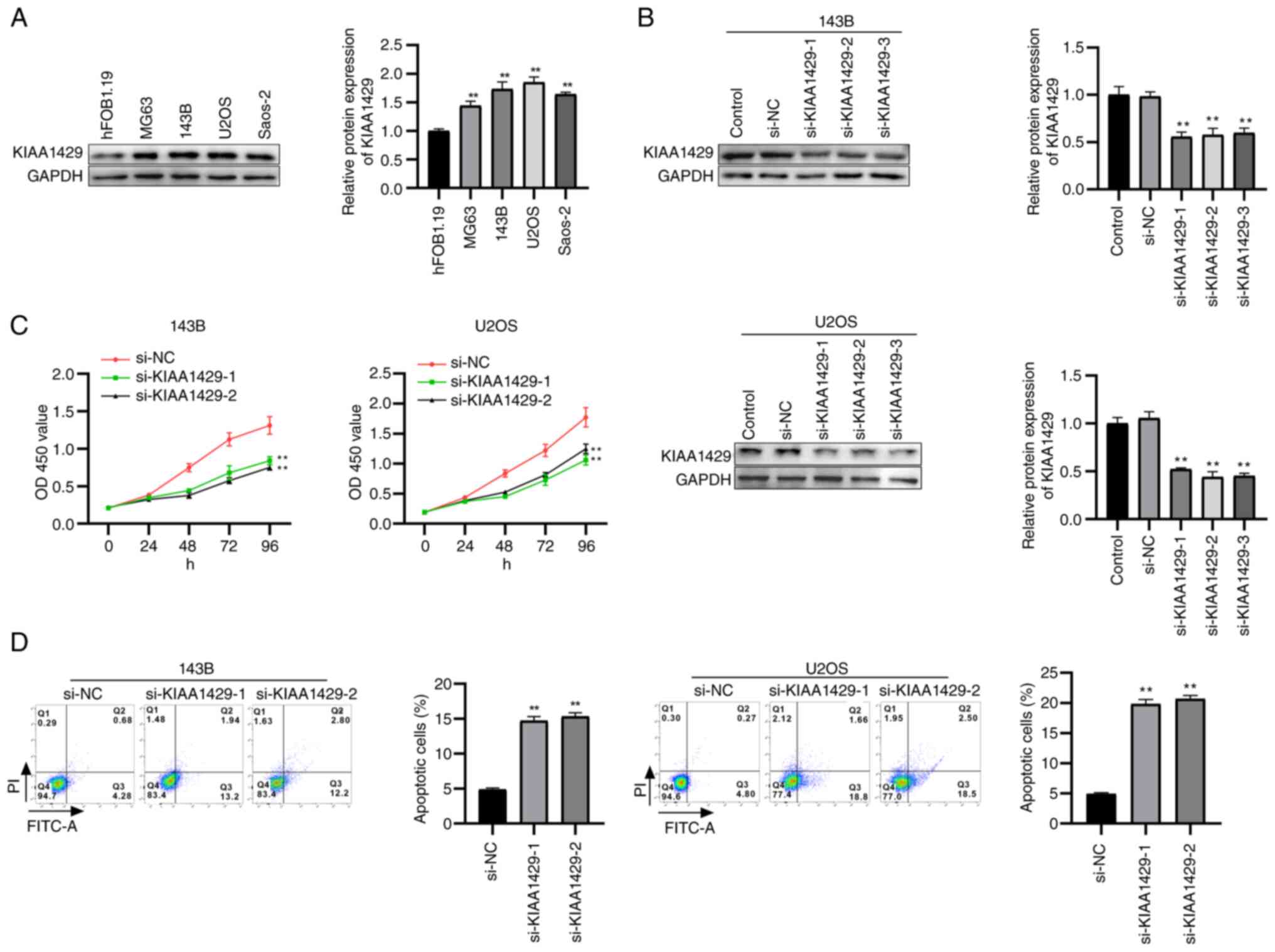
Western blot analysis was performed to determine
KIAA1429 expression levels (Fig.
1A), revealing a marked increase in all OS cell lines (MG63,
143B, U2OS, and Saos-2) compared with in normal human osteoblastic
cells (hFOB1.19). Among the OS cell lines tested, 143B and U2OS
cells showed relatively high KIAA1429 expression, and were selected
for subsequent functional assays because of their high transfection
efficiency. To investigate the functional role of KIAA1429, 143B
and U2OS cells were transfected with three independent siRNAs
targeting KIAA1429, along with si-NC and untreated groups. Western
blot analysis confirmed a significant reduction in KIAA1429 protein
levels in the siRNA-transfected groups compared with those in the
si-NC group (Fig. 1B), indicating
efficient knockdown. Cell proliferation was assessed using CCK-8
assays in 143B and U2OS cells transfected with si-NC, si-KIAA1429-1
or si-KIAA1429-2; KIAA1429 knockdown significantly inhibited cell
proliferation compared with in the si-NC group (Fig. 1C). Furthermore, apoptosis was
evaluated by flow cytometry, which showed a significant increase in
apoptosis rates in both the si-KIAA1429-1 and si-KIAA1429-2 groups
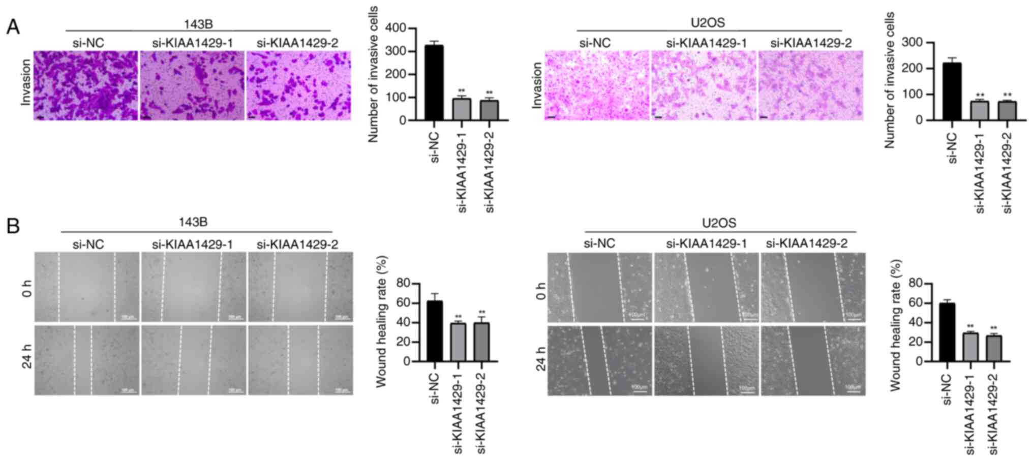
relative to si-NC-transfected cells (Fig. 1D). Cell invasion and migration
were assessed using Transwell and wound healing assays,
respectively, demonstrating that both the invasive and migratory
abilities of OS cells were markedly impaired following KIAA1429
knockdown (Fig. 2A and B).
Collectively, these results indicated that downregulation of
KIAA1429 may suppress proliferation, invasion and migration, while
promoting apoptosis in OS cells.
Downregulation of KIAA1429 promotes
ferroptosis in OS cells
To investigate whether KIAA1429 modulates
ferroptosis, 143B and U2OS cells transfected with si-NC and
si-KIAA1429 were treated with the ferroptosis inducer erastin.
CCK-8 assays showed that cell viability was significantly reduced
in the si-NC + erastin group compared with in the si-NC group
(Fig. 3A). Moreover, KIAA1429
knockdown further decreased cell viability in both the
si-KIAA1429-1 + erastin and si-KIAA1429-2 + erastin groups compared
with in the si-NC + erastin group. Western blot analysis of
ferroptosis-related proteins revealed that erastin treatment alone
(si-NC + erastin) significantly increased COX2 expression and
decreased GPX4 expression compared with in the si-NC group
(Fig. 3B). Notably, KIAA1429
knockdown synergized with erastin to further enhance COX2
expression and suppress GPX4 levels. ELISA was performed to measure
oxidative stress (MDA and GSH) and ferroptosis-related markers
(Fe2+ and ROS) levels. Erastin treatment significantly
increased MDA, iron and ROS levels, while reducing GSH
concentrations compared with in the si-NC group (Fig. 3C). KIAA1429 knockdown further
intensified these effects, resulting in more pronounced alterations
in these ferroptosis-related markers. Lipid peroxidation was
further assessed using C11-BODIPY staining. Erastin treatment
markedly increased the green/red fluorescence ratio compared with
that in the si-NC group, indicating elevated lipid peroxidation in
143B and U2OS cells (Fig. 3D).
KIAA1429 knockdown further enhanced this effect, as shown by
significantly higher green/red fluorescence ratios in both the
erastin + si-KIAA1429-1 and erastin + si-KIAA1429-2 groups compared
with in the si-NC + erastin group. Collectively, these results
indicated that KIAA1429 downregulation enhances erastin-induced
ferroptosis in OS cells.
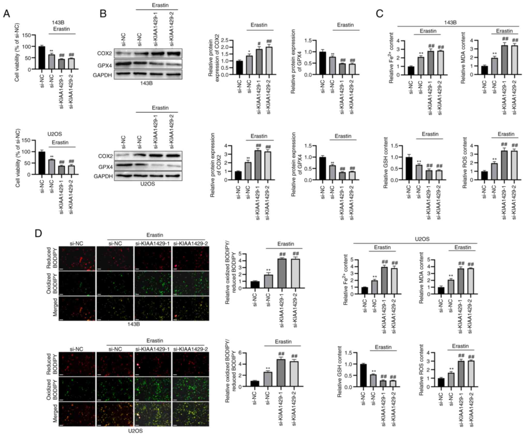 | Figure 3KIAA1429 knockdown promotes
ferroptosis in osteosarcoma cells. (A) Cell viability was evaluated
by Cell Counting Kit-8 assay. (B) Protein expression levels of
ferroptosis-related markers were measured by western blotting. (C)
ELISA was performed to evaluate levels of MDA, Fe2+, GSH
and ROS. (D) C11-BODIPY staining for lipid peroxidation
(magnification, ×200; scale bar, 100 μm).
*P<0.05, **P<0.01 vs. si-NC;
#P<0.05, ##P<0.01 vs. si-NC group.
COX2, cytochrome c oxidase 2; GPX4, GSH peroxidase 4; GSH,
glutathione; MDA, malondialdehyde; NC, negative control; ROS,
reactive oxygen species; si, small interfering. |
Downregulation of KIAA1429 inhibits
SLC7A11 expression via m6A modification
Given that KIAA1429 has been reported to bind to m6A
sites on the SLC7A11 transcript and affect the progression of
hepatocellular carcinoma (21),
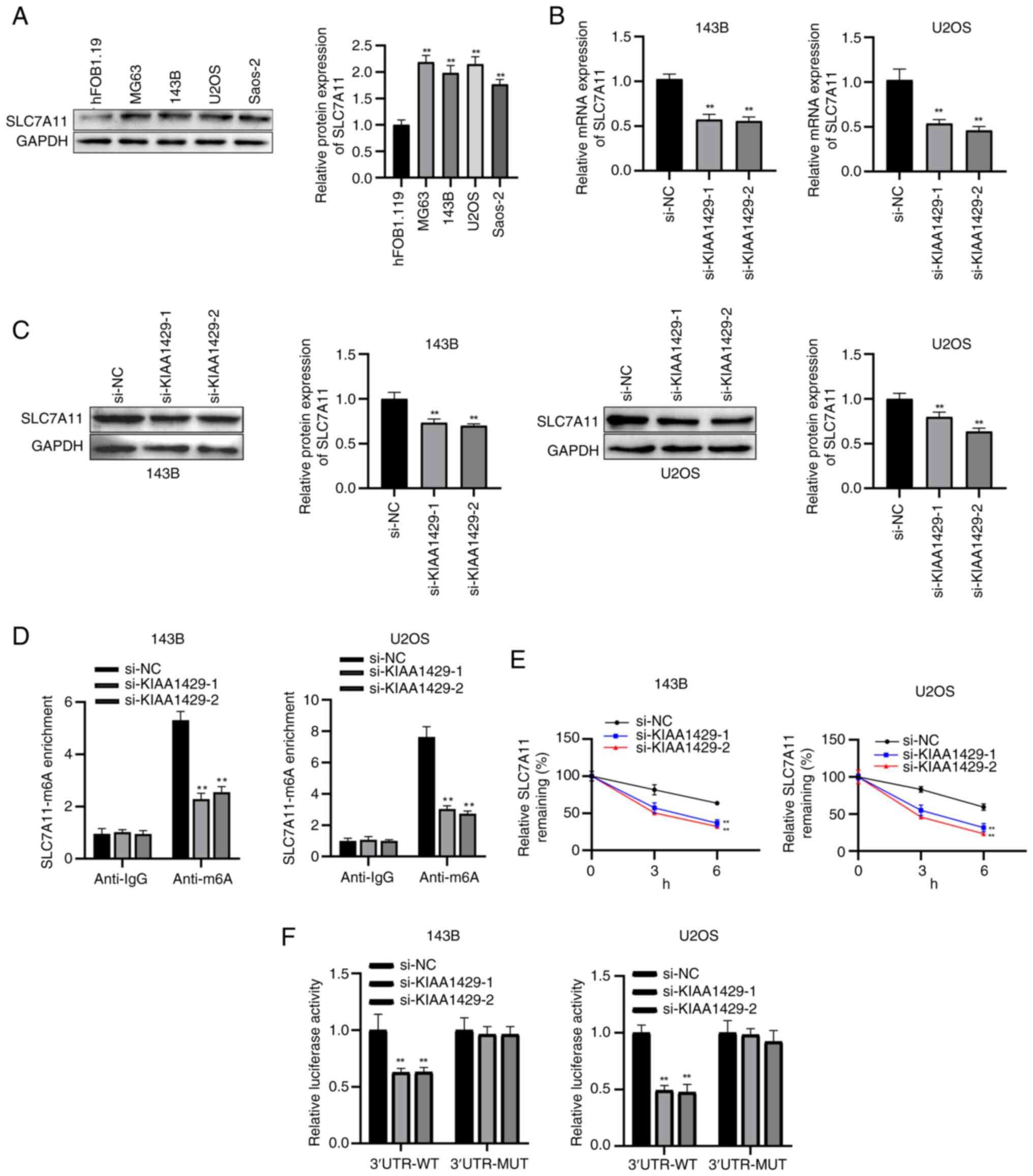
the current study aimed to elucidate its role in OS. Western blot
analysis confirmed that SLC7A11 expression was significantly higher
in human OS cell lines (MG63, 143B, U2OS and Saos-2) than in the
normal osteoblastic cell line hFOB1.19 (Fig. 4A). Both RT-qPCR and western blot
analysis showed that KIAA1429 knockdown significantly reduced
SLC7A11 expression at both the mRNA and protein levels compared
with in the si-NC group (Fig. 4B and
C). MeRIP-qPCR assays further demonstrated that m6A enrichment
of SLC7A11 mRNA was markedly decreased following KIAA1429 knockdown
(Fig. 4D). To assess mRNA
stability, cells were treated with actinomycin D (5 μg/ml)
and qPCR was performed at different time points. The results showed
that SLC7A11 mRNA decayed more rapidly in KIAA1429-knockdown cells
than in control cells (Fig. 4E).
Using a dual-luciferase reporter assay, it was revealed that
KIAA1429 knockdown significantly reduced luciferase activity of
SLC7A11-WT, whereas no significant effect was observed on
SLC7A11-MUT (Fig. 4F).
Collectively, these findings indicated that KIAA1429 knockdown
could suppress SLC7A11 expression through an m6A-dependent
mechanism, thereby reducing mRNA stability and translational
regulation.
KIAA1429 knockdown alleviates OS
progression by regulating SLC7A11 expression
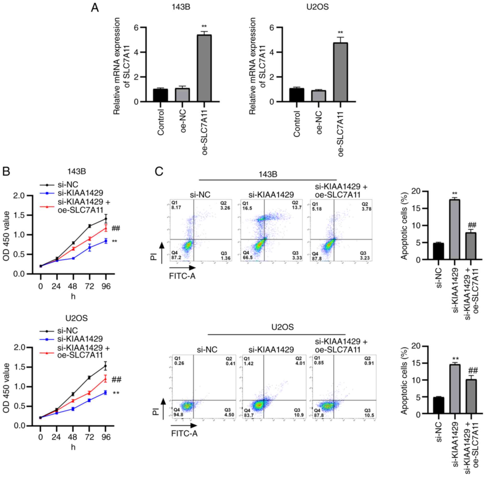
To investigate the functional role of KIAA1429 and
its interaction with SLC7A11 in OS, SLC7A11 was overexpressed in
143B and U2OS cells. RT-qPCR analysis was performed to evaluate the
expression of SLC7A11 in 143B and U2OS cells, confirming the
successful overexpression of SLC7A11 in cells transfected with
oe-SLC7A11 (Fig. 5A). CCK-8
assays showed that KIAA1429 knockdown significantly inhibited cell
proliferation compared with that in the si-NC group (Fig. 5B). By contrast, SLC7A11
overexpression partially rescued this inhibitory effect in the
rescue group (si-KIAA1429 + oe-SLC7A11). Flow cytometric analysis
demonstrated that KIAA1429 knockdown significantly increased
apoptosis compared with in the si-NC group, whereas SLC7A11
overexpression partially reversed this pro-apoptotic effect
(Fig. 5C). Transwell and wound
healing assays revealed that KIAA1429 silencing markedly reduced
the invasive and migratory abilities of OS cells compared with in
the si-NC group; however, these effects were significantly
attenuated by SLC7A11 overexpression (Fig. 6A and B). Furthermore, western blot
analysis of epithelial-mesenchymal transition (EMT)-related
proteins showed that, compared with in the si-NC group, the
si-KIAA1429 group exhibited increased E-cadherin expression and
decreased Vimentin expression (Fig.
6C). Notably, SLC7A11 overexpression partially reversed these
changes, resulting in decreased E-cadherin and increased Vimentin
levels compared with in the si-KIAA1429 group (Fig. 6C). Thus, KIAA1429 knockdown may
suppress EMT, and this effect was partially dependent on SLC7A11
downregulation. Collectively, these findings indicated that
KIAA1429 may promote OS cell proliferation, invasion and migration
while inhibiting apoptosis, largely through regulation of SLC7A11
expression.
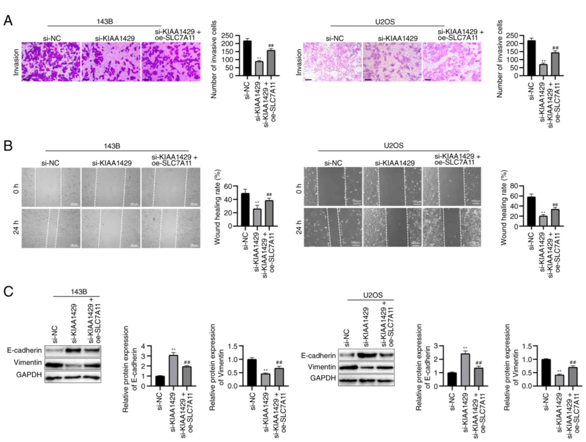 | Figure 6KIAA1429 knockdown suppresses
invasion, migration and epithelial-mesenchymal transition in OS
cells via SLC7A11. (A) Invasive potential was measured by Transwell
assay (magnification, ×200; scale bar, 100 μm). (B)
Migratory capacity was evaluated by wound healing assay
(magnification, ×200; scale bar, 100 μm). (C) Western blot
analysis of E-cadherin and Vimentin expression.
**P<0.01 vs. si-NC; ##P<0.01 vs.
si-KIAA1429. NC, negative control; OD, optical density; oe,
overexpression vector; OS, osteosarcoma; si, small interfering;
SLC7A11, solute carrier family 7 member 11. |
KIAA1429 modulates ferroptosis in OS via
regulation of SLC7A11
CCK-8 assays showed that erastin treatment
significantly reduced cell viability in the si-NC + erastin group
compared with in the si-NC group (Fig. 7A). KIAA1429 knockdown further
enhanced erastin-induced cytotoxicity, resulting in an additional
decrease in cell viability; however, SLC7A11 overexpression
partially reversed the reduction in cell viability caused by
KIAA1429 knockdown (Fig. 7A).
ELISA was used to detect the levels of ferroptosis-related markers
and showed that erastin treatment increased MDA, Fe2+
and ROS levels, while decreasing GSH concentrations; these effects
were further enhanced by KIAA1429 knockdown but were significantly
attenuated by SLC7A11 overexpression (Fig. 7B). Western blot analysis of
ferroptosis-related proteins revealed that erastin treatment
upregulated COX2 and downregulated GPX4 compared with in the si-NC
group, and KIAA1429 silencing further amplified these effects;
however, simultaneous overexpression of SLC7A11 counteracted the
effects of KIAA1429 knockdown, leading to decreased COX2 and
increased GPX4 expression (Fig.
7C). The expression of EMT-related markers was also evaluated
under ferroptosis-inducing conditions. Western blot analysis showed
that erastin treatment alone increased E-cadherin and decreased
Vimentin levels compared with those in the si-NC group, and
KIAA1429 knockdown (erastin + si-KIAA1429 group) further enhanced
these changes (Fig. 7D). However,
SLC7A11 overexpression (erastin + si-KIAA1429 + oe-SLC7A11)
significantly reversed these effects, as evidenced by decreased
E-cadherin and increased Vimentin expression levels (Fig. 7D). Collectively, these results
demonstrated that KIAA1429 knockdown could sensitize OS cells to
erastin-induced ferroptosis and suppress EMT, likely through
downregulation of SLC7A11, whereas SLC7A11 overexpression mitigates
this effect.
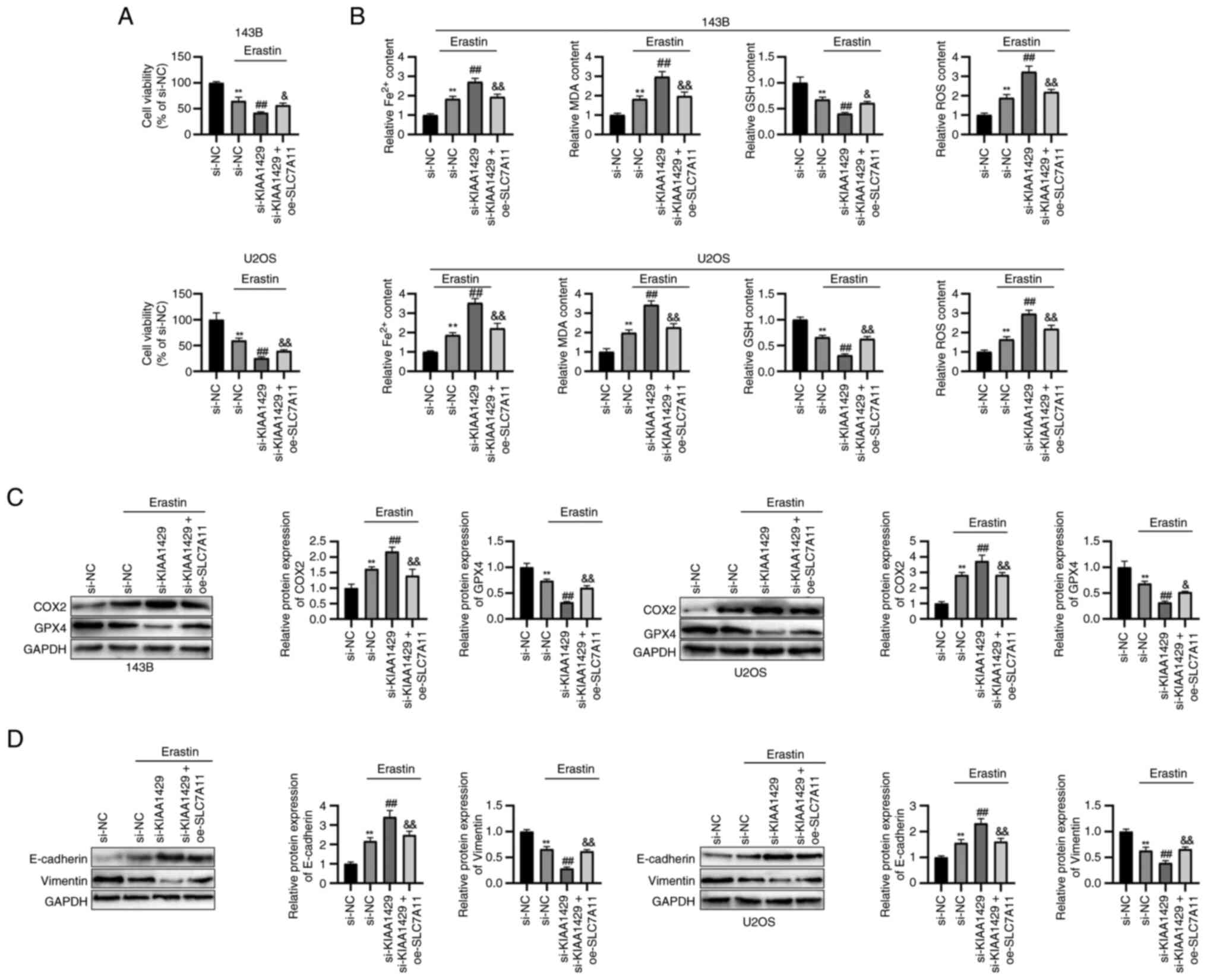 | Figure 7KIAA1429/SLC7A11 axis regulates
ferroptosis in osteosarcoma. (A) Cell viability was assessed by
Cell Counting Kit-8 assay. (B) Levels of oxidative stress
parameters (Fe2+, MDA, GSH, and ROS) were assessed by
ELISA. (C) Ferroptosis markers (COX2 and GPX4) were measured by
western blotting. (D) Western blot analysis of E-cadherin and
Vimentin expression. **P<0.01 vs. si-NC;
##P<0.01 vs. si-NC + erastin;
&P<0.05, &&P<0.01 vs.
si-KIAA1429 + erastin. COX2, cytochrome c oxidase 2; GPX4,
GSH peroxidase 4; GSH, glutathione; MDA, malondialdehyde; NC,
negative control; oe, overexpression vector; ROS, reactive oxygen
species; si, small interfering; SLC7A11, solute carrier family 7
member 11. |
KIAA1429 knockdown suppresses OS growth
and ferroptosis in vivo
To evaluate the role of KIAA1429 in OS progression
and ferroptosis in vivo, BALB/c nude mice were
subcutaneously injected with U2OS cells stably transduced with
lentiviral constructs. Successful overexpression of SLC7A11 by
lv-SLC7A11 was confirmed in U2OS cells prior to injection into mice
(Fig. S1). Compared with in the
lv-sh-NC group, KIAA1429 expression was significantly reduced in
the tumor tissues of the lv-sh-KIAA1429 group, indicating
successful knockdown of KIAA1429 in vivo; no significant
difference in KIAA1429 expression was observed between the
lv-sh-KIAA1429 and lv-sh-KIAA1429 + lv-SLC7A11 groups (Fig. 8A). Tumors were excised at the
endpoint (Fig. 8B), and tumor
weight and volume were measured. Compared with in the lv-sh-NC
group, tumors in the lv-sh-KIAA1429 group exhibited significantly
reduced weight and volume (Fig.
8C). By contrast, SLC7A11 overexpression partially restored
tumor growth compared with in the lv-sh-KIAA1429 group.
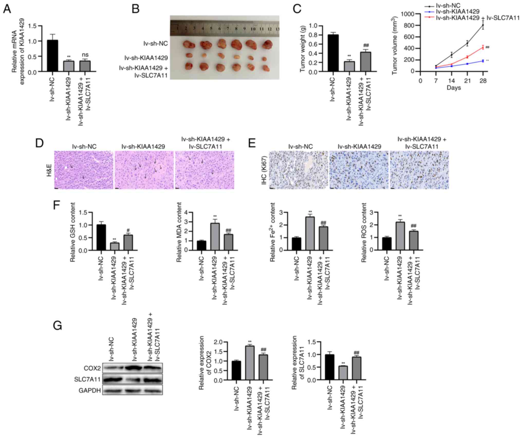 | Figure 8KIAA1429 knockdown suppresses
osteosarcoma growth and ferroptosis in vivo. (A) Reverse
transcription-quantitative PCR was performed to detect the mRNA
levels of KIAA1429 in tumor tissues. (B) Subcutaneous tumor
formation. (C) Tumor volume and weight quantification. (D)
Histopathological analysis was observed by H&E staining
(magnification, ×400; scale bar, 20 μm). Arrows indicate
inflammatory infiltration. (E) Proliferation marker Ki67 was
observed by IHC (magnification, ×400; scale bar, 20 μm). (F)
Levels of oxidative stress parameters (MDA, GSH, Fe2+
and ROS) were evaluated by ELISA. (G) Protein expression levels of
ferroptosis biomarkers were assessed by western blotting.
**P<0.01 vs. lv-sh-NC; #P<0.05,
##P<0.01 vs. lv-sh-KIAA1429. COX2, cytochrome
c oxidase 2; GPX4, GSH peroxidase 4; GSH, glutathione;
H&E, hematoxylin and eosin; IHC, immunohistochemistry; lv,
lentivirus; MDA, malondialdehyde; NC, negative control; ns, not
significant; ROS, reactive oxygen species; sh, short hairpin;
SLC7A11, solute carrier family 7 member 11. |
H&E staining of tumor tissues revealed that the
lv-sh-NC group exhibited densely arranged tumor cells with minimal
inflammatory infiltration (Fig.
8D). By contrast, the lv-sh-KIAA1429 group exhibited a
disrupted tissue architecture with pronounced inflammatory
infiltration. The lv-sh-KIAA1429 + lv-SLC7A11 group displayed
partially restored cellular density and reduced inflammatory
infiltration compared with the lv-sh-KIAA1429 group. IHC analysis
of Ki67 expression showed a marked reduction in Ki67-positive cells
in the lv-sh-KIAA1429 group compared with in the lv-sh-NC group,
whereas this effect was partially reversed by SLC7A11
overexpression (Fig. 8E). ELISA
of ferroptosis-related markers showed that KIAA1429 knockdown
increased MDA, iron and ROS levels, while decreasing GSH
concentrations compared with in the lv-sh-NC group; however, these
changes were attenuated by SLC7A11 overexpression (Fig. 8F). Western blot analysis further
demonstrated that KIAA1429 knockdown increased COX2 expression and
decreased SLC7A11 expression, whereas SLC7A11 overexpression
partially reversed these effects (Fig. 8G). Collectively, these in
vivo findings confirmed that KIAA1429 downregulation may
suppress tumor growth and enhance ferroptosis in OS, largely
through regulation of SLC7A11.
Discussion
OS poses severe clinical risks, including
debilitating pain, pathological fractures, limb dysfunction and
frequent pulmonary metastasis, which collectively account for its
high mortality rate (22,23). The present study demonstrated that
KIAA1429 was highly expressed in OS cell lines, and that its
knockdown markedly suppressed malignant tumor progression.
Mechanistically, KIAA1429 stabilized SLC7A11 mRNA via m6A
methylation, thereby inhibiting ferroptosis and facilitating tumor
progression. These findings indicated that KIAA1429 may be a
critical oncogenic driver in OS.
As the predominant internal modification within
eukaryotic mRNAs, m6A orchestrates gene expression dynamics through
its cognate 'writer', 'eraser' and 'reader' protein machinery,
exerting regulatory control over RNA splicing, stability,
translation efficiency and decay pathways (24). Its dysregulation is increasingly
implicated in the oncogenesis of various types of cancer (25). In OS, m6A methylation affects
tumor invasion and metastasis through various signaling pathways
(26). As an m6A
methyltransferase, methyltransferase-like 3 drives OS cell
progression through dual mechanisms: Regulating the m6A
modification status of lymphoid enhancer-binding factor 1 and
activating the Wnt/β-catenin signaling cascade (27). The demethylase AlkB homologue 5
facilitates tumor growth and metastasis by erasing m6A marks on
targets such as ubiquitin-specific peptidase 22, thereby disrupting
histone ubiquitination and enhancing DNA replication and repair
processes (28). By revealing the
critical contribution of m6A to OS initiation and progression,
these findings simultaneously position it as a viable therapeutic
target.
The m6A methyltransferase complex harbors KIAA1429,
a critical regulatory component involved in the pathogenesis of
numerous types of human cancer. In hepatocellular carcinoma,
acetylated KIAA1429 (by TIP60) facilitates immune evasion by
stabilizing lysine-specific demethylase 5B mRNA via m6A
modification, thereby promoting programmed death ligand 1
expression and tumor metastasis (29). In pancreatic adenocarcinoma,
KIAA1429-mediated m6A modification enhances AKT2 mRNA stability,
driving malignant progression and autophagy suppression (30). In diffuse large B-cell lymphoma,
KIAA1429 exerts oncogenic functions through m6A-dependent
regulation of carbohydrate sulfotransferase 11, thereby activating
the Hippo-YAP pathway and sustaining malignant progression
(31). In colorectal cancer,
KIAA1429 promotes oncogenesis through m6A-independent
transcriptional repression of WEE1, and its clinicopathological
relevance is underscored by its notable association with poor
patient survival (32).
Similarly, in OS, KIAA1429-mediated m6A modifications of the
ZFPM2-AS1 transcript promote malignant progression, manifested as
increased cellular proliferation, motility and invasiveness
(33). Luo et al (34) established that KIAA1429 knockdown
can exert anti-oncogenic effects in OS by disrupting Janus kinase
2/signal transducer and activator of transcription 3 signaling. The
current study further revealed that downregulation of KIAA1429 led
to pronounced suppression of OS cell proliferation, invasion and
migration, providing strong evidence of its oncogenic role in
OS.
Ferroptosis, an iron-dependent form of regulated
cell death, has garnered attention as a treatment strategy against
therapy-resistant cancer (11).
SLC7A11, a core subunit of system Xc−, imports cystine
for GSH synthesis, thereby protecting cells against oxidative
stress and ferroptosis (35).
SLC7A11 overexpression is associated with chemoresistance and poor
survival in OS, as it maintains redox balance and suppresses
iron-dependent lipid peroxidation (36). Beyond OS, SLC7A11 dysregulation
has also been observed in bladder and ovarian cancer, where it
promotes tumor resilience (37,38). In non-small cell lung cancer,
tripartite motif-containing 3 acts as a tumor suppressor and
inhibits tumorigenesis by promoting SLC7A11 degradation (39). CCAAT/enhancer binding protein γ
suppresses ferroptosis in ovarian cancer through transcriptional
regulation of SLC7A11 (40).
Targeting p62 with sulforaphane enhances autolysosomal degradation
of SLC7A11 in OS, as demonstrated in both in vivo and in
vitro models, ultimately inducing ferroptosis and inhibiting
malignant progression (41).
Similarly, lysine-specific demethylase 4A depletion induces
ferroptosis in OS cells by increasing H3K9me3 at the SLC7A11
promoter, thereby transcriptionally regulating SLC7A11 expression
(42). In the present study,
KIAA1429 silencing amplified erastin-induced ferroptosis, as
evidenced by enhanced lipid peroxidation, elevated MDA, ROS and
iron levels, and reduced GSH activity. Additionally, KIAA1429
knockdown reduced the m6A modification of SLC7A11 mRNA,
accelerating its decay and impairing its translation. This
reduction amplified ferroptotic susceptibility, which was reversed
by SLC7A11 overexpression. These results are consistent with the
present in vivo findings, showing that SLC7A11 inhibition
can trigger ferroptosis and suppresses OS growth. Thus, targeting
the KIAA1429/SLC7A11 axis may disrupt redox adaptation in OS.
In conclusion, the current study demonstrated that
KIAA1429 was significantly upregulated in OS and that KIAA1429
knockdown suppressed tumor progression. Mechanistically, KIAA1429
can stabilize SLC7A11 mRNA via m6A modification, thereby
suppressing ferroptosis. Rescue experiments confirmed that SLC7A11
overexpression reversed the antitumor effects of KIAA1429
knockdown, both in vitro and in vivo. These findings
reveal that the KIAA1429/SLC7A11 axis is a critical regulator of
ferroptosis and OS progression, providing a promising therapeutic
target for OS treatment.
Supplementary Data
Availability of data and materials
The data generated in the present study may be
requested from the corresponding author.
Authors' contributions
HZ made substantial contributions to study
conception and design, data acquisition and drafting the article.
YZ, DZ and SX performed data acquisition and drafted the article.
SZ performed data acquisition and reviewed the article. HZ and SZ
confirm the authenticity of all the raw data. All the authors took
part in the experiments. All the authors read and approved the
final manuscript.
Ethical approval and consent to
participate
All methods are reported in accordance with ARRIVE
guidelines for the reporting of animal experiments. The animal
experiments conformed to the Guide for the Care and Use of
Laboratory Animals and were approved by the Animal Ethics Committee
of Ganzhou People's Hospital (approval no. PJD2026-025-01).
Patient consent for publication
Not applicable.
Competing interests
The authors declare that they have no competing
interests.
Acknowledgements
Not applicable.
Funding
The present study was funded by the Ganzhou Science and
Technology + National Regional Medical Center 'Joint Program
Project - General Project of Key Research and Development Program'
(no. 2022-YB1446), the Jiangxi Provincial Health Commission S&T
Program (no. SKJP220212369) and the Medical Leading Discipline of
Ganzhou City.
References
|
1
|
Cersosimo F, Lonardi S, Bernardini G,
Telfer B, Mandelli GE, Santucci A, Vermi W and Giurisato E:
Tumor-associated macrophages in osteosarcoma: From mechanisms to
therapy. Int J Mol Sci. 21:52072020. View Article : Google Scholar : PubMed/NCBI
|
|
2
|
Shoaib Z, Fan TM and Irudayaraj JMK:
Osteosarcoma mechanobiology and therapeutic targets. Br J
Pharmacol. 179:201–217. 2022. View Article : Google Scholar :
|
|
3
|
Chen C, Xie L, Ren T, Huang Y, Xu J and
Guo W: Immunotherapy for osteosarcoma: Fundamental mechanism,
rationale, and recent breakthroughs. Cancer Lett. 500:1–10. 2021.
View Article : Google Scholar
|
|
4
|
Jafari F, Javdansirat S, Sanaie S, Naseri
A, Shamekh A, Rostamzadeh D and Dolati S: Osteosarcoma: A
comprehensive review of management and treatment strategies. Ann
Diagn Pathol. 49:1516542020. View Article : Google Scholar : PubMed/NCBI
|
|
5
|
Li S, Zhang H, Liu J and Shang G: Targeted
therapy for osteosarcoma: A review. J Cancer Res Clin Oncol.
149:6785–6797. 2023. View Article : Google Scholar : PubMed/NCBI
|
|
6
|
Huang J, Guo J and Jia R:
N6-methyladenosine methyltransferase component KIAA1429 is a
potential target of cancer therapy. Biomolecules. 14:13192024.
View Article : Google Scholar : PubMed/NCBI
|
|
7
|
Zhang X, Li MJ, Xia L and Zhang H: The
biological function of m6A methyltransferase KIAA1429 and its role
in human disease. PeerJ. 10:e143342022. View Article : Google Scholar : PubMed/NCBI
|
|
8
|
Ren M, Pan H, Zhou X, Yu M and Ji F:
KIAA1429 promotes gastric cancer progression by destabilizing RASD1
mRNA in an m6A-YTHDF2-dependent manner. J Transl Med.
22:5842024. View Article : Google Scholar
|
|
9
|
Li N, Zhu Z, Deng Y, Tang R, Hui H, Kang Y
and Rana TM: KIAA1429/VIRMA promotes breast cancer progression by
m6 A-dependent cytosolic HAS2 stabilization. EMBO Rep.
24:e555062023.
|
|
10
|
Li J, Rao B, Yang J, Liu L, Huang M, Liu
X, Cui G, Li C, Han Q, Yang H, et al: Dysregulated m6A-related
regulators are associated with tumor metastasis and poor prognosis
in osteosarcoma. Front Oncol. 10:7692020. View Article : Google Scholar : PubMed/NCBI
|
|
11
|
Zhou Q, Meng Y, Li D, Yao L, Le J, Liu Y,
Sun Y, Zeng F, Chen X and Deng G: Ferroptosis in cancer: From
molecular mechanisms to therapeutic strategies. Signal Transduct
Target Ther. 9:552024. View Article : Google Scholar : PubMed/NCBI
|
|
12
|
Liu J, Kang R and Tang D: Signaling
pathways and defense mechanisms of ferroptosis. FEBS J.
289:7038–7050. 2022. View Article : Google Scholar
|
|
13
|
Zhao J, Zhao Y, Ma X, Zhang B and Feng H:
Targeting ferroptosis in osteosarcoma. J Bone Oncol. 30:1003802021.
View Article : Google Scholar : PubMed/NCBI
|
|
14
|
Lin W, Wang C, Liu G, Bi C, Wang X, Zhou Q
and Jin H: SLC7A11/xCT in cancer: Biological functions and
therapeutic implications. Am J Cancer Res. 10:3106–3126.
2020.PubMed/NCBI
|
|
15
|
Rochette L, Dogon G, Rigal E, Zeller M,
Cottin Y and Vergely C: Lipid peroxidation and iron metabolism: Two
corner stones in the homeostasis control of ferroptosis. Int J Mol
Sci. 24:4492022. View Article : Google Scholar
|
|
16
|
Zhang L, Luo D, Ren H, Fan W, Yu Z, Wang
S, Wu X, Xu Y and Li S: Brusatol induced ferroptosis in
osteosarcoma cells by modulating the Keap1/Nrf2/SLC7A11 signaling
pathway. Phytomedicine. 144:1569122025. View Article : Google Scholar : PubMed/NCBI
|
|
17
|
Xie C, Xiao Y, Yang L and Luo J: KIAA1429
silencing ameliorates osteosarcoma progression through promoting
ferroptosis via Nrf2/NQO1 axis. Inflamm Res. 74:1112025. View Article : Google Scholar : PubMed/NCBI
|
|
18
|
Livak KJ and Schmittgen TD: Analysis of
relative gene expression data using real-time quantitative PCR and
the 2(-Delta Delta C(T)) method. Methods. 25:402–408. 2001.
View Article : Google Scholar
|
|
19
|
Wang X, Chen Q, Bing Z, Zhou S, Xu Z, Hou
Y, Zhao Y, Zhao S and Wang T: Low expression of m6A reader YTHDC1
promotes progression of ovarian cancer via PIK3R1/STAT3/GANAB axis.
Int J Biol Sci. 19:4672–4688. 2023. View Article : Google Scholar : PubMed/NCBI
|
|
20
|
Ji FH, Fu XH, Li GQ, He Q and Qiu XG: FTO
prevents thyroid cancer progression by SLC7A11 m6A methylation in a
ferroptosis-dependent manner. Front Endocrinol (Lausanne).
13:8577652022. View Article : Google Scholar : PubMed/NCBI
|
|
21
|
Wang H, Chen W, Cui Y, Gong H and Li H:
KIAA1429 protects hepatocellular carcinoma cells from ferroptotic
cell death with a m6 A-dependent posttranscriptional
modification of SLC7A11. J Cell Mol Med. 27:4118–4132. 2023.
View Article : Google Scholar : PubMed/NCBI
|
|
22
|
Haynes L, Kaste SC, Ness KK, Wu J,
Ortega-Laureano L, Bishop M, Neel M, Rao B and Fernandez-Pineda I:
Pathologic fracture in childhood and adolescent osteosarcoma: A
single-institution experience. Pediatr Blood Cancer. 64: View Article : Google Scholar : 2017.
|
|
23
|
Liu H, Tian L, Mu J, He X, Zhuangzhuang L,
Gong T, Wang J, Min L, Lu M and Tu C: The resection of extraosseous
osteosarcoma was accompanied by the occurrence of pulmonary
metastasis and distal metastasis at the primary site: A case
report. Front Oncol. 15:15497222025. View Article : Google Scholar : PubMed/NCBI
|
|
24
|
Uddin MB, Wang Z and Yang C: The
m6A RNA methylation regulates oncogenic signaling
pathways driving cell malignant transformation and carcinogenesis.
Mol Cancer. 20:612021. View Article : Google Scholar
|
|
25
|
An Y and Duan H: The role of m6A RNA
methylation in cancer metabolism. Mol Cancer. 21:142022. View Article : Google Scholar : PubMed/NCBI
|
|
26
|
Hu T, Wang G, Wang D, Deng Y and Wang W:
m6A methylation modification: Potential pathways to suppress
osteosarcoma metastasis. Int Immunopharmacol. 145:1137592025.
View Article : Google Scholar
|
|
27
|
Miao W, Chen J, Jia L, Ma J and Song D:
The m6A methyltransferase METTL3 promotes osteosarcoma progression
by regulating the m6A level of LEF1. Biochem Biophys Res Commun.
516:719–725. 2019. View Article : Google Scholar : PubMed/NCBI
|
|
28
|
Yadav P, Subbarayalu P, Medina D, Nirzhor
S, Timilsina S, Rajamanickam S, Eedunuri VK, Gupta Y, Zheng S,
Abdelfattah N, et al: M6A RNA methylation regulates histone
ubiquitination to support cancer growth and progression. Cancer
Res. 82:1872–1889. 2022. View Article : Google Scholar : PubMed/NCBI
|
|
29
|
Quan H, Zhou H, Chen F, Chen J, He Y, Xiao
H, Liu J, Shi L, Xie W, Chen P and Luo J: Acetylated KIAA1429 by
TIP60 facilitates metastasis and immune evasion of hepatocellular
carcinoma via N6-methyladenosine-KDM5B-mediated regulation of
FoxO1. Cell Death Discov. 11:2102025. View Article : Google Scholar : PubMed/NCBI
|
|
30
|
Li P, Lu Y, Zheng Z, Lv J and Hu J: The
role and mechanism of KIAA1429-mediated m6A modification in
pancreatic adenocarcinoma. Mol Carcinog. 64:1667–1682. 2025.
View Article : Google Scholar : PubMed/NCBI
|
|
31
|
Chen X, Lu T, Cai Y, Han Y, Ding M, Chu Y,
Zhou X and Wang X: KIAA1429-mediated m6A modification of CHST11
promotes progression of diffuse large B-cell lymphoma by regulating
Hippo-YAP pathway. Cell Mol Biol Lett. 28:322023. View Article : Google Scholar : PubMed/NCBI
|
|
32
|
Ma L, Lin Y, Sun SW, Xu J, Yu T, Chen WL,
Zhang LH, Guo YC, Wang YW, Chen T, et al: KIAA1429 is a potential
prognostic marker in colorectal cancer by promoting the
proliferation via downregulating WEE1 expression in an
m6A-independent manner. Oncogene. 41:692–703. 2022. View Article : Google Scholar
|
|
33
|
Zhang Y, Bao Y, Zhai H, Li C, Shi H, Gong
K and Bao X: KIAA1429-mediated ZFPM2-AS1 m6A
modification promotes the proliferation, migration and invasion of
osteosarcoma cells. Oncol Lett. 30:4532025.
|
|
34
|
Luo J, Wang X, Chen Z, Zhou H and Xiao Y:
The role and mechanism of JAK2/STAT3 signaling pathway regulated by
m6A methyltransferase KIAA1429 in osteosarcoma. J Bone Oncol.
39:1004712023. View Article : Google Scholar :
|
|
35
|
Koppula P, Zhuang L and Gan B: Cystine
transporter SLC7A11/xCT in cancer: Ferroptosis, nutrient
dependency, and cancer therapy. Protein Cell. 12:599–620. 2021.
View Article : Google Scholar :
|
|
36
|
Guo W, Wang X, Lu B, Yu J, Xu M, Huang R,
Cheng M, Yang M, Zhao W and Zou C: Super-enhancer-driven MLX
mediates redox balance maintenance via SLC7A11 in osteosarcoma.
Cell Death Dis. 14:4392023. View Article : Google Scholar : PubMed/NCBI
|
|
37
|
Shen L, Zhang J, Zheng Z, Yang F, Liu S,
Wu Y, Chen Y, Xu T, Mao S, Yan Y, et al: PHGDH inhibits ferroptosis
and promotes malignant progression by upregulating SLC7A11 in
bladder cancer. Int J Biol Sci. 18:5459–5474. 2022. View Article : Google Scholar : PubMed/NCBI
|
|
38
|
Fantone S, Piani F, Olivieri F, Rippo MR,
Sirico A, Di Simone N, Marzioni D and Tossetta G: Role of
SLC7A11/xCT in ovarian cancer. Int J Mol Sci. 25:5872024.
View Article : Google Scholar : PubMed/NCBI
|
|
39
|
Wang Z, Shen N, Wang Z, Yu L, Yang S, Wang
Y, Liu Y, Han G and Zhang Q: TRIM3 facilitates ferroptosis in
non-small cell lung cancer through promoting SLC7A11/xCT K11-linked
ubiquitination and degradation. Cell Death Differ. 31:53–64. 2024.
View Article : Google Scholar
|
|
40
|
Zhang X, Zheng X, Ying X, Xie W, Yin Y and
Wang X: CEBPG suppresses ferroptosis through transcriptional
control of SLC7A11 in ovarian cancer. J Transl Med. 21:3342023.
View Article : Google Scholar : PubMed/NCBI
|
|
41
|
Zou Q, Zhou X, Lai J, Zhou H, Su J, Zhang
Z, Zhuang X, Liu L, Yuan R, Li S, et al: Targeting p62 by
sulforaphane promotes autolysosomal degradation of SLC7A11,
inducing ferroptosis for osteosarcoma treatment. Redox Biol.
79:1034602025. View Article : Google Scholar
|
|
42
|
Chen M, Jiang Y and Sun Y: KDM4A-mediated
histone demethylation of SLC7A11 inhibits cell ferroptosis in
osteosarcoma. Biochem Biophys Res Commun. 550:77–83. 2021.
View Article : Google Scholar : PubMed/NCBI
|