Introduction
Botulinum neurotoxin (BoNT), produced by the
bacterium Clostridium botulinum, is widely recognized as one
of the most potent biological toxins. Discovered in the late 19th
century, its mechanism of action involves the inhibition of
acetylcholine release at the neuromuscular junction, resulting in
flaccid paralysis (1). Despite its
high toxicity, BoNT has been utilized for therapeutic purposes,
particularly in the fields of neurology, ophthalmology, dermatology
and urology, owing to its ability to induce temporary muscle
relaxation (1).
BoNT consists of a heavy chain (HC; 100 kDa) and a
light chain (LC; 50 kDa), which are connected by a disulfide bond.
The HC selectively binds to synaptic vesicle protein 2 on neuronal
membranes, leading to endocytosis (2,3),
whereas the LC cleaves specific snap receptor proteins, inhibiting
acetylcholine release (4). There
are seven serotypes of BoNT (A-G) (5), each with distinct molecular targets.
BoNT/A, C and E cleave synaptosomal-associated protein 25 kDa
(SNAP-25), while BoNT/B, D, F and G cleave vesicle-associated
membrane proteins, and BoNT/C also targets syntaxin-1 (6). Among these, the BoNT/A serotype has
the longest duration of action, lasting ≤6 months, and is commonly
used for both therapeutic and cosmetic applications (7,8).
Meanwhile, BoNT/E has a rapid onset and short duration of action
compared with other Botulinum toxin serotypes, making it
suitable for specific clinical scenarios, such as transient muscle
relaxation in neurological diagnostics or short-term therapeutic
trials requiring rapid reversibility (9).
The therapeutic applications of BoNT have
revolutionized the management of various medical conditions,
ranging from neuromuscular disorders such as dystonia and
spasticity to cosmetic treatments for reducing facial wrinkles
(7). Previous research has also
unveiled its potential roles in alleviating chronic pain, migraines
and hyperhidrosis (10–12). However, several considerations must
be assessed when using BoNT, including the development of
resistance, immunogenicity, variable patient responses and the need
for precise dosing regimens (13).
While extending the half-life of BoNT may enhance therapeutic
efficacy and reduce the frequency of injections, it also raises
potential concerns such as prolonged muscle paralysis, unintended
diffusion to non-target tissues and the formation of neutralizing
antibodies. These risks must be carefully considered when
developing BoNT variants with enhanced stability (14,15).
Various strategies have been employed to extend the
half-life of therapeutic proteins in vivo, including
PEGylation, Fc fusion, albumin fusion and glycosylation, all of
which are widely used (16).
However, these methods often involve structural modifications that
can affect protein size, immunogenicity or functional activity,
potentially limiting their clinical utility and compromising
protein activity or structural integrity (17,18).
By contrast, residue-specific lysine-to-arginine substitution
offers a more precise strategy to enhance protein stability without
substantially modifying the native structure (19,20).
Additionally, modulation of the ubiquitin-proteasome system (UPS)
has emerged as a promising approach for regulating protein
half-life. These strategies may ultimately support more durable
therapeutic effects with reduced dosing frequency, which are
particularly beneficial in chronic or repetitive treatment
settings. The UPS is a key cellular pathway in eukaryotic cells and
is responsible for the degradation of the majority of intracellular
proteins. It regulates essential processes such as cell cycle
progression, DNA repair and oxidative stress response, thereby
maintaining cellular homeostasis. Dysregulation of this system has
been implicated in various diseases, including cancer,
neurodegenerative disorders and immune dysfunctions (21,22).
Proteins targeted for degradation via the UPS are tagged with
ubiquitin molecules in a cascade involving E1 (activating enzyme),
E2 (conjugating enzyme) and E3 (ligase enzyme). The ubiquitin
chains are recognized and degraded by the 26S proteasome (23). Ubiquitin molecules are primarily
attached to the lysine residues on proteins, forming polyubiquitin
chains by linking multiple ubiquitin units (24,25).
In contrast to ubiquitination, deubiquitination occurs when
deubiquitinating enzymes (DUBs) remove these ubiquitin molecules
(26). Together, these processes
regulate protein stability and various cellular functions,
maintaining cellular homeostasis (26).
In the present study, an innovative approach was
applied to BoNT, aiming to extend its half-life. The primary
objective was to enhance the duration of its therapeutic efficacy
while minimizing the required dosage. The present study aimed to
enhance the stability and half-life of BoNT by introducing
lysine-to-arginine mutations at key ubiquitination sites, while
also assessing the potential effects on its enzymatic activity.
Furthermore, this approach provides new possibilities for broader
applications of BoNT in both therapeutic and cosmetic fields.
Materials and methods
Cell culture and transfection
293T (cat. no. CRL-11268; American Type Culture
Collection), cells were cultured in Dulbecco's Modified Eagle
Medium (Gibco; Thermo Fisher Scientific, Inc.) with high glucose,
supplemented with 10% fetal bovine serum (Gibco; Thermo Fisher
Scientific, Inc.) and 1% penicillin-streptomycin (Gibco; Thermo
Fisher Scientific, Inc.). The cells were maintained at 37°C in a
humidified atmosphere with 5% CO2.
Plasmid DNA and antibodies
Wild type (WT)-BoNT/A1, BoNT/A2 and
BoNT/E cDNA were synthesized and subcloned into the
pCS4-3XFlag-tagged vector (Invitrogen; Thermo Fisher Scientific,
Inc.). The pRK5-hemagglutinin (HA)-tagged ubiquitin (HA-Ub)
plasmid was provided by Professor Byung Joon Hwang (Kangwon
National University, Chuncheon, South Korea) and used for
co-transfection to assess ubiquitination levels. For transfection
(100 mm dish), 1–3 µg each plasmid DNA was used
(Flag-BoNT/A1-WT and -mutants, Flag-BoNT/A2-WT and
K335R, Flag-BoNT/E-WT and -mutants and HA-Ub).
Transfection of each construct was carried out using
polyethyleimine (cat. no. 23966-1; Polysciences, Inc.) according to
the manufacturer's protocol. Cells were incubated at 37°C during
transfection. After 24 h, the culture medium was replaced with
fresh medium, and cells were harvested 48 h post-transfection for
downstream experiments. cDNAs (Flag-BoNT/A1,
Flag-BoNT/A2, Flag-BoNT/E and HA-Ub) were
transformed into the Escherichia coli DH5α (cat. no. RH617;
RBC Bioscience Corp.) bacterial strain and the bacteria were grown
for 16 h at 37°C in Luria-Bertani broth (cat. no. MB-L4488; Kisan
Bio Co., Ltd.). Anti-β-actin (1:3,000; cat. no. sc-4778; Santa Cruz
Biotechnology, Inc.), anti-Flag (1:4,000; cat. no. M185-3L; MBL
International Co.) and anti-HA (1:1,000; cat. no. sc-53516; Santa
Cruz Biotechnology, Inc.) antibodies were used for western blotting
and immunoprecipitation.
Site-directed mutagenesis
Site-directed mutagenesis was performed using WT
Flag-BoNT/A1, Flag-BoNT/A2 and Flag-BoNT/E
plasmids as a template to generate lysine-to-arginine substitution
mutants and amplify the mutant plasmids. The PCR conditions were as
follows: 95°C for 30 sec, 54°C for 40 sec and 68°C for 6 min, for a
total of 15 cycles. The primers used for each substitution are
listed in Table I. The amplified
products were subsequently subjected to DpnI digestion to
remove the template plasmid, followed by transformation into
competent cells for propagation and sequence verification.
 | Table I.Primers for site-directed mutagenesis
of BoNT-LC. |
Table I.
Primers for site-directed mutagenesis
of BoNT-LC.
| Target | Mutations | Sequence,
5′-3′ |
|---|
|
BoNT/A1-LC | K212R | F:
GGTGCAGGCAGATTTGCT |
|
|
| R:
AGCAAATCTGCCTGCACC |
|
| K301R | F:
CTTAATAAAGCTAGATCAATAGTAGGT |
|
|
| R:
ACCTACTATTGATCTAGCTTTATTAAG |
|
| K320R | F:
TTTTAAAGAGAGATATCTCCTA |
|
|
| R:
TAGGAGATATCTCTCTTTAAAA |
|
| K330R | F:
CATCTGGAAGATTTTCGG |
|
|
| R:
CCGAAAATCTTCCAGATG |
|
| K335R | F:
TTTCGGTAGATAGATTAAATT |
|
|
| R:
AATTTTAATCTATCTACCGAAA |
|
| K340R | F:
TTTTGTATAACCTATCAAATT |
|
|
| R:
ACTAAACTAGAAATTTTACTGG |
|
| K417R | F:
ACTAAACTAGAAATTTTACTGG |
|
|
| R: CCAGTAAAATTTC
TTAGTTTAGT |
|
BoNT/A2-LC | K335R | F:
TCGGTAGATAGATTAAATTTGATAAGTTATACAAAATGTTAACAGAG |
|
|
| R:
ATCAAATTTTAATCTATCTACCGAAAATTTTCCAGATGTATC |
|
BoNT/E-LC | K3R | F:
TGCCAAGAATTAATAGTTTTAATTATAATGATCCTGTTAATGATAGAAC |
|
|
| R:
AAAACTATTAATTCTTGGCATGGATCCGTCC |
|
| K62R | F:
ACTTCATTAAGAAATGGAGATAGTAGTTATTATGACCC |
|
|
| R:
ATCTCCATTTCTTAATGAAGTAGGCGGATGAAAATC |
|
| K85R | F:
TTTTAAGAATAGTCACAAAAATATTTAATAGAATAAATAATAATCTTTC |
|
|
| R:
TTTTGTGACTATTCTTAAAAATCTATCCTTTTCTTCATCACTTTG |
|
| K230R | F:
ATTACTACAAGGTATACTATAACACAAAAACAAAATCC CCTAATAAC |
|
|
| R:
TATAGTATACCTTGTAGTAATCCCTTTAGCCCCATATAG |
|
| K281R | F:
GATTATAAAAGAATAGCGTCTAAACTTAGCAAAGTAC |
|
|
| R:
CGCTATTCTTTTATAATCAGCTAGAAGATTAGTATAGATATCATTGGAC |
|
| K288R | F:
AAACTTAGCAGAGTACAAGTATCTAATCCACTACTTAATCC |
|
|
| R: TACTTGT
ACTCTGCTAAGTTTAGACGCTATTTTTTTATAATCAG |
|
| K342R | F:
AACTTGAAATCTAGTTGCTAAATCAAATTCCGTAAAGCTG |
|
|
| R:
TTAGCAACTAGATTTCAAGTTAAATGTAGGCAAACTTATATTG |
|
| K413R | F:
TAGATTTTGTAGAAATATTGTTTCTGTAAAAGGCATAAGG |
|
|
| R:
ACAATATTTCTACAAAATCTAATGATTTTTTTTACTAGTCCTCTAC |
|
| K419R | F:
TATTGTTTCTGTAAGAGGCATAAGGTCTAGAGGGC |
|
|
| R:
TGCCTCTTACAGAAACAATATTTTTACAAAATCTAATGATTTTTTTTAC |
Bioinformatics analysis of potential
ubiquitination sites
To identify potential ubiquitination sites in the LC
of BoNT/A1 and BoNT/E, bioinformatics tools were employed.
Predicted ubiquitinated lysine residues in BoNT protein were
identified using the BDM ubiquitination prediction tool (BDM-PUB)
(27), which was analyzed in March
2022 for BoNT/A1, and in December 2022 for BoNT/A2 and BoNT/E. The
analysis conducted using PyMOL (https://www.pymol.org/; Ver.3.1; Schrödinger Inc.) and
AlphaFold (Ver. 2) via Google Colaboratory (https://colab.research.google.com/), which enabled
GPU-based computation due to limited local hardware resources
(28), revealed K212, K301, K320,
K330, K335, K340 and K417 for BoNT/A1, and K3, K62, K85, K230,
K281, K288, K343, K413 and K419 for BoNT/E as major ubiquitination
sites. Subsequently, protein structure files for BoNT/A1, BoNT/A2
and BoNT/E, were obtained through the Protein Data Bank (PDB;
http://www.rcsb.org/), were analyzed using the
PyMOL program (PDB ID: 2ISE for BoNT/A1; PDB ID: 5MOY for BoNT/A2;
and PDB ID: 7K7Y for BoNT/E). Among the identified lysine residues,
those located on the protein surface were considered potential
ubiquitination sites. Site-directed mutagenesis was carried out to
substitute the selected lysine residues with arginine, generating
lysine-to-arginine mutants. These mutants were experimentally
validated to assess their effects on ubiquitination levels and
protein stability.
Western blotting and
immunoprecipitation
Harvested cells were lysed using RIPA lysis buffer
[1% Triton X-100, 150 mM NaCl, 50 mM Tris-HCl (pH 8.0), 1 mM EDTA,
10% glycerol, 100 mM PMSF and protease inhibitor cocktail]. Cell
lysates were centrifuged at 16,000 × g for 20 min at 4°C, and the
supernatants were mixed with 2X SDS buffer and boiled at 100°C.
Protein concentration was determined using the Bio-Rad Protein
Assay Dye Reagent (cat. no. 5000006; Bio-Rad Laboratories, Inc.).
For western blotting, 20–30 µg protein was loaded per lane and was
separated by SDS-PAGE on 6 or 8% gels, depending on the molecular
weight of the target protein. Subsequently, the proteins were
transferred onto PVDF membranes (cat. no. IPVH00010;
MilliporeSigma), which were blocked with 5% skim milk in TBS-0.5%
Tween 20 (TBST) for 30 min at room temperature. The membranes were
incubated with the primary antibodies overnight at 4°C, and after
washing three times (7 min each) with TBST at room temperature,
they were incubated with the HRP-conjugated secondary antibodies
[1:30,000; cat. no. 5220-0341 (074–1806); KPL, Inc.; and 1:10,000;
cat. no. 31470; Thermo Fisher Scientific, Inc.] for 1 h at room
temperature. The membranes were then washed a three times with TBST
(7 min each) and developed using an enhanced chemiluminescence
reagent (cat. no. LF-QC0103; Abfrontier). The intensity of each
western blot band was measured and semi-quantified using ImageJ
software (Version 1.53t; National Institutes of Health) to assess
relative protein expression levels.
For immunoprecipitation, cells were lysed using RIPA
lysis buffer [1% Triton X-100, 150 mM NaCl, 50 mM Tris-HCl (pH
8.0), 1 mM EDTA, 10% glycerol, 100 mM PMSF and protease inhibitor
cocktail]. 1.5–2 mg protein lysates were incubated with 2 µg
anti-Flag antibody at 4°C overnight on a rotator. Subsequently, 35
µl Protein A/G PLUS-agarose beads (cat. no. sc-2003; Santa Cruz
Biotechnology Inc.) were added and the samples were incubated for 2
h at 4°C. The immunocomplexes were pelleted by centrifugation at
800 × g for 5 min at 4°C and washed three times with ice-cold RIPA
buffer. After the final wash, the beads were resuspended in 2X SDS
sample buffer and boiled at 100°C for 7 min. The supernatants were
then analyzed by SDS-PAGE followed by western blotting.
Ubiquitination assay
For the ubiquitination assay, 293T cells
(2×10⁶) were co-transfected with Flag-tagged
BoNT-A1 (WT or K212R, K301R, K320R, K330R, K335R,
K340R and K417R mutants), BoNT-A2 (WT or
K335R mutant) or BoNT-E (WT or K3R, K62R,
K85R, K230R, K281R, K288R, K342R, K413R and K419R
mutants) along with HA-Ub. After 4 h of treatment with MG132
at 37°C (10 µM cat. no. F1100; Ubiquitin-Proteasome
Biotechnologies, LLC), the cells were harvested and lysed using
RIPA lysis buffer supplemented with protease inhibitors.
Immunoprecipitation was carried out on the cell lysates (1.5–2 mg
total protein) using an anti-Flag antibody as aforementioned, and
the ubiquitination levels of Flag-BoNT-LC were analyzed by western
blotting with anti-HA antibody to detect ubiquitinated
proteins.
Sequence homology analysis
Amino acid sequences of BoNT serotypes (A-G)
were obtained from the National Center for Biotechnology
Information database, (https://www.ncbi.nlm.nih.gov/) GenBank (https://www.ncbi.nlm.nih.gov/genbank/)
using their respective accession numbers. Sequence alignment was
performed using Clustal X 2.1 (http://www.clustal.org/clustal2/) with default
parameters to calculate the percentage identity of each serotype
relative to BoNT/A1. The resulting pairwise percentage
homologies were tabulated and used to evaluate sequence similarity
across serotypes (Table II).
| Table II.Comparison of sequence homology
percentages among different types of Botulinum
neurotoxin. |
Table II.
Comparison of sequence homology
percentages among different types of Botulinum
neurotoxin.
| GenBank protein
ID | Description | Percentage homology
with type A1 | Type |
|---|
| AUN16864.1 | Botulinum
neurotoxin type A (Clostridium botulinum) | NA | A1 |
| CAA51824.1 | Botulinum
neurotoxin type A2 (Clostridium botulinum) | 89.66% | A2 |
| AAA23211.1 | Botulinum
neurotoxin type B (Clostridium botulinum) | 40.35% | B |
| BAC22064.1 | Botulinum
neurotoxin type C (Clostridium botulinum) | 39.88% | C |
| CAA38175.1 | Botulinum
neurotoxin type D (Clostridium botulinum) | 39.29% | D |
| CAA44558.1 | Botulinum
neurotoxin type E (Clostridium botulinum) | 38.77% | E |
| CAA57358.1 | Botulinum
neurotoxin type F (Clostridium botulinum) | 40.15% | F |
| CAA52275.1 | Botulinum
neurotoxin type G (Clostridium botulinum) | 33.28% | G |
Protein stability assay
To evaluate the stability of the BoNT/A1-, BoNT/A2-
and BoNT/E-LCs, cycloheximide (CHX; cat. no. 01810; Sigma-Aldrich;
Merck KgaA) chase experiments were conducted. 293T cells were
transfected with plasmids encoding either WT LCs or the
lysine-to-arginine mutants (K212R, K301R, K320R, K330R, K335R,
K340R and K417R for A1, K335R for A2 and
K3R, K62R, K85R, K230R, K281R, K288R, K343R, K413R and
K419R for E). Following transfection, CHX was added
at a final concentration of 100 µg/ml to inhibit de novo
protein synthesis. Cells were harvested at designated time points
(0, 12, 18 or 0, 6, 12 h post-CHX treatment at 37°C in a 5%
CO2 incubator).
Statistical analysis
Data were obtained from at least three independent
experiments. Mean values were calculated, and all quantitative data
are presented as the mean ± SD unless otherwise specified in the
figure legends. Statistical analysis was performed using two-way
ANOVA followed by Tukey's post hoc test for multiple group
comparisons. All analyses were conducted using GraphPad Prism
(Ver.9; Dotmatics). P<0.05 was considered to indicate a
statistically significant difference.
Results
Sequence homology between BoNT
types
There are seven known types of BoNT (A-G). Sequence
homology among these types was compared and is summarized in
Table II. BoNT/A2
exhibited the highest similarity to BoNT/A1, with a sequence
homology of 89.66%. By contrast, the other BoNT types
displayed 33–40% sequence similarity to BoNT/A1. These
sequence variations are not associated with differences in their
cleavage targets. For example, BoNT/A, BoNT/C and BoNT/E all target
SNAP-25, yet BoNT/C and BoNT/E share only 39.88 and
38.77% sequence homology with BoNT/A1, respectively
(Table II).
293T cells were transfected with Flag-tagged
BoNT/A1-LC, BoNT/A2-LC and BoNT/E-LC
constructs. To confirm the ubiquitination of BoNT/A1, BoNT/A2 and
BoNT/E, 293T cells were co-transfected with HA-Ub and
Flag-BoNT/A1-LC, BoNT/A2-LC and BoNT/E-LC
constructs. Following treatment with the proteasome inhibitor
MG132, immunoprecipitation was performed using anti-Flag antibody.
Compared with the control condition lacking HA-Ub or MG132
treatment, increased ubiquitin chain signal was detected for all
three BoNT subtypes, indicating that BoNT/A1, BoNT/A2 and BoNT/E
undergo UPS-mediated degradation (Fig.
1A-C).
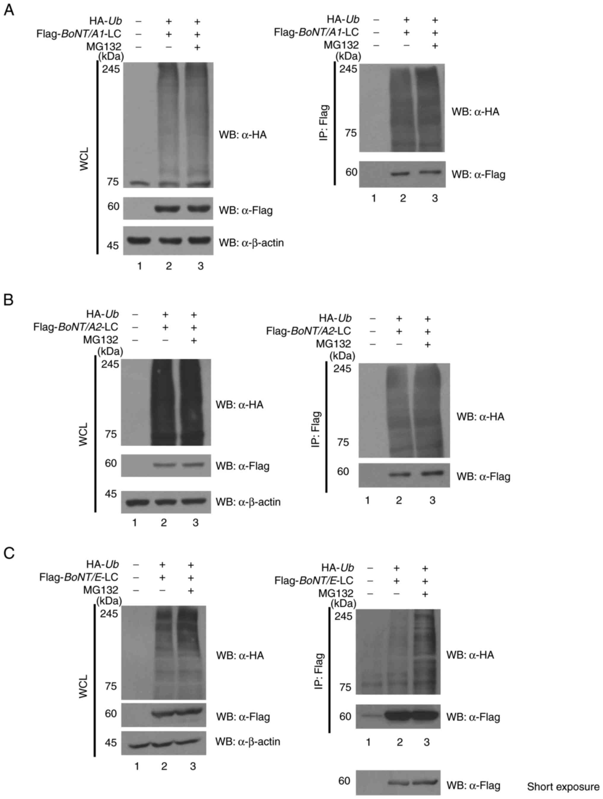 | Figure 1.Ubiquitination of BoNT/A1, BoNT/A2
and BoNT/E-LCs. (A) BoNT/A1-LC, (B) BoNT/A2-LC and (C) BoNT/E-LC
were analyzed in the presence of the proteasome inhibitor MG132.
Cells were transfected with the indicated BoNT-LC constructs and
HA-tagged ubiquitin, followed by IP using the anti-Flag antibody
and WB with anti-HA to detect ubiquitinated proteins. Enhanced
ubiquitination was observed for all BoNT types under proteasome
inhibition. WB, western blotting; BoNT, Botulinum
neurotoxin; WCL, whole cell lysate; IP, immunoprecipitation; HA,
hemagglutinin; Ub, ubiquitin; LC, light chain. |
Reduced ubiquitination in lysine
mutants compared with WT BoNT/A1, A2 and E
To predict ubiquitinated lysine residues in BoNT/A1
and BoNT/E, a ubiquitination prediction tool BDM, analyzed in March
2022 (BoNT/A1) and December 2022 (BoNT/A2, BoNT/E)] was used
(27). The identified lysine sites
were further analyzed using the PyMOL program and structural data
from the PDB for each BoNT subtype. Surface-exposed lysines were
selected as potential ubiquitination sites, while those buried
within the structure of the protein were excluded. Based on these
predictions (Fig. 2A), lysine
residues K212, K301, K320, K330, K335, K340 and K417 in BoNT/A1
were substituted with arginine to block ubiquitination at these
positions. 293T cells were co-transfected with mutant plasmids and
HA-Ub. Immunoprecipitation was carried out using anti-Flag
antibodies to isolate the mutant proteins. As shown in Fig. 2B (right panel), HA-Ub immunoblots
showed substantially decreased polyubiquitin chains in the lanes
corresponding to BoNT/A1-K330R, -K335R and -K417R mutants, compared
with that in the WT group, supporting their identification as key
ubiquitination sites in BoNT/A1 (Fig.
2B).
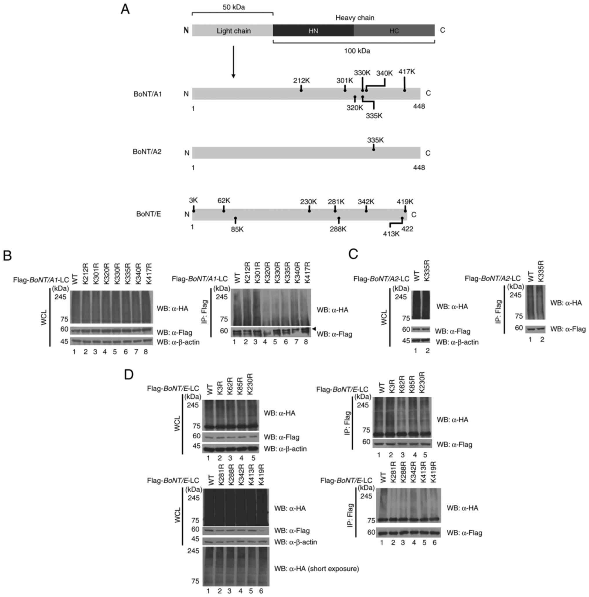 | Figure 2.Ubiquitination levels of WT and
lysine mutant forms of BoNT-LCs. (A) Protein is composed of an LC
(gray) and heavy chain (black and dark gray). The heavy chain is
further divided into HN (black) and HC (C-terminal of heavy chain)
(dark gray) regions. Mutation sites were selected based on
ubiquitin prediction analysis and mutated accordingly. BoNT/A1,
BoNT/A2 and BoNT/E are represented. (B) Ubiquitination levels of WT
and K212R, K301R, K320R, K330R, K335R, K340R and K417R mutants of
BoNT/A1-LC. (C) Ubiquitination levels of WT and the K335R mutant of
BoNT/A2-LC. (D) Ubiquitination levels of WT and K3R, K62R, K85R,
K230R, K281R, K288R, K342R, K413R and K419R mutants of BoNT/E-LC.
Short exposure images were used to avoid signal saturation and
clearly visualize differences in ubiquitination levels between
wild-type and mutant proteins. WT, wild type; BoNT,
Botulinum neurotoxin; LC, light chain; HN, N-terminus of
heavy chain HC; C-terminus of heavy chain; WCL, whole cell lysate;
IP, immunoprecipitation; HA, hemagglutinin; Ub, ubiquitin. |
Given the high sequence similarity (89.66%) between
BoNT/A2 and BoNT/A1, lysine 335 was also analyzed in BoNT/A2.
Prediction site analysis confirmed a similar ubiquitination profile
between the two subtypes. Using site-directed mutagenesis, the
K335R mutant for BoNT/A2 was generated, and ubiquitination assays
revealed reduced ubiquitin binding compared with the WT BoNT/A2,
consistent with the findings for BoNT/A1 (Fig. 2C).
By contrast, BoNT/E exhibits only ~40% sequence
similarity with BoNT/A1, necessitating new predictions for
ubiquitinated lysine residues. Bioinformatics analysis identified
lysine residues K3, K62, K85, K230, K281, K288, K342, K413 and K419
as potential ubiquitination sites. These residues were individually
substituted with arginine, and ubiquitination assays revealed that
the K62R and K288R mutants reduced ubiquitin binding in the HA
blot, Fig. 2D (right panel); HA-Ub
immunoblots showed markedly reduced polyubiquitin chains signal in
lanes corresponding to K62R and K288R mutants, compared with that
in the WT group. This highlights K62R and K288R as key
ubiquitination sites in BoNT/E (Fig.
2D). Overall, the results of the present study identified key
ubiquitination sites in BoNT/A1, BoNT/A2 and BoNT/E. Further
experiments aimed to assess whether targeting these residues could
extend the half-life these proteins, thereby enhancing their
therapeutic potential.
Extending protein half-life through
lysine mutations
A CHX chase assay was conducted to determine whether
lysine-to-arginine mutations, which reduce ubiquitination compared
with WT serotypes, could enhance the stability of BoNT proteins by
decreasing their degradation rates (Fig. 3A-C) In BoNT/A1 proteins, the K212
mutation exhibited a trend toward reduced protein stability
compared with WT. Conversely, mutations at K335 and K417
demonstrated increased protein stability, with K335 showing
significant stabilization at 12 h (1.516-fold) and at 18 h
(2.05-fold). Similarly, the K417 mutation exhibited significant
stabilization at both 12 h (1.528-fold) and 18 h (1.727-fold;
Fig. 3A). For BoNT/A2, the K335R
mutant displayed significantly enhanced stability compared with WT
at both 12 h (1.37-fold) and 18 h (1.58-fold), consistent with the
findings for BoNT/A1 (Fig. 3B). To
avoid signal saturation and to better resolve subtle differences in
protein degradation rates, short exposure blots were included for
CHX chase assays. These allow more accurate comparison of stability
between WT and mutant forms. In BoNT/E, lysine residues K62 and
K288 were analyzed. Both mutations revealed improved protein
stability relative to WT. K62 revealed a 1.34-fold increase in
stability at 12 h, while K288 demonstrated a 1.32-fold increase at
12 h and a significant 2.23-fold increase at 12 h (Fig. 3C). These results highlight the
ability of these lysine residues in regulating protein
stability.
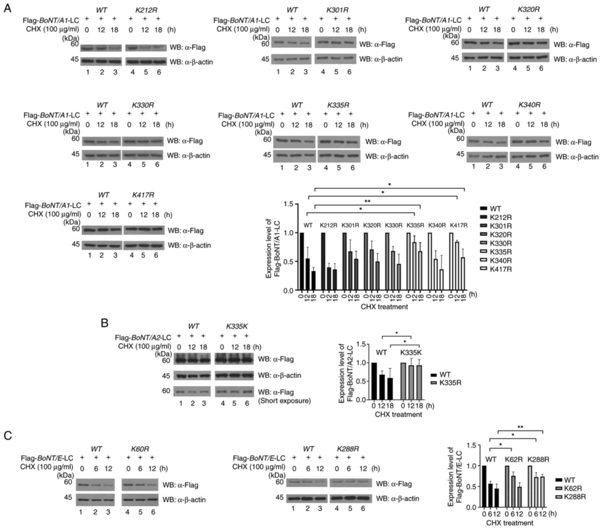 | Figure 3.Protein stability of WT and lysine
mutant forms of BoNT-LCs. (A) CHX chase assay comparing protein
stability of BoNT/A1-LC WT and K212R, K301R, K320R, K330R, K335R,
K340R and K417R mutants. Protein levels were analyzed by WB. (B)
CHX chase assay of BoNT/A2-LC WT and K335R mutant. (C) CHX chase
assay of BoNT/E-LC WT, K62R and K288R mutants. WB was carried out
to compare protein expression levels of WT and mutant forms under
identical experimental conditions. Short exposure images were used
to avoid signal saturation and clearly visualize differences in
Flag-BoNT transfected cells. *P<0.05 and **P<0.01. WT, wild
type; BoNT, Botulinum neurotoxin; LC, light chain; CHX,
cycloheximide; WB, western blotting. |
These findings underscored the potential of
targeting specific lysine residues through lysine-to-arginine
mutations to extend the half-life of BoNT proteins, providing a
foundation for developing more durable therapeutic
formulations.
Discussion
The therapeutic and cosmetic applications of BoNT
have considerably advanced over the past few decades, offering
effective solutions for treating various diseases and enhancing
cosmetology (29–32). Among the seven serotypes, BoNT/A is
the most widely used due to its prolonged half-life (~6 months) and
stable action, whereas BoNT/E is preferred in specific cases for
its rapid onset and shorter duration of action (33–36).
Despite these benefits, the limited half-life and short duration of
efficacy of BoNTs remain notable challenges in clinical
applications. In the present study, the role of the UPS in BoNT
degradation was investigated strategies to enhance the stability of
BoNTs by inhibiting ubiquitination were explored (8,37).
The UPS is a primary pathway for intracellular
proteolysis, carrying out a key role in regulating protein
stability and maintaining cellular homeostasis (21,22).
The BoNT-LC undergoes degradation primarily through ubiquitination,
a significant factor contributing to its limited half-life
(38,39). Previous studies have demonstrated
that the BoNT/E-LC interacts with the RING finger protein TNF
receptor associated factor 2 to facilitate its degradation
(38,40). These findings suggest that
targeting the ubiquitination pathway could effectively extend the
half-life of BoNTs.
The UPS also regulates the degradation of BoNT/A,
and alterations in UPS activity have been shown to influence the
persistence of this toxin (39,40).
One study screened 32 UPS inhibitors to identify compounds that
modulate BoNT/A degradation. This previous study aimed to mitigate
BoNT poisoning by exploring compounds that regulate ubiquitination
and deubiquitination. Among the inhibitors tested, 10 were found to
effectively modulate ubiquitination, with WP1130, PR-619 and
celastrol being particularly effective in reducing the persistence
of BoNT/A. These compounds also demonstrated the ability to inhibit
SNAP-25 cleavage even after toxin exposure (38), further supporting their potential
utility in managing BoNT activity. These compounds function as
broad-spectrum DUB inhibitors, disrupting the removal of ubiquitin
chains and thereby promoting proteasomal degradation of target
proteins (41). However, due to
their lack of specificity, they may inadvertently affect various
cellular processes, including oxidation stress responses and
proteostasis, leading to potential off-target effects.
Consequently, while their therapeutic potential is promising,
further optimization is required to enhance their specificity and
reduce toxicity (38,42).
Collectively, these studies highlight the potential
of regulating the UPS pathway not only for managing BoNT toxicity
but also for optimizing therapeutic strategies. By targeting the
processes of ubiquitination and deubiquitination, which antagonize
each other in regulating protein expression, it may be possible to
reduce toxin dosage requirements, prolong its persistence and
enhance its therapeutic utility.
The present study revealed structural differences
among BoNT serotypes and demonstrated the importance of specific
lysine residues in their degradation through ubiquitination. While
BoNT/A1 and BoNT/A2 were revealed to share ~89.99% sequence
similarity, BoNT/E shares only ~40% sequence similarity with
BoNT/A1. This variability results in differences in predicted and
identified ubiquitination sites, suggesting that each BoNT subtype
has distinct mechanisms of ubiquitination and degradation.
Previous studies have suggested that BoNT
degradation is mediated by the ubiquitination of its LC (29,30,38);
however, the specific lysine residues involved and their functional
roles have not been clearly defined. In the present study, primary
ubiquitination sites were identified in BoNT/A1, BoNT/A2 and
BoNT/E, and protein stability was enhanced by inhibiting
ubiquitination through site-specific mutations. K335 and K417 in
BoNT/A1 were identified as major ubiquitination sites, with the
mutations reducing ubiquitination. Similarly, the K335R mutation in
BoNT/A2 demonstrated increased protein stability. For BoNT/E, K62
and K288 were identified as key ubiquitination sites, with the K62R
and K288R mutations effectively inhibiting ubiquitination. Notably,
the lysine-to-arginine mutations at K335 and K417 in BoNT/A1, K335R
in BoNT/A2, and K62 and K288 in BoNT/E each contributed to
increased protein half-life, indicating that site-specific
inhibition of ubiquitination can effectively enhance the stability
of BoNT LC.
BoNTs are internalized into nerve cells by binding
specific receptors on the neuronal membrane (43). After internalization, the BoNT/A-LC
translocates into the cytoplasm, where it directly interacts with
SNAP-25 (43,44). This interaction is mediated by the
α and β-exosite in the LC, which determine the substrate
specificity for SNAP-25. The cleavage of SNAP-25 between Gln197 and
Arg198 is the primary mechanism of BoNT/A1 action. The low homology
domain (LHD) in the BoNT/A1-LC (residues 268–357) is key for stable
interactions with SNAP-25 and anchoring to the plasma membrane
(44). Structurally, the LHD is
divided into three regions: R1 (275–300), R2 (302–334) and R3. R1
forms an α-helix structure that mediates indirect interaction with
SNAP-25, whereas R2 adopts a loop-α-helix-loop structure that
establishes strong ionic and hydrogen bonds with SNAP-25 (43,44).
Since the activity of BoNT relies on its precise interaction with
SNAP-25, it is important to evaluate whether lysine-to-arginine
mutations, while extending the protein stability of LCs, might
affect its neurotoxin function. Given the key role of the LHD and
exosite regions in mediating SNAP-25 recognition, it is essential
to consider whether lysine-to-arginine substitutions affect
residues within or adjacent to these functional domains. In the
present study, the substituted lysine residues K335 and K417 in
BoNT/A1 are located outside the core motifs for SNAP-25
interaction, indicating that direct interference with substrate
binding is unlikely. Previous structural studies have also revealed
that mutations distal to catalytic or binding interfaces can
enhance protein stability without compromising enzymatic function
(45,46). Since BoNT is recombinantly
expressed in Escherichia coli, its sequence can be
rationally engineered and screened to optimize the balance between
structural stability and functional integrity. Nevertheless, to
fully assess the therapeutic relevance of these mutations, future
studies should directly compare the enzymatic activity and
functional efficacy of WT and mutant BoNTs under physiologically
relevant conditions.
By extending the half-life of BoNTs through K335R,
K417R, K62R and K288R mutations, the present study introduced the
potential for prolonged therapeutic effects with lower doses,
reducing the frequency of administration. This approach is
particularly beneficial in therapeutic settings, improving patient
convenience and compliance. Moreover, extending the half-life of
BoNTs could mitigate immunogenicity issues associated with repeated
administration (47,48). Repeated administration of BoNT can
lead to the formation of neutralizing antibodies, reducing the
effectiveness of the drug. Extending the half-life of BoNT could
reduce administration frequency, thereby minimizing this risk
(47,48).
Existing technologies for extending protein
half-life, such as PEGylation, Fc fusion, albumin fusion and
glycosylation, often compromise the biological activity or
structural stability of proteins (47). In contrast to conventional
strategies, the approach taken in the present study utilizes
residue-specific lysine-to-arginine substitutions, offering a
minimally invasive alternative that preserves the native structure
of the protein. Conventional strategies, such as PEGylation or
fusion with Fc or albumin domains, extend serum half-life by
promoting steric hindrance or receptor recycling. However, these
strategies often reduce biological activity, increase
immunogenicity and pose manufacturing challenges due to their bulky
modifications (49–51). By comparison, lysine substitution
directly inhibits ubiquitin-dependent degradation without
structural enlargement, offering the potential to preserve
functional activity and mitigate the risk of immune responses.
Nonetheless, it should be noted that each method has inherent
trade-offs, and the optimal choice may vary depending on the
therapeutic context and specific protein characteristics.
Clinically, prolonging the intracellular half-life of BoNT through
reduced ubiquitination may extend its duration of neuromuscular
blockade, thereby reducing the frequency of injections required for
therapeutic efficacy. This could be particularly advantageous in
chronic indications such as spasticity or dystonia, where repeated
administration is often necessary. Furthermore, fewer injections
may help lower the risk of immunogenicity and patient discomfort,
both of which are key considerations in long-term treatment. Thus,
by interfering with the degradation pathway rather than altering
the delivery or formulation, this strategy may preserve the native
function of BoNT while supporting improved therapeutic
outcomes.
While the present study demonstrated a promising
strategy to stabilize BoNT through lysine-to-arginine
substitutions, several unresolved limitations should be taken into
account when interpreting the results. To further evaluate the
physiological relevance of these findings, additional studies using
in vivo models are needed. Comparative experiments between
WT and mutant BoNTs could assess functional persistence through
measurements of muscle response duration, evaluate neurotoxicity
profiles via hemidiaphragm assays and determine enzymatic activity
by quantifying SNAP-25 cleavage in neural tissues. These approaches
would help to establish whether the introduced mutations extend
protein stability without compromising biological efficacy. To
address these limitations, more precise evaluations of BoNT
stability and functionality should be conducted through structural
simulations and comprehensive in vivo experiments.
Additionally, this approach must be evaluated for its effectiveness
in other subtypes (BoNT/B, BoNT/C, BoNT/D, BoNT/F, BoNT/G and
BoNT/H) beyond BoNT/A1, BoNT/A2 and BoNT/E. The potential of lysine
mutations to reduce immunogenicity also warrants further
exploration. While the present study focused on the role of the
UPS, BoNTs may also be degraded through other pathways, such as
autophagy. Therefore, a comprehensive investigation of the
mechanisms involved in BoNT degradation is essential for fully
understanding and optimizing the stability and therapeutic
potential of these neurotoxins (52,53).
Acknowledgements
Not applicable.
Funding
This research was supported by a research fund from Alkemier
Inc. (grant no. Alkemier-2024-1).
Availability of data and materials
The data generated in the present study may be
requested from the corresponding author.
Authors' contributions
HSC, HRS, ML and KHB conceptualized and designed the
present study. Material preparation, data collection and analysis
were conducted by HSC, HRS, MC, HYK and SKJ. The initial draft of
the manuscript was prepared by HSC, HRS and KHB. All authors
contributed to the review and revision of the manuscript, and the
final version was reviewed and approved by KHB. HSC and KHB confirm
the authenticity of all the raw data. All authors read and approved
the final manuscript.
Ethics approval and consent to
participate
Not applicable.
Patient consent for publication
Not applicable.
Competing interests
The authors declare that they have no competing
interests.
Glossary
Abbreviations
Abbreviations:
|
BoNT
|
Botulinum neurotoxin
|
|
UPS
|
ubiquitin-proteasome system
|
|
HC
|
heavy chain
|
|
LC
|
light chain
|
|
SNAP-25
|
synaptosomal-associated protein 25
kDa
|
|
LHD
|
low homology domain
|
|
WT
|
wild type
|
|
CHX
|
cycloheximide
|
|
HA
|
hemagglutinin
|
|
PDB
|
Protein Data Bank
|
|
DUB
|
deubiquitinating enzyme
|
References
|
1
|
Bach K and Simman R: The multispecialty
toxin: A literature review of botulinum toxin. Plast Reconstr Surg
Glob Open. 10:e42282022. View Article : Google Scholar : PubMed/NCBI
|
|
2
|
Yeo JC, Tay FP, Bennion R, Loss O, Maignel
J, Pons L, Foster K, Beard M and Bard F: Botulinum toxin
intoxication requires retrograde transport and membrane
translocation at the ER in RenVM neurons. Elife. 12:RP928062024.
View Article : Google Scholar : PubMed/NCBI
|
|
3
|
Joensuu M, Syed P, Saber SH, Lanoue V,
Wallis TP, Rae J, Blum A, Gormal RS, Small C, Sanders S, et al:
Presynaptic targeting of botulinum neurotoxin type A requires a
tripartite PSG-Syt1-SV2 plasma membrane nanocluster for synaptic
vesicle entry. EMBO J. 42:e1120952023. View Article : Google Scholar : PubMed/NCBI
|
|
4
|
Matak I, Bölcskei K, Bach-Rojecky L and
Helyes Z: Mechanisms of Botulinum toxin type A action on pain.
Toxins (Basel). 11:4592019. View Article : Google Scholar : PubMed/NCBI
|
|
5
|
Montecucco C, Schiavo G and Rossetto O:
The mechanism of action of tetanus and botulinum neurotoxins. Arch
Toxicol Suppl. 18:342–354. 1996. View Article : Google Scholar : PubMed/NCBI
|
|
6
|
Eleopra R, Tugnoli V, Quatrale R, Rossetto
O and Montecucco C: Different types of botulinum toxin in humans.
Mov Disord. 19 (Suppl 8):S53–S59. 2004. View Article : Google Scholar : PubMed/NCBI
|
|
7
|
Dressler D and Johnson EA: Botulinum toxin
therapy: Past, present and future developments. J Neural Transm
(Vienna). 129:829–833. 2022. View Article : Google Scholar : PubMed/NCBI
|
|
8
|
Pirazzini M, Rossetto O, Eleopra R and
Montecucco C: Botulinum neurotoxins: Biology, pharmacology, and
toxicology. Pharmacol Rev. 69:200–235. 2017. View Article : Google Scholar : PubMed/NCBI
|
|
9
|
Wang J, Meng J, Lawrence GW, Zurawski TH,
Sasse A, Bodeker MO, Gilmore MA, Fernández-Salas E, Francis J,
Steward LE, et al: Novel chimeras of botulinum neurotoxins A and E
unveil contributions from the binding, translocation, and protease
domains to their functional characteristics. J Biol Chem.
283:16993–17002. 2008. View Article : Google Scholar : PubMed/NCBI
|
|
10
|
Jackson JL, Kuriyama A and Hayashino Y:
Botulinum toxin A for prophylactic treatment of migraine and
tension headaches in adults: A meta-analysis. JAMA. 307:1736–1745.
2012. View Article : Google Scholar : PubMed/NCBI
|
|
11
|
Atraszkiewicz D, Ito R and Bahra A: The
efficacy of botulinum toxin type-A for intractable chronic migraine
patients with no pain-free time. Br J Pain. 16:41–49. 2022.
View Article : Google Scholar : PubMed/NCBI
|
|
12
|
Lang H, Peng C, Wu K, Chen X, Jiang X, He
L and Chen N: Efficacy and safety of onabotulinumtoxinA in the
treatment of medication overuse headache: A systematic review.
Front Neurol. 15:14531832024. View Article : Google Scholar : PubMed/NCBI
|
|
13
|
Morra ME, Elgebaly A, Elmaraezy A, Khalil
AM, Altibi AM, Vu TL, Mostafa MR, Huy NT and Hirayama K:
Therapeutic efficacy and safety of Botulinum toxin A therapy in
trigeminal neuralgia: A systematic review and meta-analysis of
randomized controlled trials. J Headache Pain. 17:632016.
View Article : Google Scholar : PubMed/NCBI
|
|
14
|
Naumann M, Boo LM, Ackerman AH and
Gallagher CJ: Immunogenicity of botulinum toxins. J Neural Transm
(Vienna). 120:275–290. 2013. View Article : Google Scholar : PubMed/NCBI
|
|
15
|
Mejia NI, Vuong KD and Jankovic J:
Long-term botulinum toxin efficacy, safety, and immunogenicity. Mov
Disord. 20:592–597. 2005. View Article : Google Scholar : PubMed/NCBI
|
|
16
|
Yu M, Wu J, Shi J and Farokhzad OC:
Nanotechnology for protein delivery: Overview and perspectives. J
Control Release. 240:24–37. 2016. View Article : Google Scholar : PubMed/NCBI
|
|
17
|
Strohl WR: Fusion proteins for half-life
extension of biologics as a strategy to make biobetters. BioDrugs.
29:215–239. 2015. View Article : Google Scholar : PubMed/NCBI
|
|
18
|
Kontermann RE: Strategies to extend plasma
half-lives of recombinant antibodies. BioDrugs. 23:93–109. 2009.
View Article : Google Scholar : PubMed/NCBI
|
|
19
|
Sokalingam S, Raghunathan G, Soundrarajan
N and Lee SG: A study on the effect of surface lysine to arginine
mutagenesis on protein stability and structure using green
fluorescent protein. PLoS One. 7:e404102012. View Article : Google Scholar : PubMed/NCBI
|
|
20
|
Austerberry JI, Thistlethwaite A, Fisher
K, Golovanov AP, Pluen A, Esfandiary R, van der Walle CF, Warwicker
J, Derrick JP and Curtis R: Arginine to lysine mutations increase
the aggregation stability of a single-chain variable fragment
through unfolded-state interactions. Biochemistry. 58:3413–3421.
2019. View Article : Google Scholar : PubMed/NCBI
|
|
21
|
Hershko A and Ciechanover A: The ubiquitin
system. Annu Rev Biochem. 67:425–479. 1998. View Article : Google Scholar : PubMed/NCBI
|
|
22
|
Park J, Cho J and Song EJ:
Ubiquitin-proteasome system (UPS) as a target for anticancer
treatment. Arch Pharm Res. 43:1144–1161. 2020. View Article : Google Scholar : PubMed/NCBI
|
|
23
|
Kim Y, Kim EK, Chey Y, Song MJ and Jang
HH: Targeted protein degradation: Principles and applications of
the proteasome. Cells. 12:18462023. View Article : Google Scholar : PubMed/NCBI
|
|
24
|
Sun M and Zhang X: Current methodologies
in protein ubiquitination characterization: From ubiquitinated
protein to ubiquitin chain architecture. Cell Biosci. 12:1262022.
View Article : Google Scholar : PubMed/NCBI
|
|
25
|
He M, Zhou Z, Shah AA, Zou H, Tao J, Chen
Q and Wan Y: The emerging role of deubiquitinating enzymes in
genomic integrity, diseases, and therapeutics. Cell Biosci.
6:622016. View Article : Google Scholar : PubMed/NCBI
|
|
26
|
Wang J, Zhou Q, Ding J, Yin T, Ye P and
Zhang Y: The conceivable functions of protein ubiquitination and
deubiquitination in reproduction. Front Physiol. 13:8862612022.
View Article : Google Scholar : PubMed/NCBI
|
|
27
|
Cai B and Jiang X: Computational methods
for ubiquitination site prediction using physicochemical properties
of protein sequences. BMC Bioinformatics. BMC Bioinformatics.
17:1162016. View Article : Google Scholar : PubMed/NCBI
|
|
28
|
Jumper J, Evans R, Pritzel A, Green T,
Figurnov M, Ronneberger O, Tunyasuvunakool K, Bates R, Žídek A,
Potapenko A, et al: Highly accurate protein structure prediction
with AlphaFold. Nature. 596:583–589. 2021. View Article : Google Scholar : PubMed/NCBI
|
|
29
|
Serrera-Figallo MA, Ruiz-de-León-Hernández
G, Torres-Lagares D, Castro-Araya A, Torres-Ferrerosa O,
Hernández-Pacheco E and Gutierrez-Perez JL: Use of botulinum toxin
in orofacial clinical practice. Toxins (Basel). 11:1122020.
View Article : Google Scholar
|
|
30
|
Wollmer MA, Magid M, Kruger THC and Finzi
E: Treatment of depression with botulinum toxin. Toxins (Basel).
14:3832022. View Article : Google Scholar : PubMed/NCBI
|
|
31
|
Park J and Park HJ: Botulinum toxin for
the treatment of neuropathic pain. Toxins (Basel). 9:2602017.
View Article : Google Scholar : PubMed/NCBI
|
|
32
|
Anandan C and Jankovic J: Botulinum toxin
in movement disorders: An update. Toxins (Basel). 13:422021.
View Article : Google Scholar : PubMed/NCBI
|
|
33
|
Cohen JL, Green LJ, Beer KR, Liu Y and
Gallagher CJ: Prior botulinum toxin treatment does not impact
efficacy or safety in clinical trials: Analysis of
daxibotulinumtoxinA for injection in the SAKURA program. Dermatol
Surg. 47:511–515. 2021. View Article : Google Scholar : PubMed/NCBI
|
|
34
|
Ascher B, Rzany B, Kestemont P, Hilton S,
Heckmann M, Bodokh I, Noah EM, Boineau D, Kerscher M, Volteau M, et
al: Liquid formulation of abobotulinumtoxinA: A 6-month, phase 3,
double-blind, randomized, placebo-controlled study of a single
treatment, ready-to-use toxin for moderate-to-severe glabellar
lines. Aesthet Surg J. 40:93–104. 2020. View Article : Google Scholar : PubMed/NCBI
|
|
35
|
Nestor MS, Arnold D and Fischer DL: The
mechanisms of action and use of botulinum neurotoxin type A in
aesthetics: Key clinical postulates II. J Cosmet Dermatol.
11:2785–2804. 2020. View Article : Google Scholar : PubMed/NCBI
|
|
36
|
Yoelin SG, Dhawan SS, Vitarella D, Ahmad
W, Hasan F and Abushakra S: Safety and efficacy of EB-001, a novel
type E botulinum toxin, in subjects with glabellar frown lines:
Results of a phase 2, randomized, placebo-controlled,
ascending-dose study. Plast Reconstr Surg. 142:847e–855e. 2018.
View Article : Google Scholar : PubMed/NCBI
|
|
37
|
Swaminathan S and Eswaramoorthy S:
Structural analysis of the catalytic and binding sites of
Clostridium botulinum neurotoxin B. Nat Struct Biol.
7:693–699. 2000. View
Article : Google Scholar : PubMed/NCBI
|
|
38
|
Tsai YC, Maditz R, Kuo CL, Fishman PS,
Shoemaker CB, Oyler GA and Weissman AM: Targeting botulinum
neurotoxin persistence by the ubiquitin-proteasome system. Proc
Natl Acad Sci USA. 107:16554–16559. 2010. View Article : Google Scholar : PubMed/NCBI
|
|
39
|
Sen E, Kota KP, Panchal RG, Bavari S and
Kiris E: Screening of a focused ubiquitin-proteasome pathway
inhibitor library identifies small molecules as novel modulators of
Botulinum neurotoxin type A toxicity. Front Pharmacol.
12:7639502021. View Article : Google Scholar : PubMed/NCBI
|
|
40
|
Shoemaker CB and Oyler GA: Persistence of
Botulinum neurotoxin inactivation of nerve function. Curr Top
Microbiol Immunol. 364:179–196. 2013.PubMed/NCBI
|
|
41
|
Kapuria V, Peterson LF, Fang D, Bornmann
WG, Talpaz M and Donato NJ: Deubiquitinase inhibition by
small-molecule WP1130 triggers aggresome formation and tumor cell
apoptosis. Cancer Res. 70:9265–9276. 2010. View Article : Google Scholar : PubMed/NCBI
|
|
42
|
Larbret F, Biber P, Dubois N, Ivanov S,
Lafanechere L, Tartare-Deckert S and Deckert M: Deubiquitinase
inhibitors impair leukemic cell migration through cofilin oxidation
and alteration of actin reorganization. Front Pharmacol.
12:7782162022. View Article : Google Scholar : PubMed/NCBI
|
|
43
|
Breidenbach MA and Brunger AT: Substrate
recognition strategy for botulinum neurotoxin serotype A. Nature.
432:925–929. 2004. View Article : Google Scholar : PubMed/NCBI
|
|
44
|
Gardner AP, Barbieri JT and Pellett S: How
Botulinum neurotoxin light chain A1 maintains stable association
with the intracellular neuronal plasma membrane. Toxins (Basel).
14:8142022. View Article : Google Scholar : PubMed/NCBI
|
|
45
|
Lacy DB, Tepp W, Cohen AC, DasGupta BR and
Stevens RC: Crystal structure of botulinum neurotoxin type A and
implications for toxicity. Nat Struct Biol. 5:898–902. 1998.
View Article : Google Scholar : PubMed/NCBI
|
|
46
|
Kumaran D, Rawat R, Ahmed SA and
Swaminathan S: Substrate binding mode and its implication on drug
design for botulinum neurotoxin A. PLoS Pathog. 4:e10001652008.
View Article : Google Scholar : PubMed/NCBI
|
|
47
|
Atassi MZ and Oshima M: Structure,
activity, and immune (T and B cell) recognition of botulinum
neurotoxins. Crit Rev Immunol. 19:219–260. 1999.PubMed/NCBI
|
|
48
|
Cai F, Adrion CB and Keller JE: Comparison
of extracellular and intracellular potency of Botulinum
neurotoxins. Infect Immun. 74:5617–5624. 2006. View Article : Google Scholar : PubMed/NCBI
|
|
49
|
Gao Y, Joshi M, Zhao Z and Mitragotri S:
PEGylated therapeutics in the clinic. Bioeng Transl Med.
9:e106002023. View Article : Google Scholar : PubMed/NCBI
|
|
50
|
Sleep D: Albumin and its application in
drug delivery. Expert Opin Drug Deliv. 12:793–812. 2015. View Article : Google Scholar : PubMed/NCBI
|
|
51
|
Kim A, Oh MS, Lee GH, Song S, Byun MS,
Choi D, Yu BY and Lee H: Understanding the pharmacokinetic journey
of Fc-fusion protein, rhIL-7-hyFc using complementary approach of
two analytical methods, accelerator mass spectrometry and ELISA.
Antib Ther. 7:105–113. 2024.PubMed/NCBI
|
|
52
|
Finley D: Recognition and processing of
ubiquitin-protein conjugates by the proteasome. Annu Rev Biochem.
78:477–513. 2009. View Article : Google Scholar : PubMed/NCBI
|
|
53
|
Mizushima N and Levine B: Autophagy in
human diseases. N Engl J Med. 383:1564–1576. 2020. View Article : Google Scholar : PubMed/NCBI
|

















