


Triiodothyronine lowers the potential of colorectal cancer stem cells in vitro
- Authors:
- Published online on: December 8, 2022 https://doi.org/10.3892/or.2022.8458
- Article Number: 21
-
Copyright: © Rostkowska et al. This is an open access article distributed under the terms of Creative Commons Attribution License.
Abstract
Introduction
Colorectal cancer (CRC) is the second leading cause of cancer-related death worldwide with 916,000 deaths in 2020 according to the WHO and studies of CRC markedly attract attention in both basic and clinical sciences. The discovery of novel therapeutic targets in cancer cells and increase of anticancer drug choices have significantly improved CRC treatment strategies in recent years. Several studies demonstrated that hormonal context of the patient plays a crucial role in carcinogenesis, progression and treatment outcome of breast, lung, colon, liver and prostate cancers (1–4). It was revealed that hyperthyroidism can increase the risk of cancer transformation and promote tumor cells proliferation and migration. Concurrently, hypothyroidism was found to exert opposite effects and may induce apoptosis of cancer cells (5,6).
Despite the fact that cancer stem cells (CSCs) major functions include driving the incidence, mortality and metastasis of cancers (7), the relationship between thyroid gland function, thyroid hormones activity and CSCs properties remains controversial and requires further studies. Since the existence of CSCs was demonstrated for most of cancer types, including CRC (8), the analysis of relations between thyroid hormones and CSCs transformation appears to be crucial in order to improve success rate of therapeutic strategies and survival rates of CRC patients.
The aim of the present study was to investigate the impact of triiodo-L-thyronine (T3) on the features of CRC CSCs since these aspects of CRC microenvironment are still not fully understood. Analysis of phenotype, cell cycle, survival and proliferation abilities of CSCs was conducted in a 3D model of CSCs cultures. Colonospheres are considered to represent a widely used reliable surrogate assay to assess CSCs ability as it has been previously reported by the authors (9,10). The evaluation of the influence of cancer microenvironment elements on cancer cells, their proliferative and metastatic capabilities is a challenge to be addressed.
Materials and methods
Study design
Freshly isolated CRC tissues from CRC patients were used to isolate and expand CSCs. CSCs were also isolated and expanded from two commercially available CRC cell lines, HCT116 and HT29, that were cultured in spherical forms. All subpopulations of CSCs were treated in vitro with T3 thyroid hormone and following 3 day-incubation certain biological properties were determined. All results were compared with untreated control cells. All human CRC fragments involved into the current study were obtained from surgically resected specimens from CRC patients. Written informed consent for receiving samples from the specimens was obtained from the patients. Ethical approval (approval no. (NKBBN/203/2020) was obtained from The Independent Bioethics Committee of Medical University of Gdansk (Gdansk, Poland). The development of a second neoplastic disease and previous chemo- and/or radiotherapy were exclusion criteria in the present study. All included patients underwent cancer staging and treatment according to EURECCA guidelines (11). Additionally, cancers were staged according to the tumor, node, metastases (TNM) classification (8th edition) (12).
For the purpose of this study data were analyzed in two groups: early (stages I + II) and advanced CRC stage (stages III + IV). The rationale for this stratification is based on differing prognosis and treatment guidelines between early (predominantly surgery) and advanced cancers (surgery with adjuvant chemotherapy or neoadjuvant chemoradiation in rectum) (11).
CRC primary cell lines isolation and expansion
A total of 27 CRC patients were enrolled into the study in the Department of General, Endocrine and Transplant Surgery, Medical University of Gdansk, Poland. The samples were collected between July 2020 and April 2021. The solid tumor fragments were placed into sterile PBS supplemented with Antibiotic-antimycotic solution and immediately processed in culture. All chemical supplements and compounds were purchased from Sigma-Aldrich; Merck KGaA. The growth factors were purchased from R&D Systems, Inc. CRC fragments were washed in serum-free Dulbecco's modified Eagle medium (DMEM)-F12 with antibiotic-antimycotic agent. Afterwards, all specimens were sectioned into smaller pieces (1–2 mm3) to improve impregnation with collagenase (20 ng/ml) and hyaluronidase (20 ng/ml) for 90 min. Next, cells were filtrated through a 70-µm cell strainer. Primary colon spheroid cultures (SC) were obtained in serum-free stem cell medium (SCM): DMEM-F12 medium supplemented with ITS Liquid Media Complement (1X), BSA (4 mg/ml), glucose (3 ml/ml), Hepes (5 mM), L-glutamine (2 nM), progesterone (20 nM), putrescine (9.6 µg/ml), Heparin (4 µg/ml), EGF (20 ng/ml), bFGF (20 ng/ml), and Antibiotic-antimycotic solution (1X). The composition of SCM was previously established by our group (9,10). Only early passaged SCs were used for analysis in the current study.
HCT116 and HT29 cell lines expansion
Two human CRC cell lines (HT29 and HCT116; (obtained from American Type culture Collection) were used. Cells were tested for Mycoplasma contamination. The adherent form of these cell lines was expanded routinely in McCoy's medium supplemented with 10% FBS, 1% penicillin-streptomycin and 2 mM L-glutamine. The cells were passaged with the use of trypsin 2–3 times/week when they achieved 80% confluency. For the need of the current study, CRC cell lines were cultured as colonospheres (spherical 3D forms, tumorospheres) in SCM of the same composition as medium used for CSCs from CRC patients. HCT116 and HT29 cell lines needed at least three passages to obtain the appropriate spherical forms to be included into further analyses.
Cell treatment
HCT116 and HT29 cells (8×10⁵/ml) and cancer cells isolated from CRC patients were seeded in 24-well ultra-low attachment plates and maintained in SCM. Spheres were treated for 3 days with thyroid hormone T3 (3,3′,5-triiodo-L-thyronine; Sigma-Aldrich; Merck KGaA) and incubated at 37°C under a humidified atmosphere of 5% CO2. To prepare T3 solutions, 1.0 ml of 1.0 N NaOH to 1 mg of T3 (powder form) was added, gently swirled to dissolve the powder and 49 ml of sterile medium was then added, according to the manufacturer's instructions. All T3 solutions were prepared shortly before use.
Due to limited number of cells (particularly obtained from CRC patients) certain experiments were conducted with only chosen T3 concentrations. The cells maintained in the SCM without T3 were used as control cells. Initially, T3 concentrations for the present study were established according to literature data (0.5, 1 or 2 nM) (13–16). Our first analyses indicated that T3 in concentrations higher than 1 nM causes cell death, thus if a selection had to be made lower T3 concentrations (0.5 or 1 nM) were used. Specifically, the T3 concentrations used are clearly presented on each Fig. and in figure legends.
The analysis of CRC cells' phenotype with flow cytometry
CRC cell lines and cancer cells isolated from CRC patients were stained with monoclonal antibodies (BD Biosciences) characteristic for some CSC-specific antigens: anti-CD29-APC (clone MAR4, IgG1κ; cat. no 559883), anti-CD44-FITC (clone C26, IgG2bκ; cat. no 555478). Additionally, anti-CD133/2-PE (clone 293C3, IgG2bκ; cat. no 130-113-186) monoclonal antibody (Miltenyi Biotec, Inc.) was used. Cells were incubated for 30 min in the dark at room temperature (RT), washed and resuspended with PBS containing 1 mM EDTA for FACS analyses which were performed using FACS Calibur flow cytometer (BD Biosciences) and BD CellQuest Pro 6.0 software. The frequency of positive cells was compared with untreated control cells. To set a threshold of positive signal, unstained cells were used. FACS data are presented as a percentage count of cells with particular phenotype within population set on FSC/SSC plot with excluded small and dead cells.
Cell death assay (7AAD)
Following the T3 treatment of HCT116 and HT29 cells and cancer cells isolated from CRC patients the proportion of dead cells in our samples was evaluated. For this purpose, 7AAD Via Probe (BD Biosciences) was used. The cells were incubated for 30 min at RT with 10 µm of a dye, washed with PBS and prepared for FACS analysis. To set a threshold of positive signal, unstained cells were used. The frequency of 7AAD− positive cells was compared with untreated control cells.
Cell cycle analysis
After T3 treatment, 5–10×106 HCT116 and HT29 cells and cancer cells isolated from CRC patients were washed twice in PBS (Sigma-Aldrich; Merck KGaA), fixed with 70% ethanol on ice and stored at −20°C until analysis (up to 7 days). Next, cells were treated with ribonuclease to remove any contaminating RNA molecules. Afterwards, the DNA of cells was stained with propidium iodide (50 µg/ml) (PI; Sigma-Aldrich; Merck KGaA). The fluorescence of the PI-stained cells was analyzed by flow cytometry (FACSCalibur™; BD Biosciences) and the internal control was untreated cells. To set a threshold of positive signal, unstained cells were used.
Proliferation assay
HCT116 and HT29 cells (1.5×10⁴/ml were seeded in 96-well low attachment plates in SCM, then newly formed spheroids were treated with T3 (0.5, 1 or 2 nM) in 3 replicates for each option. The whole experiment was repeated twice. Non-treated cells were negative control. After 3 days, cell proliferation was assessed by colorimetric Cell Tilter 96® Aqueous One Solution Cell Proliferation Assay (Promega Corporation), according to the manufacturer's instructions. Briefly, 20 µl of Cell Titer 96® Aqueous One Solution Reagent were added into each well of the 96-well assay plate containing the samples in 200 µl of culture medium. After 4 h of incubation at 36°C the absorbance at 490 nm was recorded using a microplate reader (BioTek Instruments, Inc.).
Viability assay
HCT116 and HT29 cells (1.5×10⁴/ml) were seeded in 96-well low attachment plates in SCM, then colonospheres were treated with T3 (0.5, 1 or 2 nM) in 3 replicates for each option. The whole experiment was repeated twice. Non-treated cells were used as negative control. After 3 days total levels of cellular ATP were assessed to analyze the cell viability with the use of Luminescent ATP Detection Assay kit (Abcam). The luminescent ATP assay protocol involves lysis of the cell sample, addition of luciferase enzyme and luciferin, and measurement of the emitted light using a microplate-based luminometer (BioTek Instruments, Inc.). This kit irreversibly inactivates ATPases during the lysis step, ensuring that the luminescent signal obtained truly corresponds to the endogenous levels of ATP. The procedure was conducted according to the manufacturer's instructions. Briefly, detergent solution was added to each well and incubated for 5 min at RT to lyse and stabilize ATP. Subsequently, substrate solution was added and after 10 min incubation in dark at RT the luminescence signal was detected. The concentration of ATP in each sample was estimated according to the standard curves acquired after analysis of standard dilutions. Finally, results were presented in µM units.
Colonosphere formation and quantification
The colonospheres derived from HCT116 and HT29 cells or cells isolated from CRC patients were cultured in sphere-forming media and treated with T3 for 3 days. Then the diameter of at least 50 spheres of each experimental group was measured with an inverted light microscope (CKX53) coupled with a digital camera Olympus SC50 (Olympus Corporation). Untreated cells were used as internal control.
Secondary sphere formation ability
After 3 days of treatment with T3, cells (5×10⁴/ml) were pooled, suspended and seeded in fresh SCM in 96-well low attachment plates. Spheres derived from HCT116 and HT29 cells or cells isolated from CRC patients were monitored by measuring the maximal outgrowth of sphere's diameter after 1 week. Untreated cells were used as internal control. Images were captured using an inverted microscope (CKX53) coupled with digital camera Olympus SC50 (Olympus Corporation).
Western blot analysis
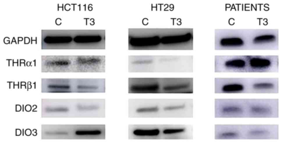
Cell lysates were prepared from colonospheres obtained from both treated and control HCT116 and HT29 cells and cancer cells isolated from CRC patients. After treatment, cells were incubated for 30 min on ice in a RIPA lysis buffer (Sigma-Aldrich; Merck KGaA) supplemented with protease and phosphatase inhibitor cocktail; then centrifuged at 16,000 × g for 10 min at 4°C. Protein concentration in the lysates was measured with Bradford reagent (Sigma-Aldrich; Merck KGaA). Protein samples (10 µg) were loaded to 4–20% Mini-PROTEAN® TGX™ Precast Protein Gels (Bio-Rad Laboratories, Inc.) and electroblotted onto a PVDF membrane with the use of the Trans-Blot Turbo system (Bio-Rad Laboratories, Inc.). Membranes were incubated with 5% non-fat milk in 1% TBST buffer for 1 h at RT. Afterwards, the membranes were incubated overnight at 4°C with the following primary antibodies (purchased from Thermo Fisher Scientific, Inc.): rabbit polyclonal anti-THRα1 antibody (1:500; cat. no BS-6221R), rabbit polyclonal anti-THRβ1 antibody (1:100; cat. no PA1213A), rabbit polyclonal anti-DIO2 antibody (1:1,000; cat. no PA549631) and rabbit polyclonal anti-DIO3 antibody (1:1,000; cat. no PA526537). On the next day, blots were washed with TBST and incubated for 1.5 h at RT with HRP-conjugated anti-rabbit IgG antibody (1:10,000; cat. no 1706515; Bio-Rad Laboratories, Inc.). Anti-GAPDH peroxidase-conjugated IgM antibody (1:50,000; cat. no G9295; Sigma-Aldrich; Merck KGaA; 1 h at RT) was used as the loading control. The membranes were washed and subsequently subjected to luminol reagents (Bio-Rad Laboratories, Inc.). The chemiluminescence signal was measured with ImageQuant LAS 500 (Cytiva). Changes in protein level were assessed by densitometric scanning of the bands (ImageQuant™ TL 10.1 analysis software (Cytiva) and corrected for GAPDH loading control. Each experiment was performed in triplicate. Proteins with molecular weights 51, 43, 60, 34 and 37 kDa for THRα1, THRβ1, DIO2 in dimeric form, DIO3 and GAPDH, respectively, were analyzed.
Statistical analysis
Statistical analysis was performed using GraphPad Prism (version 6.05; GraphPad Software, Inc.). Data were subjected to non-parametric Mann-Whitney U test or Kruskal-Wallis test followed by Dunn's test as a post hoc procedure and Spearman's rank correlation analysis. P<0.05 was considered to indicate a statistically significant difference. Data in figures are presented as the median ± interquartile range or median with min-max values.
Results
In the present study, early stage CRC consisted of 2 (7.5%) stage I cancer patients and 11 (40.5%) stage II cancer patients. Group of advanced stage consisted of 12 (44.5%) stage III cancer patients and 2 (7.5%) stage IV cancer patients. In our study population, cancer was localized in cecum (11%), ascending colon (15%), transverse colon (7.5%), descending colon (7.5%), sigmoid (33%), rectum (22%) and one of the patients (4%) had two CRC in two different segments of large bowel. In detail 14 patients had lymph node metastases (stage III or IV) and 2 patients had distant metastases at the time of diagnosis (stage IV).
Patient-derived spherical cultures contained variable proportion of CSCs bearing CD133 marker (ranging from 7 to 91%), thus the samples were divided according to CD133 median value (which was 20%) into 2 groups: with high (43±19%) and low (12±4.4%) CD133+ cells count. Apparently, the CD133+ cells count was significantly different between these groups (P<0.0001).
T3 eliminates colorectal CSCs in vitro culture
Fresh CRC specimens were collected from 27 patients and it was possible to establish spherical cultures from the cancer tissue of 21 (77.8%) patients, in the remaining cultures cancer cells did not survive ex vivo due to bacterial contamination. All functional tests were conducted with cells obtained after the first passage since it has been already revealed that the phenotype of CSCs along the expansion was stable (9). The additional clinicopathological information concerning patients included into the present study are presented in Table I.
Both HCT116 and HT29 CRC cell lines and cancer cells from patients were cultured in a form of colonospheres which are highly enriched with CSCs, as previously reported by the authors (10,17). All cell populations were subjected to cytometric analysis of phenotype following the treatment with T3 thyroid hormone. The proportion of CSCs with commonly used CSC-like markers, namely CD133, CD44 and CD29, was evaluated. The number of CD133+ cells in culture of spheroids obtained from all cancer cells populations did not change significantly (Figs. 1 and 2 and S1), with two exceptions (Fig. 1A).
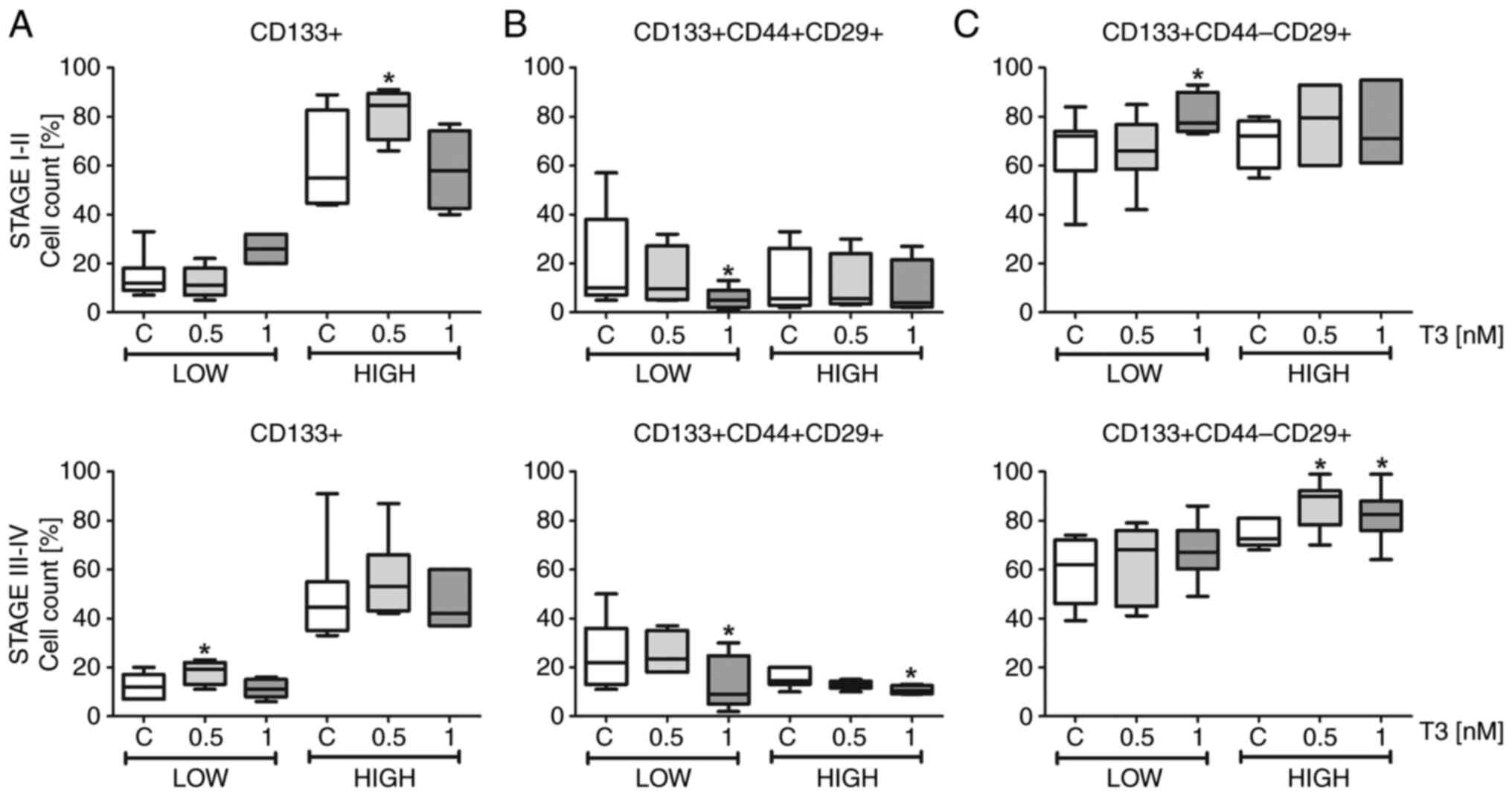
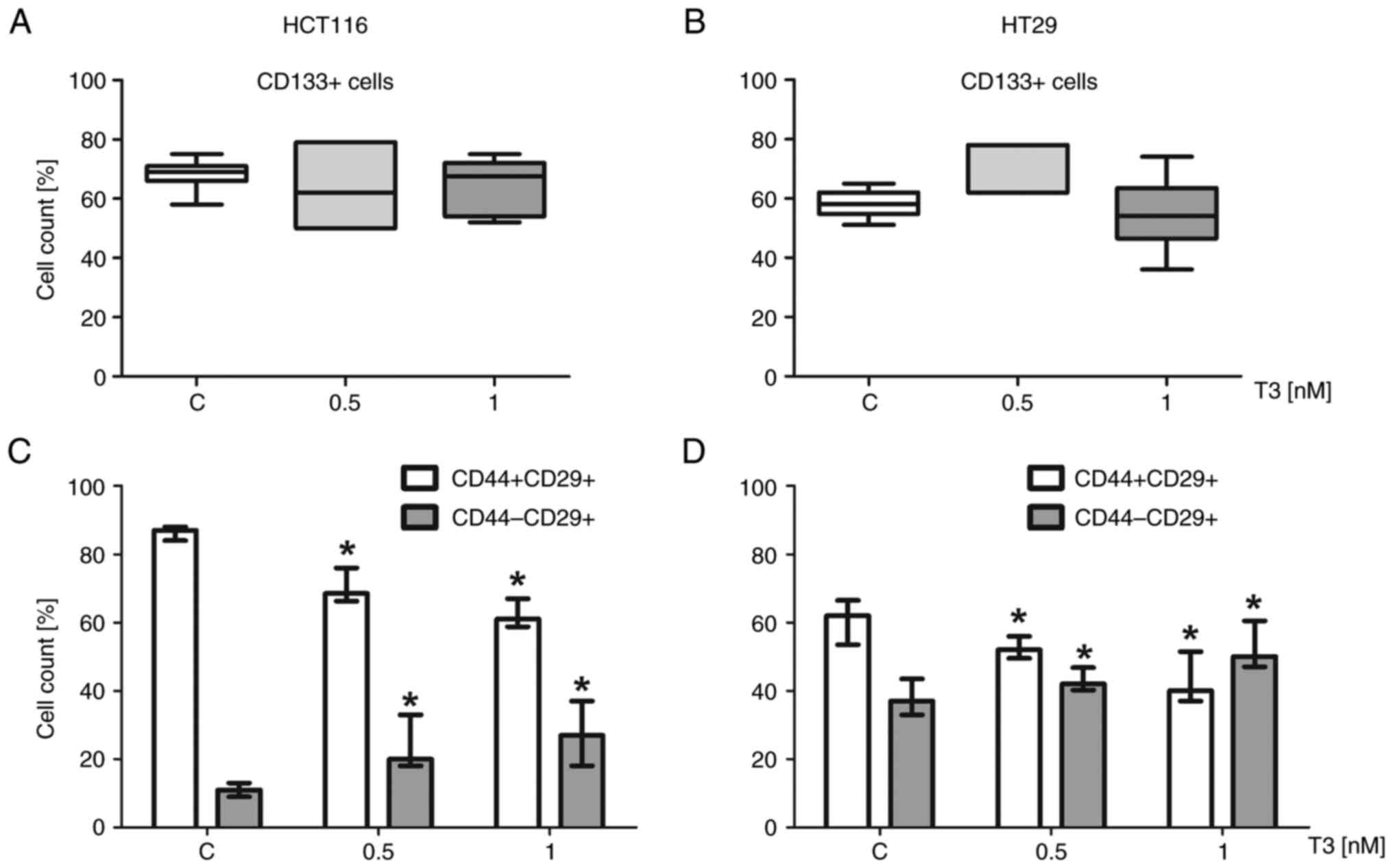
The general count of CD133+ CSCs in colonospheres representing tissue of patients with CRC of stages I and II did not differ significantly from colonospheres derived from tissues of advanced cancer patients (stages III and IV), thus these groups were divided according to the number of CD133+ median value (into Low and High groups). The statistically significant difference in the percentage of CD133+ expressing cells in colonospheres derived from patients representing both Low and High groups was confirmed (P<0.05; Fig. 1). The presented division of patients' samples (Fig. 1) demonstrated that both groups with early and advanced cancer contained samples with high and low count of these primitive cells. This information is of interest since CSCs are known to be responsible for the most serious events of carcinogenesis. Additionally, the analysis of correlation revealed that the amount of untreated and treated CD133+ cells did not depend directly on clinical stage of CRC (R=0.249; P=0.25) (Fig. S2). Furthermore, all spherical populations were analyzed with respect to the presence of CD29 and CD44 integrins. It was observed that CSCs in colonospheres were more likely to change the percentage of cells bearing another analyzed marker in response to T3 treatment. It was noted that CRC colonospheres treated with T3 had increased count of CD133+CD44−CD29+ cells (P<0.05). The alterations in count of CD133+CD44−CD29+ cancer cells were a mirror image (Fig. 1C) compared with CD44+ counterparts (Figs. 1B and C and 2C and D).
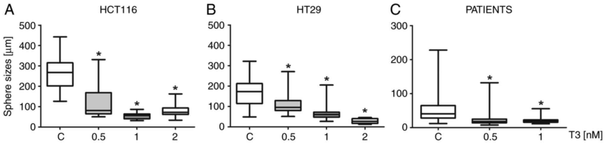
T3 in all concentrations caused visible changes in the morphology of spheres derived from both examined CRC cell lines and cells of patients with CRC. The sizes of colonospheres were significantly reduced when the cells were treated (Fig. 3) with T3 (P<0.05) and the number of dead cells and debris in the medium increased following the treatment.

Additionally, another assay was carried out to determine the ability of CRC cells to form colonospheres following 72-h-long incubation of T3-treated cells. Following T3 treatment cells were transferred into fresh medium without T3 (Fig. 4). After 7 days, the effects of T3 on the ‘secondary sphere’ formation ability were examined, reflecting the tumor-initiating capacity and susceptibility of the CSC-like cells to T3. Both CRC cell lines and cells of patients with CRC presented similar results. Pretreatment with T3 exerted the constant influence on CRC cells. After one week of incubation in fresh medium without T3, CRC cells should form colonospheres with the typical morphology and size, but it was found that the T3 pre-treated colonospheres were evidently smaller (P<0.05) in comparison to primary spheres. Additionally, numerous freely floating cells or small aggregates were observed.
T3 decreases the viability of colorectal CSCs
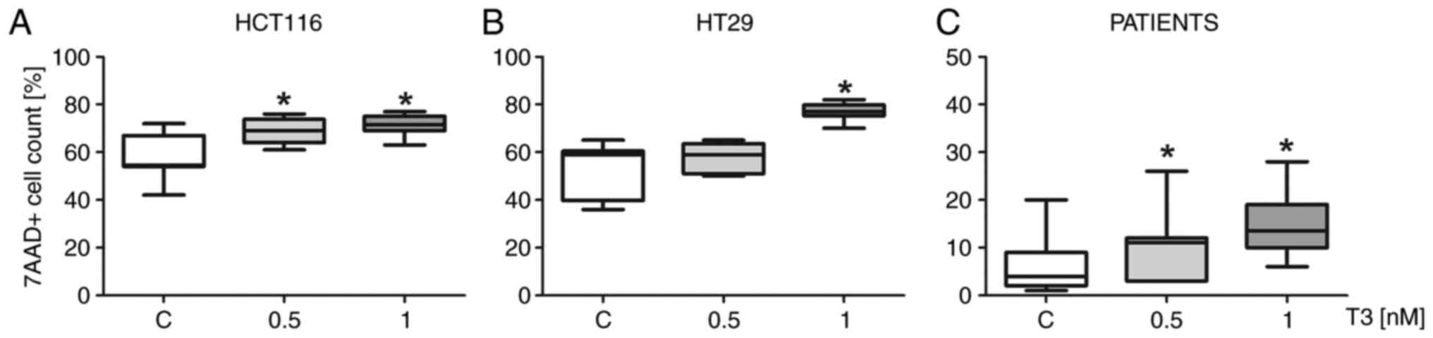
The percentage of non-viable cells following T3 treatment was evaluated by flow cytometric assay using 7-AAD dye (Figs. 5 and S3), which is excluded by living cells, but binds selectively to the DNA of damaged permeabilized cells. It was revealed that the number of 7AAD-positive cells among cultured cells increased after incubation with T3 in concentration-dependent manner (P<0.05). Similar results were observed for colonospheres derived from both CRC cell lines and cells isolated from patients with CRC. These results were confirmed by the analysis of cell cycle using PI (Figs. 6D-F and S4). The increased proportion of cells in subG1 phase was noted after the incubation of cells with T3 in different concentrations in comparison to untreated control. At both concentrations of T3 the fraction of cells undergoing apoptosis (in subG1 phase) increased significantly up to 22±4.53% (median ± SD) for 1 nM T3-treated cells of patients (Fig. 6).
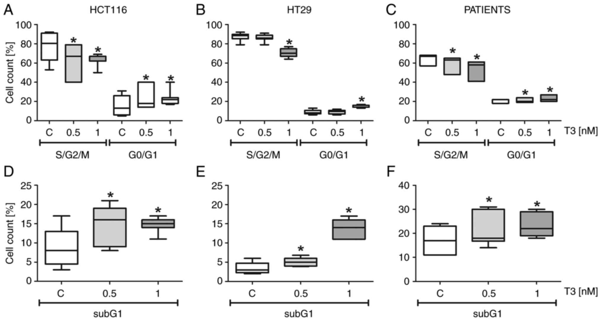
The distribution of cells in various phases of the cell cycle was determined in colonospheres' cultures of HCT116 and HT29 cell lines and cancer cells obtained from CRC patients. Generally, the proportion of cells in active phases (S/G2/M) was higher in comparison to cells representing quiescence pool (G0/G1) (Fig. 6A-C) of colonospheres obtained from all populations included into the present study. G0/G1 cell cycle growth arrest was observed after the incubation of spheres with T3, while the number of cells in S and G2-M phases (active phases of cell cycle) was markedly reduced in comparison to untreated control.
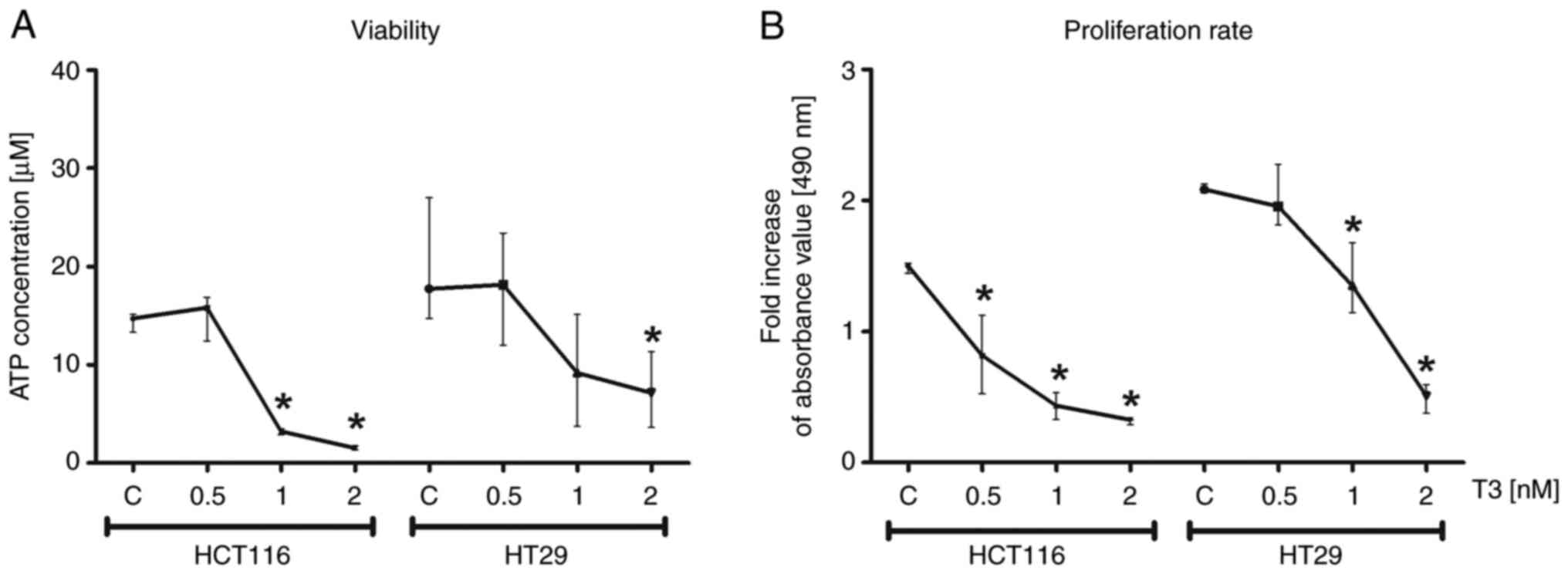
The viability of CRC cells of HCT116 and HT29 cell lines was analyzed by the assessment of total levels of cellular ATP. The test assumed the decrease of ATP level in samples as an indicator of cell death. The results confirmed that the increased concentration of T3 in culture reduced viability of colonospheres (P<0.05; Fig. 7A). In addition, colorimetric test was performed relaying on MTS reagent to determine the proliferative capacity of HCT116 and HT29-derived colonospheres treated with T3. It was revealed that the proliferation rate was significantly decreased following T3 treatment in comparison to control cells (P<0.05; Fig. 7B).
T3 influences the expression of proteins associated with T3 activity
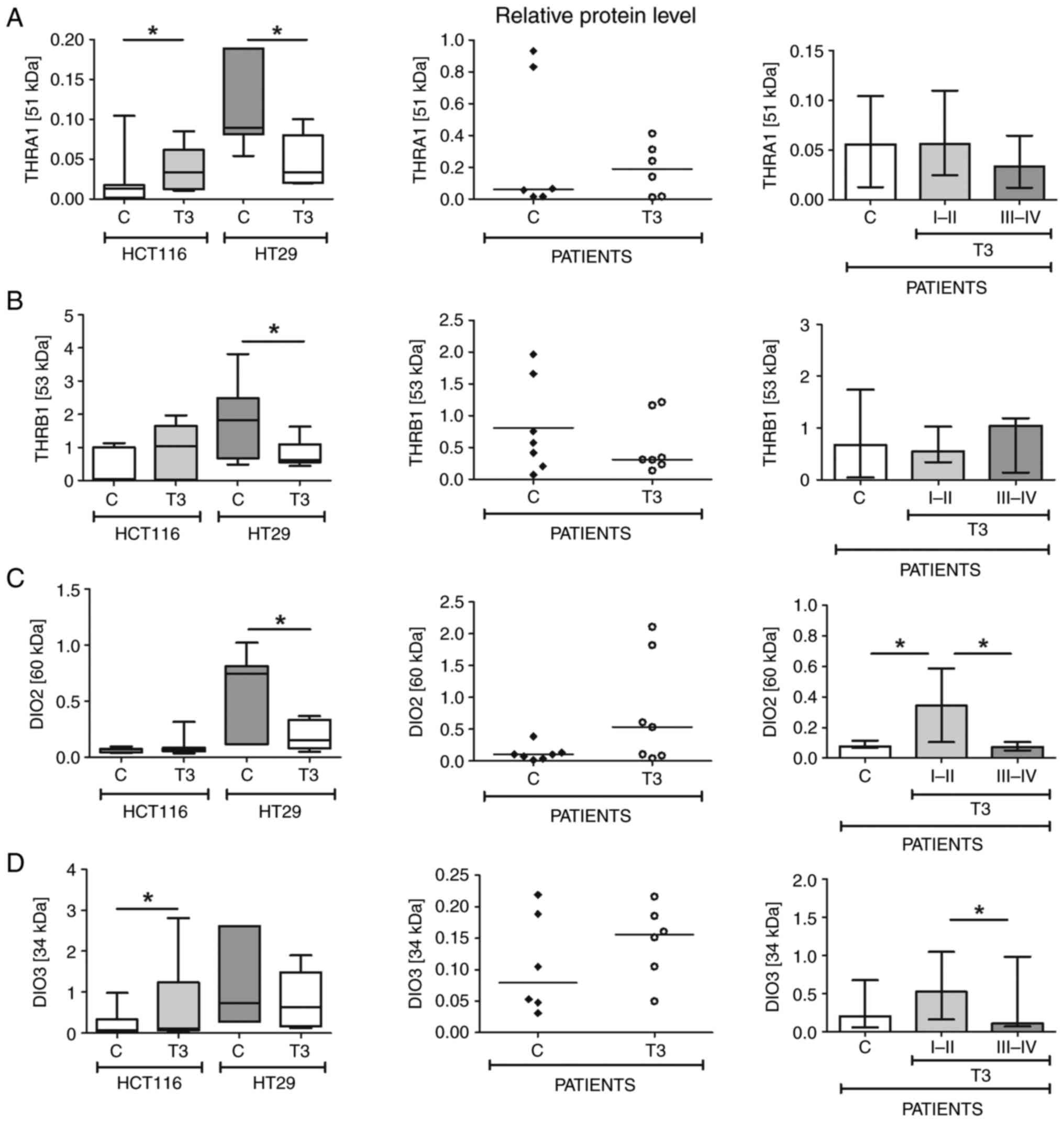
Since the effects of thyroid hormones depend on the activity of some proteins engaged in their activation or binding, the impact of T3 on protein level of two thyroid hormone receptors (THRα1 and THRβ1) and two deiodinases (DIO2 and DIO3) was evaluated by western blotting.
As demonstrated in Figs. 8 and 9, the results obtained for CRC cell lines-derived colonospheres were not fully consistent with changes observed in cancer cells obtained from patients. Western blotting of colonospheres originated from HCT116 cells treated with T3 demonstrated increased relative levels of almost all analyzed proteins. The elevations of THRα1 and DIO3 levels were statistically significant in comparison to control (P<0.05). The level of DIO2 remained at the same level after T3 treatment. Concurrently, the expression of all proteins in colonospheres obtained from HT29 cells was lower in treated samples than in control spheres and only DIO3 change did not reach statistical significance. The western blot analysis of colonospheres from patients with CRC revealed no substantial alterations in the expression of proteins assessed in the present study, however, when samples of patients were divided according to cancer clinical stages, the expression of DIOs was found lower in cancer cells obtained from patients with stage III–IV CRC in comparison to samples collected from patients with stage I–II CRC. Furthermore, the DIO2 and DIO3 protein relative levels were mostly upregulated in patients representing group with early cancer (stage I–II).
Discussion
Thyroid hormones play crucial role in the regulation of multiple physiological activities including differentiation, growth and metabolism and are required for normal function of nearly all tissues (18,19). However, thyroid hormones attract attention since their regulatory functions were found crucial for both physiological processes in normal cells of healthy tissues and also have a great impact on the proliferative abilities and cancer growth of cancer cells. Clinical hypothyroidism was found to be associated with delayed cancer development. Concurrently, hyperthyroidism is correlated with increased cancer growth, including breast, thyroid, lung, brain, liver and CRC (1,18,20–22).
The current study provided novel evidence that T3 can be an important modulator of CRC CSC properties. A model of expansion of colonospheres derived from CRC cell lines and CRC cells isolated from cancer tissue was used. These procedures were previously exercised by the authors (10). The experimental conditions mimicked the microenvironment with locally increased T3 concentration where it was monitored how this crucial thyroid hormone altered features of CSCs or colonospheres. It was found that the proportion of CD133+CD29+CD44+ stem-like cells was significantly decreased following the 3-day treatment with T3, suggesting that treated colonospheres displayed a lower content of CSCs in comparison to untreated control. Indeed, it was identified that T3 significantly inhibited the ability of cancer cells to form colonospheres by increased apoptosis and elevation of G1/G0 silenced pool within cultures. Similarly, the treatment of MCF breast cancer cell line with T3 resulted in decreased proliferative and migratory potential of these cells and reduced number of CSCs (23).
The actions of T3 are initiated by binding to nuclear thyroid receptors (TRs), encoded by two genes, α and β, which give rise to different receptor isoforms (24). TRs are widely distributed in mammalian tissues, but transformed or immortalized cells in general express very low levels of TRs. In addition, there is increasing evidence that alterations in TRs are common events in cancer and it has been suggested that TR genes may function as tumor suppressors (25–28), although the role of these receptors in the pathogenesis and progression of neoplastic processes is currently unclear (28) and results in different types of cancer are not fully consistent (20,22,29–32). The present results are concomitant with this general concept since lower THRα1 and THRβ1 relative protein levels were observed in untreated colonospheres derived from HCT116 cell line which represent CRC with higher progression status in comparison to HT29 cells (TNM3/Dukes' D vs. TNM2/Dukes' C, respectively; P<0.05) (33). Tissue samples of patients presented more diverse results which can be partially explained by heterogeneity of individuals recruited to the present study. CRC cells appeared to sustain both receptors at low but constant level for the potential need to activate the expression of T3 target genes (34). Similar results concerning expression of thyroid hormone receptors were described by Wang et al (2), who reported that thyroid hormones increase cells' self-renewal capacity and the percentage of CD90+ CSCs and promote drug resistance of primary human HCC cells. It was suggested that THRβ1 acts as tumor suppressor in a number of cancers and one of the proposed mechanism relay on upregulation of the nuclear receptor co-repressor 1 and suppression of invasion, tumor growth, and metastasis in human as it was demonstrated for hepatocellular carcinomas (35) and neuroblastomas (36). In addition, breast cancer mammospheres presented reduced tumorigenesis upon stimulation of THRβ1 (37). The possible mechanisms are downregulation of cyclin D1 expression and modulation of the TNFα-NFκB signaling (23,37). Certain of the present results supported these previous observations since it was observed that the expression of THRβ1 receptor was significantly lower in HT29 cells following T3 treatment. In addition, an increased level of THRβ1 protein was demonstrated along with clinical advancement of CRC.
The primary secretory product of the thyroid gland, 3,5,3′,5′ tetra iodothyronine (T4) or thyroxine, must be converted to T3 for its activation. DIO2 (activating DIO) converts the prohormone thyroxine to the active thyroid hormone T3, whereas DIO3 (deactivator DIO), by inactivating both T4 and T3, terminates thyroid hormone action (38). This pre-receptor process is an essential mechanism that regulates thyroid hormone function at the intracellular level. In colorectal adenomas and carcinomas, DIO3 expression is significantly higher than in normal tissues and negatively correlates with the histologic grade of the lesions suggesting that DIO3 could be a marker of early stages of carcinogenesis (38). That appears to be consistent with the present results conducted with both CRC cell lines and cells of patients. Higher DIO3 level was observed in samples derived from patients with lower CRC stage (I–II) in comparison to patients with III–IV stage. The parallel increased expression of both DIOs in cancer cells was assumed to rapidly customize the thyroid hormone signal if necessary what was previously presented for BCC cells as well (39). The alterations in protein levels of both enzymes depended on cancer clinical stage and it was supposed to protect cells from anti-proliferative and anti-survival effect of T3 in vitro cultures. Similar conclusions may be drawn from the analysis of levels of DIOs in the samples of the present study. Higher DIO2 and DIO3 was revealed in stage I–II CRC samples compared with untreated control and stage III–IV CRC cells. These results are in accordance with observations of increased stromal DIO2 level of intestinal polyps of ApcΔ716 mice, a mouse model of familial adenomatous polyposis and early stage sporadic CRC (40).
Furthermore, it was identified that DIO3 is a direct transcriptional target of the β-catenin/TCF complex and its expression was higher in human intestinal adenomas and carcinomas than in healthy intestinal mucosa. Experimental attenuation of β-catenin reduced DIO3 levels and induced DIO2 thereby increasing T3-dependent transcription. In the absence of DIO3, the T3 excess reduced cell proliferation and promoted differentiation in cultured cells and in xenograft mouse models (38). Increased level of DIO3 in samples after treatment with T3 may be an evidence that the overabundance of active thyroid hormone can compromise the fragile niche homeostasis. The increasing T3 concentration inhibited proliferation of treated cells which can be an effect of insufficient DIO3 level. The decline was more significant for HCT116 cells with lower initial DIO3 expression. The CSCs tended toward silencing their activity after treatment with elevated T3 concentration which was revealed with the analysis of phenotype and cell cycle.
It was also observed that the incubation of colonospheres with T3 decreased viability, proliferative and spherogenic potential of cancer cells. In addition, increased apoptotic rate of CRC cells was revealed. Concurrently, cells of treated colonospheres were more likely to move into silenced pool in G0/G1 phase of the cell cycle. Additionally, the smaller sizes of colonospheres following the treatment with T3 suggested that T3 can lower the proportion of primitive cells actively supplying the pool which proliferate within spheres. It was hypothesized that this could be a protective mechanism aimed to avoid the elimination of all cancer-initiating cells from colonospheres. In this context it was confirmed that the hormonal constituents of cancer niche have a crucial role for the fate of CSCs.
The technical limitation is the use of 1.0 N NaOH-based solvent to prepare T3 solutions. Although our initial study demonstrated that the buffer did not influence cells features, the use of clear medium to dissolve T3 powder may have provided more in vivo-like results.
The authors are conscious that the present study presents only a part of cancer-related interactions between certain micro-niche components. The analysis of a narrow fragment of thyroid hormone homeostasis was conducted, rather than the whole TSH-T4-T3-rT3 axis. These hormones may be considered biologically active for healthy cells; however, more efforts are needed to evaluate the role of T4 and T3 in cancer cells. One of the limitations of the present study was the insufficient number of patients to correlate some more clinical parameters with obtained laboratory results. Individuals with CRC are a heterogeneous group of patients, for instance, according to the location of the disease. Cancers localized in rectum and colon are biologically different. All criteria could not be included because our study groups would be too small and subsequent analyses of subsets would be underpowered. Exclusion of patients after neoadjuvant chemoradiation renders comparisons easier and more homogenous but it does not analyze the real-life scenario. Particularly that certain hormones and drugs are known to have a specific interplay with chemotherapy or radiosensitvity. Nevertheless, the present study proposed a novel function of thyroid hormone signaling in the regulation of CRC CSCs fate and the following projects by the authors may extend the patients group to analyze the effect of other clinical parameters on thyroid hormone axis in CSCs in CRC.
In conclusion, considering the present results, it could be hypothesized that thyroid hormones exert a great impact on the fate of colorectal CSCs. Since CRC is one of the most prevalent cancers worldwide the unveiling novel aspects of CRC niche seems to be extremely important. It was found that the T3 role in cancer biology depends on clinical stage of CRC and in near future such data may be taken into consideration in therapeutic decision making. Proteins involved in thyroid hormone homeostasis by their concerted actions in cancer niche cooperate in the synchronized manner with cellular internal elements associated with next steps of carcinogenesis. However, the signals regulating functions of all these elements during cancer development are largely unknown. CSCs which are considered to be responsible for the most serious events in carcinogenesis were found to be highly sensitive to T3. The cellular balance of thyroid hormone associated proteins can be considered as a potential therapeutic target for designing anticancer drugs. The results of the present study suggested that thyroid hormone homeostasis may have potential prognostic and therapeutic value in human CRC. Further research evaluating the detailed functions of thyroid hormone axis on molecular level and their exact biological mechanism may enable introduction of specific chemotherapeutic protocols in the future.
Considering that T3 was found to target CSCs it can be considered to be supplemented to CRC patients to influence the content of tumor microenvironment. Previous studies demonstrated that depletion of endogenous T4 to increase T3 can improve clinical outcomes of patients with advanced cancer. In patients with pre-existent primary hypothyroidism, T3 administration was observed to lower the T4 level and improve survival of patients (4,41). The adjunctive therapy with thyroid hormones can give a hope to improve the efficacy of conventional chemotherapy, however, analyses concerning different type of cancer arises numerous controversies and further research is needed.
Supplementary Material
Supporting Data
Acknowledgements
Not applicable.
Funding
The present study was supported by the National Science Centre of Poland (grant no. DEC-2019/03/X/NZ3/00434).
Availability of data and materials
The datasets generated during and/or analyzed during the current study are available from the corresponding author on reasonable request.
Authors' contributions
OR, MS, PS and JK conceived the study. OR, MS, AO-K, PS and JK developed methodology. OR, MS and JK performed software analysis. OR, MS and JK validated the data. OR, MS, AO-K and PS performed formal analysis. MS, AO-K and PS conducted investigation. MS provided resources. RO, MS and AO-K curated the data. RO and MS prepared the original draft. AO-K, PS and JK wrote reviewed and edited the manuscript. MS and JK supervised the study. JK acquired funding. All authors have read and approved the final version of the manuscript. OR, AO-K, MS, PS and JK confirm the authenticity of all the raw data.
Ethics approval and consent to participate
The present study was conducted according to the guidelines of the Declaration of Helsinki, and was approved (approval no. NKBBN/203/2020) by The Independent Bioethics Committee of Medical University of Gdansk (Gdansk, Poland) from 24.04.2020. Informed consent was obtained from all subjects involved in the present study.
Patient consent for publication
Not applicable.
Competing interests
The authors declare that they have no competing interests.
References
|
Hellevik AI, Asvold BO, Bjøro T, Romundstad PR, Nilsen TI and Vatten LJ: Thyroid function and cancer risk: A prospective population study. Cancer Epidemiol Biomarkers Prev. 18:570–574. 2009. View Article : Google Scholar : PubMed/NCBI | |
|
Wang T, Xia L, Ma S, Qi X, Li Q, Xia Y, Tang X, Cui D, Wang Z, Chi J, et al: Hepatocellular carcinoma: Thyroid hormone promotes tumorigenicity through inducing cancer stem-like cell self-renewal. Sci Rep. 6:251832016. View Article : Google Scholar : PubMed/NCBI | |
|
Chung IH, Chen CY, Lin YH, Chi HC, Huang YH, Tai PJ, Liao CJ, Tsai CY, Lin SL, Wu MH, et al: Thyroid hormone-mediated regulation of lipocalin 2 through the Met/FAK pathway in liver cancer. Oncotarget. 6:15050–15064. 2015. View Article : Google Scholar : PubMed/NCBI | |
|
Hercbergs A, Johnson RE, Ashur-Fabian O, Garfield DH and Davis PJ: Medically induced euthyroid hypothyroxinemia may extend survival in compassionate need cancer patients: An observational study. Oncologist. 20:72–76. 2015. View Article : Google Scholar : PubMed/NCBI | |
|
Moeller LC and Führer D: Thyroid hormone, thyroid hormone receptors, and cancer: A clinical perspective. Endocr Relat Cancer. 20:R19–R29. 2013. View Article : Google Scholar : PubMed/NCBI | |
|
Conde SJ, Luvizotto Rde A, de Síbio MT and Nogueira CR: Thyroid hormone status interferes with estrogen target gene expression in breast cancer samples in menopausal women. ISRN Endocrinol. 2014:3173982014. View Article : Google Scholar : PubMed/NCBI | |
|
Szaryńska M, Olejniczak A and Kmieć Z: The role of cancer stem cells in pathogenesis of colorectal cancer. Postepy Hig Med Dosw (Online). 70:1469–1482. 2016. View Article : Google Scholar : PubMed/NCBI | |
|
Todaro M, Francipane MG, Medema JP and Stassi G: Colon cancer stem cells: Promise of targeted therapy. Gastroenterology. 138:2151–2162. 2010. View Article : Google Scholar : PubMed/NCBI | |
|
Szarynska M, Olejniczak A, Wierzbicki P, Kobiela J, Laski D, Sledzinski Z, Adrych K, Guzek M and Kmiec Z: FasR and FasL in colorectal cancer. Int J Oncol. 51:975–986. 2017. View Article : Google Scholar : PubMed/NCBI | |
|
Olejniczak A, Szarynska M and Kmiec Z: In vitro characterization of spheres derived from colorectal cancer cell lines. Int J Oncol. 52:599–612. 2018.PubMed/NCBI | |
|
van de Velde CJ, Boelens PG, Borras JM, Coebergh JW, Cervantes A, Blomqvist L, Beets-Tan RG, van den Broek CB, Brown G, Van Cutsem E, et al: EURECCA colorectal: Multidisciplinary management: European consensus conference colon & rectum. Eur J Cancer. 50:1.e1–1.e34. 2014. View Article : Google Scholar : PubMed/NCBI | |
|
Brierley DJ, Gospodarowicz M and Wittekind C: Colon and Rectum: (ICD-O-3 C18-20). Classification of malignant tumors digestive system tumours. TNM Online. 73–76. 2017. View Article : Google Scholar | |
|
Shinderman-Maman E, Weingarten C, Moskovich D, Werner H, Hercbergs A, Davis PJ, Ellis M and Ashur-Fabian O: Molecular insights into the transcriptional regulatory role of thyroid hormones in ovarian cancer. Mol Carcinog. 57:97–105. 2018. View Article : Google Scholar : PubMed/NCBI | |
|
Weingarten C, Jenudi Y, Tshuva RY, Moskovich D, Alfandari A, Hercbergs A, Davis PJ, Ellis M and Ashur-Fabian O: The interplay between epithelial-mesenchymal transition (EMT) and the thyroid hormones-αvβ3 axis in ovarian cancer. Horm Cancer. 9:22–32. 2018. View Article : Google Scholar : PubMed/NCBI | |
|
Cohen K, Flint N, Shalev S, Erez D, Baharal T, Davis PJ, Hercbergs A, Ellis M and Ashur-Fabian O: Thyroid hormone regulates adhesion, migration and matrix metalloproteinase 9 activity via αvβ3 integrin in myeloma cells. Oncotarget. 5:6312–6322. 2014. View Article : Google Scholar : PubMed/NCBI | |
|
Hsieh MT, Wang LM, Changou CA, Chin YT, Yang YSH, Lai HY, Lee SY, Yang YN, Whang-Peng J, Liu LF, et al: Crosstalk between integrin αvβ3 and ERα contributes to thyroid hormone-induced proliferation of ovarian cancer cells. Oncotarget. 8:24237–24249. 2017. View Article : Google Scholar : PubMed/NCBI | |
|
Olejniczak-Kęder A, Szaryńska M, Wrońska A, Siedlecka-Kroplewska K and Kmieć Z: Effects of 5-FU and anti-EGFR antibody in combination with ASA on the spherical culture system of HCT116 and HT29 colorectal cancer cell lines. Int J Oncol. 55:223–242. 2019.PubMed/NCBI | |
|
Chi HC, Chen CY, Tsai MM, Tsai CY and Lin KH: Molecular functions of thyroid hormones and their clinical significance in liver-related diseases. Biomed Res Int. 2013:6013612013. View Article : Google Scholar : PubMed/NCBI | |
|
Oppenheimer JH, Schwartz HL, Mariash CN, Kinlaw WB, Wong NC and Freake HC: Advances in our understanding of thyroid hormone action at the cellular level. Endocr Rev. 8:288–308. 1987. View Article : Google Scholar : PubMed/NCBI | |
|
Kamiya Y, Puzianowska-Kuznicka M, McPhie P, Nauman J, Cheng SY and Nauman A: Expression of mutant thyroid hormone nuclear receptors is associated with human renal clear cell carcinoma. Carcinogenesis. 23:25–33. 2002. View Article : Google Scholar : PubMed/NCBI | |
|
Park JW, Zhao L and Cheng SY: Inhibition of estrogen-dependent tumorigenesis by the thyroid hormone receptor β in xenograft models. Am J Cancer Res. 3:302–311. 2013.PubMed/NCBI | |
|
Heublein S, Mayr D, Meindl A, Angele M, Gallwas J, Jeschke U and Ditsch N: Thyroid hormone receptors predict prognosis in BRCA1 associated breast cancer in opposing ways. PLoS One. 10:e01270722015. View Article : Google Scholar : PubMed/NCBI | |
|
López-Mateo I, Alonso-Merino E, Suarez-Cabrera C, Park JW, Cheng SY, Alemany S, Paramio JM and Aranda A: Thyroid hormone receptor β inhibits self-renewal capacity of breast cancer stem cells. Thyroid. 30:116–132. 2020. View Article : Google Scholar : PubMed/NCBI | |
|
Yen PM: Physiological and molecular basis of thyroid hormone action. Physiol Rev. 81:1097–1142. 2001. View Article : Google Scholar : PubMed/NCBI | |
|
Drabkin H, Kao FT, Hartz J, Hart I, Gazdar A, Weinberger C, Evans R and Gerber M: Localization of human ERBA2 to the 3p22-3p24.1 region of chromosome 3 and variable deletion in small cell lung cancer. Proc Natl Acad Sci USA. 85:9258–9262. 1988. View Article : Google Scholar : PubMed/NCBI | |
|
González-Sancho JM, García V, Bonilla F and Muñoz A: Thyroid hormone receptors/THR genes in human cancer. Cancer Lett. 192:121–132. 2003. View Article : Google Scholar : PubMed/NCBI | |
|
Lin KH, Shieh HY, Chen SL and Hsu HC: Expression of mutant thyroid hormone nuclear receptors in human hepatocellular carcinoma cells. Mol Carcinog. 26:53–61. 1999. View Article : Google Scholar : PubMed/NCBI | |
|
Furuyama K, Kawaguchi Y, Akiyama H, Horiguchi M, Kodama S, Kuhara T, Hosokawa S, Elbahrawy A, Soeda T, Koizumi M, et al: Continuous cell supply from a Sox9-expressing progenitor zone in adult liver, exocrine pancreas and intestine. Nat Genet. 43:34–41. 2011. View Article : Google Scholar : PubMed/NCBI | |
|
Guigon CJ, Kim DW, Willingham MC and Cheng SY: Mutation of thyroid hormone receptor-β in mice predisposes to the development of mammary tumors. Oncogene. 30:3381–3390. 2011. View Article : Google Scholar : PubMed/NCBI | |
|
González-Sancho JM, Figueroa A, López-Barahona M, López E, Beug H and Muñoz A: Inhibition of proliferation and expression of T1 and cyclin D1 genes by thyroid hormone in mammary epithelial cells. Mol Carcinog. 34:25–34. 2002. View Article : Google Scholar : PubMed/NCBI | |
|
Chi HC, Liao CH, Huang YH, Wu SM, Tsai CY, Liao CJ, Tseng YH, Lin YH, Chen CY, Chung IH, et al: Thyroid hormone receptor inhibits hepatoma cell migration through transcriptional activation of Dickkopf 4. Biochem Biophys Res Commun. 439:60–65. 2013. View Article : Google Scholar : PubMed/NCBI | |
|
Furuya F, Lu C, Willingham MC and Cheng SY: Inhibition of phosphatidylinositol 3-kinase delays tumor progression and blocks metastatic spread in a mouse model of thyroid cancer. Carcinogenesis. 28:2451–2458. 2007. View Article : Google Scholar : PubMed/NCBI | |
|
Ahmed D, Eide PW, Eilertsen IA, Danielsen SA, Eknæs M, Hektoen M, Lind GE and Lothe RA: Epigenetic and genetic features of 24 colon cancer cell lines. Oncogenesis. 2:e712013. View Article : Google Scholar : PubMed/NCBI | |
|
Pascual A and Aranda A: Thyroid hormone receptors, cell growth and differentiation. Biochim Biophys Acta. 1830:3908–3916. 2013. View Article : Google Scholar : PubMed/NCBI | |
|
Martínez-Iglesias OA, Alonso-Merino E, Gómez-Rey S, Velasco-Martín JP, Martín Orozco R, Luengo E, García Martín R, Ibáñez de Cáceres I, Fernández AF, Fraga MF, et al: Autoregulatory loop of nuclear corepressor 1 expression controls invasion, tumor growth, and metastasis. Proc Natl Acad Sci USA. 113:E328–E337. 2016. View Article : Google Scholar : PubMed/NCBI | |
|
García-Silva S and Aranda A: The thyroid hormone receptor is a suppressor of ras-mediated transcription, proliferation, and transformation. Mol Cell Biol. 24:7514–7523. 2004. View Article : Google Scholar : PubMed/NCBI | |
|
Park JW, Zhao L, Willingham MC and Cheng SY: Loss of tyrosine phosphorylation at Y406 abrogates the tumor suppressor functions of the thyroid hormone receptor β. Mol Carcinog. 56:489–498. 2017. View Article : Google Scholar : PubMed/NCBI | |
|
Dentice M, Luongo C, Ambrosio R, Sibilio A, Casillo A, Iaccarino A, Troncone G, Fenzi G, Larsen PR and Salvatore D: β-Catenin regulates deiodinase levels and thyroid hormone signaling in colon cancer cells. Gastroenterology. 143:1037–1047. 2012. View Article : Google Scholar : PubMed/NCBI | |
|
Miro C, Ambrosio R, De Stefano MA, Di Girolamo D, Di Cicco E, Cicatiello AG, Mancino G, Porcelli T, Raia M, Del Vecchio L, et al: The concerted action of type 2 and type 3 deiodinases regulates the cell cycle and survival of basal cell carcinoma cells. Thyroid. 27:567–576. 2017. View Article : Google Scholar : PubMed/NCBI | |
|
Kojima Y, Kondo Y, Fujishita T, Mishiro-Sato E, Kajino-Sakamoto R, Taketo MM and Aoki M: Stromal iodothyronine deiodinase 2 (DIO2) promotes the growth of intestinal tumors in ApcΔ716 mutant mice. Cancer Sci. 110:2520–2528. 2019. View Article : Google Scholar : PubMed/NCBI | |
|
Sterle HA, Hildebrandt X, Valenzuela Álvarez M, Paulazo MA, Gutierrez LM, Klecha AJ, Cayrol F, Díaz Flaqué MC, Rosemblit C, Barreiro Arcos ML, et al: Thyroid status regulates the tumor microenvironment delineating breast cancer fate. Endocr Relat Cancer. 28:403–418. 2021. View Article : Google Scholar : PubMed/NCBI |


