Introduction
Surgical extraction of an impacted third molar,
typically performed by oral and maxillofacial surgeons, frequently
causes postoperative limitation of jaw function and swelling. The
surgical trauma-initiated inflammatory process promotes these
conditions (1). Numerous approaches
have been used to suppress postoperative inflammation, including
the use of nonsteroidal anti-inflammatory drugs, corticosteroids,
drains, different types of incisions and low-level laser therapy
(LLLT) (2,3).
Since the development of laser therapy in 1971, LLLT
has been used for the management of various diseases, such as
osteoarthritis, carpal tunnel syndrome, tendinopathy, rheumatoid
arthritis, lumbago, non-healing ulcers and epicondylitis (1,4,5). The application of LLLT in dentistry
also began in the 1970s (6). Laser
therapy has been used to prevent or reduce trismus and swelling
following the extraction of impacted third molars, and for the
treatment of chronic sinusitis, herpes simplex, chronic facial
pain, gingivitis, sensory anomalies in the inferior alveolar nerve,
dentinal hypersensitivity and pain following periodontal surgery
(6). However, while LLLT has been
previously used to prevent postoperative trismus and swelling
following removal of the third molar, the outcomes are unclear.
This may be due to variations in the study plans, differences in
the determination of variables associated with postoperative
swelling, the use of diverse types of lasers and hand-pieces, and
differences in the treatment parameters in previous studies
(1).
The objective assessment of postoperative swelling
subsequent to the extraction of impacted mandibular third molars is
difficult, and evaluation of the results mainly depends on the
subjective opinion of the physician. However, facial imaging
systems are rapidly improving with the advent of three-dimensional
(3D) devices. With these systems, soft tissues of the face can be
assessed objectively in a noninvasive manner, as compared with the
conventional two-dimensional (2D) imaging technology (7–9). Imaging
methods such as traditional 2D cephalometry have disadvantages such
as exposing the patient to radiation (10). The 3dMD system provides high
precision 3D surface imaging and has several advantages over the
traditional 2D imaging systems, including ease of use, rapid image
acquisition and being a noninvasive technique. It has been
previously reported that 3D imaging affects the diagnosis,
preoperative planning and postoperative evaluation (8,11).
However, there are few studies in the literature that have
evaluated 3D imaging techniques in the assessment of postoperative
swelling following the extraction of impacted mandibular third
molars (10,12).
It is hypothesized that the postoperative swelling
of patients receiving two-dose LLLT following the removal of
impacted third molars may be reduced as compared with that
occurring after single-dose LLLT. The present study investigated
this hypothesis using a 3dMD system to objectively evaluate whether
the use of LLLT decreased the postoperative swelling following the
removal of impacted third molars. In addition, the current study
aimed to evaluate and compare the effects of single- and two-dose
LLLT on the maximum mouth opening (indicator of trismus) and the
pain level following the molar extraction.
Materials and methods
Patients
A total of 45 patients with an age of ≥16 years were
enrolled into the present study. The study was approved by the
Human Ethics Committee of Inonu University (Malatya, Turkey). All
subjects were informed of the risks of oral surgery and
experimental treatment, and informed written consent was obtained
from all patients.
The inclusion criteria were as follows: Female or
male gender, age of >16 years, absence of systemic illness,
presence of impacted mandibular third molar(s), and surgical
difficulty grade of III B according to the scales of Pell and
Gregory (13). Exclusion criteria
included: Local infection, contraindications to laser therapy,
tobacco use, systemic illness, pregnancy, oral contraceptive use
and breastfeeding. All subjects were operated by the same surgeon
using similar surgical procedures. The duration of the surgical
procedure was noted in each case. LLLT was performed by a different
operator on all patients of each group, and measurements were
performed by another operator blinded to the patient groups.
Treatment groups
Patients were randomized into three treatment groups
(n=15 in each), as follow is: Group 1, which received only routine
management with ice application and served as the control group;
Group 2, which received single-dose LLLT immediately following
surgery; and Group 3, which received two-dose LLLT, immediately
following surgery and on postoperative day 2. Ice therapy was given
for 24 h after surgery. The laser was applied extraorally at the
insertion point of the masseter muscle.
Surgical procedure and treatment
Surgery was performed under local anesthesia with 2
ml of 4% articaine with 1:100,000 epinephrine
(Ultracain® D-S Forte; Sanofi Aventis, Topkapı,
Istanbul, Turkey). A single surgeon performed all the surgical
procedures in order to avoid variations due to the different
surgeon skills, which may influence the results. Following surgery,
all patients received 500 mg paracetamol (Parol; Atabay
Pharmaceuticals and Fine Chemicals, Inc., Istanbul, Turkey) and
benzydamine hydrochloride + chlorhexidine gluconate gargle
antiseptic solution (Farhex; Santa Farma, Istanbul, Turkey) two
times per day for 7 days.
In the present experimental study, LLLT was
performed using a gallium-aluminum-arsenide (GaAlAs) diode laser
device (Cheese Dental Laser System; Wuhan Gigaa Optronics
Technology Co., Ltd., Wuhan, China) was used. Parameters of the
LLLT are given in Table I.
 | Table I.Parameters of the low-level laser
therapy performed in the present study. |
Table I.
Parameters of the low-level laser
therapy performed in the present study.
| Parameter | Value |
|---|
| Wavelength | 810 nm |
| Beam area | 3 cm2 |
| Output power | 0.3 Watts |
| Irradiation time | 40 sec |
| Energy density | 4
J/cm2 |
| Energy delivered | 12 J |
| Pulse rate | Continuous |
| Application | Non-contact |
Assessment of trismus and pain
Trismus was assessed at postoperative days 2 and 7
by determining the maximal opening between the right upper and
right lower central incisors with a compass and comparing with that
prior to surgery as described previously (14). The pain degree was assessed using a
visual analog scale (VAS) of 10 points. The scores extended between
0 (no pain) and 10 (the greatest pain). Subsequent to surgery, the
patients were directed to mark the intensity level of pain during
the postoperative period.
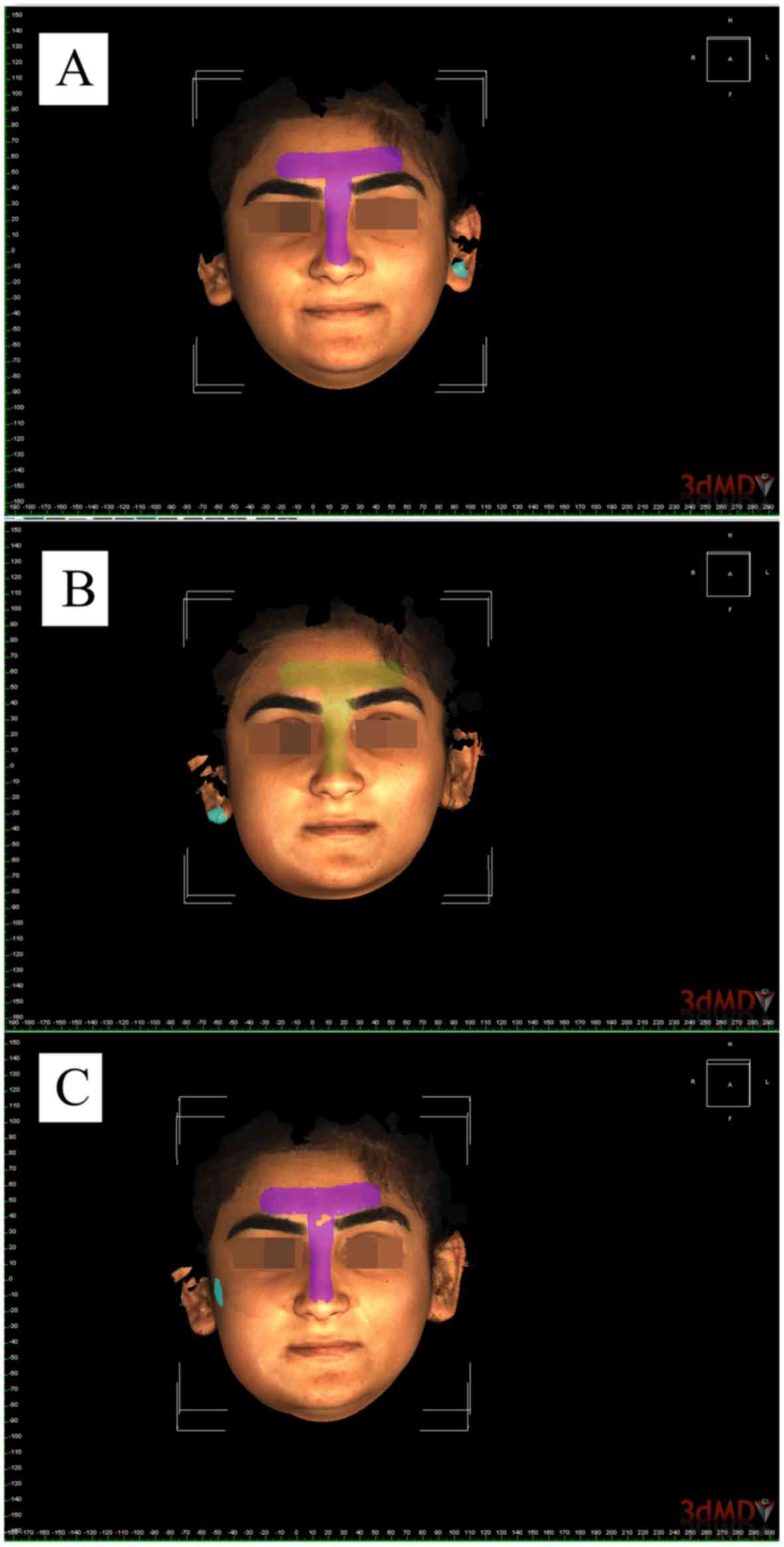
Imaging
The 3dMD face system (3dMD Inc., Atlanta, Georgia,
USA) was used to obtain a preoperative image and postoperative
images on the days 2 and 7 in all patients included in the present
study (Fig. 1). The 3dMD Vultus
software was used to analyze the images. Preoperative and
postoperative 3D stereophotogrammetric images were imported into
the 3dMD Vultus software. Using this program, two different images
can be aligned on the selected surfaces, and linear and volumetric
measurements can be performed between the aligned images. The
analysis began by transferring the images of the patients obtained
prior to the surgical procedure, and on postoperative days 2 and 7
as tsb file format into the 3dMD Vultus software. Two images were
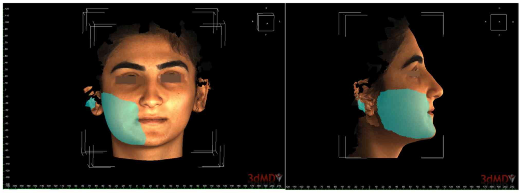
aligned on the forehead and nasofrontal area for examination
subsequent to adjustment. A quadrilateral area with the subnasale,
tragion, gonion and menton points as the corners was selected after
the images were aligned (Fig. 2),
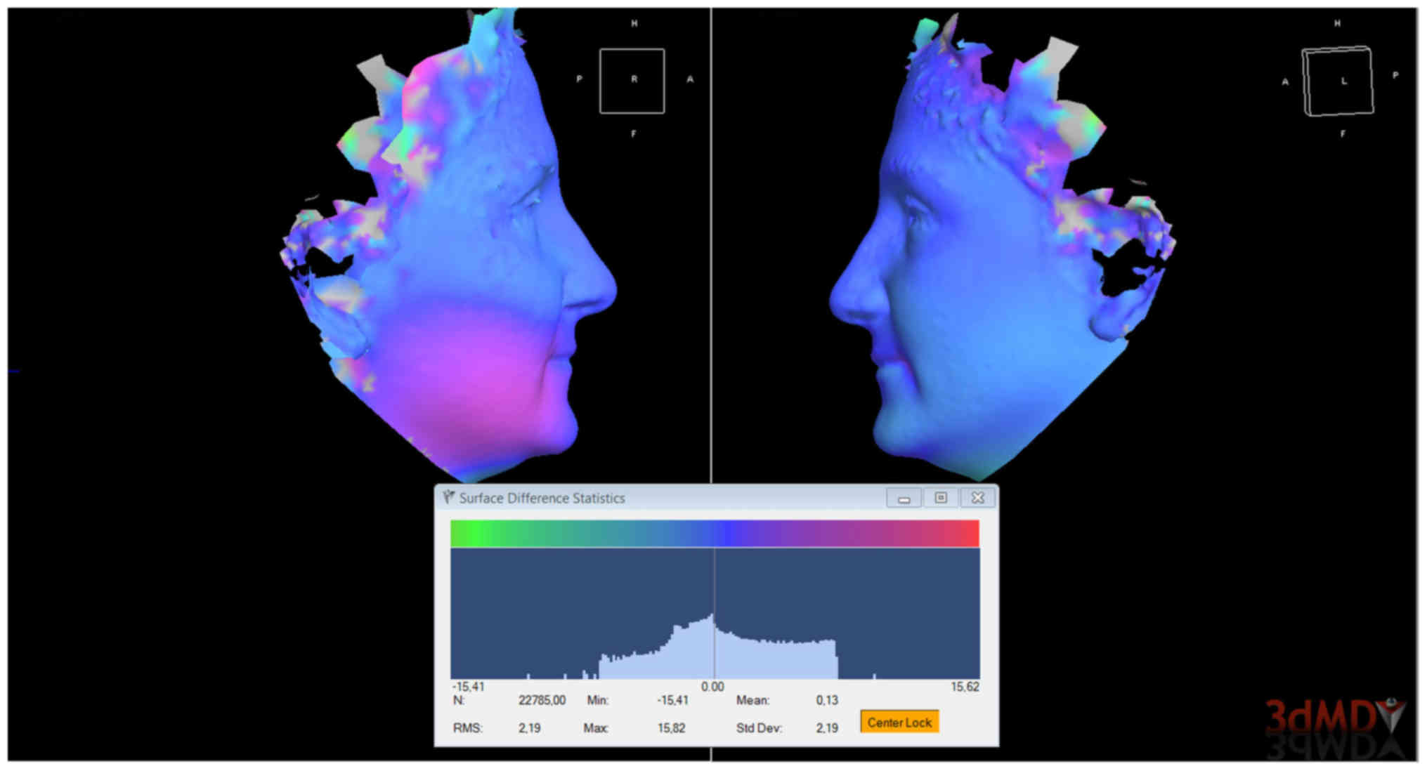
and the volumetric difference between the two surfaces was
calculated (Fig. 1). Furthermore, a
color histogram was prepared showing the relative volume change
between the preoperative and postoperative image (Fig. 3).
Statistical analysis
The IBM SPSS version 22.0 statistics program (IBM
Corp., Armonk, NY, USA) was used for statistical analyses. The data
are summarized as the mean ± standard deviation. Normal
distribution was assessed using the one-way analysis of variance
test. For non-normally distributed data, the Mann-Whitney U test
with Bonferroni correction was used to compare between two groups.
P<0.05 was considered to indicate a statistically significant
difference.
Results
Patient demographics
A total of 45 patients (18 males and 27 females) who
had asymptomatic impacted mandibular third molars extracted were
included in the study. The patients in Group 1 were 17 to 27 years
old (mean, 22.4 years); the patients in Group 2 were 16 to 21 years
old (mean, 18.4 years) and the patients in Group 3 were 17 to 27
years old (mean, 21.7 years). Mean duration of surgery was 15.3 min
in Group 1, 14.9 min in Group 2 and 12.5 min in Group 3. Regarding
the age of patients included in the study, the mean age of Group 2
patients was significantly lower compared with that in Groups 1 and
3 (P<0.001; Table II). By
contrast, there were no statistically significant differences in
the sex ratios and duration of surgery among the three groups
(P=0.757 and P=0.119, respectively; Table II).
| Table II.Patient demographics and surgery
duration. |
Table II.
Patient demographics and surgery
duration.
| Parameter | Group 1 | Group 2 | Group 3 | P-value |
|---|
| Gender, n (%) |
|
|
| 0.757 |
|
Female | 9 (60) | 8 (53.3) | 10 (66.7) |
|
|
Male | 6 (40) | 7 (46.7) | 5
(33.3) |
|
| Age, years |
22.4±5.4a | 18.4±1.4 |
21.7±3.2a | 0.002 |
| Duration of
surgery, min | 15.3±5.8 | 14.9±3.8 | 12.5±3.2 | 0.119 |
Swelling, pain level and trismus
The swelling and pain level (according to the VAS
values) were significantly reduced between days 2 and 7 in all
groups (Table III). However, no
statistically significant difference was observed in the mean
swelling among the different groups on postoperative day 2 or on
day 7 (P=0.140 and P=0.643, respectively). In addition, there was
no significant difference in the mean VAS scores among the three
treatment groups on postoperative day 2 (P=0.233). By contrast, on
day 7 following surgery, there was a statistically significant
difference in VAS values among the groups (P=0.008). The mean VAS
score of Group 1 was significantly higher compared with that of
Group 2 (P=0.005), although no significant difference was detected
between Group 3 and Groups 1 and 2 (P=0.178 and P=0.021,
respectively; Table III).
| Table III.Swelling, trismus (according to the
interincisal opening alteration) and VAS of patients. |
Table III.
Swelling, trismus (according to the
interincisal opening alteration) and VAS of patients.
| Parameter | Group 1 | Group 2 | Group 3 | P-value |
|---|
| Swelling (ml) |
|
|
|
|
| Day
2 |
20.3±11.8 | 15.5±5.4 |
22.8±13.4 | 0.140 |
| Day
7 |
6.6±8.2 |
2.3±1.8 |
3.8±3.6 | 0.643 |
|
P-value | 0.001 | 0.001 | 0.001 |
|
| VAS score |
|
|
|
|
| Day
2 |
4.1±2.0 |
3.4±1.9 |
4.7±2.4 | 0.233 |
| Day
7 |
2.1±1.4 |
0.6±1.2a |
1.5±1.1 | 0.008 |
|
P-value | 0.007 | 0.001 | 0.002 |
|
| Interincisal
opening (mm) |
|
|
|
|
| Day
0 | 44.1±6.0 | 43.3±6.9 | 45.0±4.1 | 0.730 |
| Day
2 | 26.8±4.6 |
31.1±10.7 | 28.1±7.5 | 0.441 |
| Day
7 | 37.2±6.9 | 37.1±9.7 | 38.1±6.1 | 0.918 |
|
P-value | <0.001 | <0.001 | <0.001 |
|
For the determination of alterations in trismus, the
interincisal mouth opening of patients was examined. The results
detected no statistically significant difference in the trismus
occurring subsequent to surgery in Groups 1, 2 or 3 when compared
with the interincisal opening prior to surgery (P=0.730, P=0.441
and P=0.918; Table III).
Discussion
The removal of impacted mandibular third molar teeth
is one of the most common procedures in oral surgery (15). The surgical extraction of an impacted
third molar tooth results in postoperative morbidity, which may be
divided into immediate postoperative tissue reactions and
complications (16). Pain, swelling
and limited mouth opening due to muscle spasm (also known as
trismus) are the most common complications that cause significant
postoperative discomfort (17), and
adversely affect the quality of life of patients (18). Therefore, clinicians have highlighted
the necessity for better management of these complications in
patients who undergo third molar surgery (19).
These complications can be reduced by administration
of nonsteroidal anti-inflammatory drugs (NSAIDs), local or systemic
corticosteroids, or the combination of corticosteroids and NSAIDs
(20–23). However, these drugs may be unsafe in
certain patients and may cause various side effects, such as
gastrointestinal irritation, systemic bleeding or allergic
reactions (22). Thus, there is
growing interest in establishing alternative methods without side
effects. In this regard, the use of LLLT offers promising
application possibilities (1).
LLLT was first used in the fields of dentistry and
oral surgery in the early 1970s, and has since expanded to
different medical specialties (1).
Due to variations in application, the efficacy of LLLT for the
prevention of pain, postoperative swelling and trismus subsequent
to third molar surgery remains controversial. This variation may be
due to differences in study design or methods, difficulties in the
measurement of variables associated with postoperative sequelae,
differences in the type of lasers and hand-pieces used, and
differences in irradiation parameters (1,24,25).
Numerous studies have used LLLT in dentoalveolar
surgery in order to reduce facial swelling, pain and trismus
(1,6,21,24,26).
However, there is not sufficient evidence to support that the use
of LLLT is more effective when compared with no active treatment
(placebo or no treatment) to minimize pain, swelling and trismus
following impacted mandibular third molar surgical removal
(27). Studies with positive, as
well as negative results have been reported. For instance, Carrillo
et al (26) reported no
significant differences in the levels of pain and swelling between
the laser-treated and the placebo groups. However, in the same
study, LLLT (He-Ne; 633 nm; energy density of 10 J/cm2)
provided a significant reduction in trismus in the laser-treated
group after 7 days. In addition, Aras and Güngörmüş (6) observed that LLLT (GaAlAs; 808 nm;
energy density of 4 J/cm2) significantly decreased
trismus, although there were no significant differences in the
levels of swelling between the intraoral-LLLT and the placebo
groups. By contrast, López-Ramírez et al (14) reported that LLLT (GaAlAs; 810 nm;
energy density of 5 J/cm2) had no beneficial effects in
reducing pain, swelling and trismus following removal of impacted
third molars. Røynesdal et al (28) reported that LLLT (830 nm; 40 mW; 6 J)
had similar results to those observed by the López-Ramírez et
al (14) study. Furthermore,
Ferrante et al (1)
demonstrated that LLLT (980 nm; 300 mW; 180 sec) was useful for the
reduction of postoperative trismus and swelling subsequent to third
molar surgery. According to the results of the present study,
significantly reduced pain and swelling was observed between days 2
and 7 after surgery in all three groups. However, LLLT treatment
had no significant effect on the swelling and trismus among the
three groups at either postoperative time point. In addition, while
no statistically significant differences were observed in the
levels of pain at 2 days after surgery, there was a statistically
significant difference in pain levels among the groups at 7 days
after surgery.
Amarillas-Escobar et al (29) performed a similar study to the
current study, although in order to evaluate the cumulative effect
of the therapeutic laser, LLLT (Nd-YAG; 810 nm; 4 J/cm2)
was applied as multiple daily intraoral doses immediately after
surgery and postoperatively at 24, 48 and 72 h. The results of
their study demonstrated no significant differences in the
reduction of pain, swelling or trismus between the laser-treated
and control groups. In the present study, patients in Group 3
received two doses of LLLT, immediately following surgery and
postoperatively at 48 h, and no statistically significant
differences were observed in the swelling and trismus between
groups. However, a statistically significant difference was
identified in the mean VAS levels between the groups at
postoperative day 7 (P=0.008), and the mean VAS score was
significantly higher in Group 1 compared with that in Group 2.
In the current study, the laser was used extraorally
since a previous study by Aras and Güngörmüş (6) demonstrated that extraoral LLLT was more
effective in comparison with intraoral LLLT for the reduction of
postoperative trismus and swelling following extraction of the
lower third molar.
A number of different techniques have been
previously used to measure postoperative swelling, including verbal
response scales, mechanical methods (cephalostats, calipers, and
registration of reference points or landmarks), ultrasound,
photographic techniques, computed tomography and magnetic resonance
imaging (28,30–32).
Advances in 3D imaging techniques have made it possible to capture
and superimpose facial images and measure alterations in soft
tissue position in three dimensions (33). Soft tissue images obtained with the
3dMD system provide photorealistic views and capture the texture of
the skin with better accuracy and reproducibility (34). Therefore, the present study used the
3dMD face imaging system to measure the postoperative swelling
following mandibular third molar surgery. The 3D facial images were
obtained immediately prior to the surgical procedure, as well as on
days 2 and 7 following the surgical procedure.
Clinically, 3dMD system can be used to objectively
measure volume changes in the craniomaxillofacial region and to
evaluate the effects of clinical interventions (35). Asutay et al (9) evaluated the effects of platelet rich
fibrin on swelling with the 3dMD imaging system, which was used for
the first time in lower third molar surgery. Furthermore, in our
earlier study (12), we evaluated
the effect of LLLT (two doses) on the pain, mouth opening and
swelling of patients whose bilateral impacted third molar teeth
were extracted in addition to measurement volumetrically to the
edema with 3dMD face system. A random side impacted tooth of the
patients was extracted at the first appointment, and an extraoral
laser was applied on the area of masseter muscle immediately after
surgery and at postoperative day 2. At the follow-up appointment 1
month later, the other side impacted 3rd molar tooth was then
extracted and ice was applied for the first 48 h. In this earlier
study, although the results show that the proposed method reduces
pain, swelling, and trismus, significant differences in pain level
were observed only at day 7 compared with the control group
(12). The current study aimed to
examine the effect of two different LLLT protocols (single dose and
two doses) on postoperative pain, facial swelling and trismus of
patients whose unilateral impacted third molar tooth was extracted.
The results of the present study have shown that LLLT was effective
in reducing pain level only at postoperative day 7.
In conclusion, the present study evaluated the
effect of two different LLLT protocols on postoperative pain,
facial swelling and trismus subsequent to mandibular third molar
surgery. Furthermore, a 3D method was used to objectively evaluate
volume changes in swelling. Intergroup analysis revealed no
significant differences between the groups regarding swelling and
trismus on postoperative days 2 or 7. However, single-dose LLLT
resulted in a significant reduction in pain on day 7. These data
suggested that the use of single-dose LLLT was more effective
compared with routine management for the reduction of pain
following third molar surgery. However, the application of two-dose
LLLT did not increase the beneficial effects on reducing pain,
swelling and trismus following the surgery.
Acknowledgements
This study was presented as a poster presentation at
the 21st Congress of the Balkan Stomatological Society, May 2016,
Banja Luka, Bosnia and Herzogovina.
References
|
1
|
Ferrante M, Petrini M, Trentini P,
Perfetti G and Spoto G: Effect of low-level laser therapy after
extraction of impacted lower third molars. Lasers Med Sci.
28:845–849. 2013. View Article : Google Scholar : PubMed/NCBI
|
|
2
|
Mehrabi M, Allen JM and Roser SM:
Therapeutic agents in perioperative third molar surgical
procedures. Oral Maxillofac Surg Clin North Am. 19:69–84, vi. 2007.
View Article : Google Scholar : PubMed/NCBI
|
|
3
|
Carrillo JS, Calatayud J, Manso FJ,
Barberia E, Martinez JM and Donado M: A randomized double-blind
clinical trial on the effectiveness of helium-neon laser in the
prevention of pain, swelling and trismus after removal of impacted
third molars. Int Dent J. 40:31–36. 1990.PubMed/NCBI
|
|
4
|
Mester E, Spiry T, Szende B and Tota JG:
Effect of laser rays on wound healing. Am J Surg. 122:532–535.
1971. View Article : Google Scholar : PubMed/NCBI
|
|
5
|
Silveira PC, Silva LA, Freitas TP, Latini
A and Pinho RA: Effects of low-power laser irradiation (LPLI) at
different wavelengths and doses on oxidative stress and
fibrogenesis parameters in an animal model of wound healing. Lasers
Med Sci. 26:125–131. 2011. View Article : Google Scholar : PubMed/NCBI
|
|
6
|
Aras MH and Güngörmüş M:
Placebo-controlled randomized clinical trial of the effect two
different low-level laser therapies (LLLT)-intraoral and
extraoral-on trismus and facial swelling following surgical
extraction of the lower third molar. Lasers Med Sci. 25:641–645.
2010. View Article : Google Scholar : PubMed/NCBI
|
|
7
|
Kau CH, Cronin AJ and Richmond S: A
three-dimensional evaluation of postoperative swelling following
orthognathic surgery at 6 months. Plast Reconstr Surg.
119:2192–2199. 2007. View Article : Google Scholar : PubMed/NCBI
|
|
8
|
Simanca E, Morris D, Zhao L, Reisberg D
and Viana G: Measuring progressive soft tissue change with
nasoalveolar molding using a three-dimensional system. J Craniofac
Surg. 22:1622–1625. 2011. View Article : Google Scholar : PubMed/NCBI
|
|
9
|
Asutay F, Yolcu Ü, Geçör O, Acar AH,
Öztürk SA and Malkoç S: An evaluation of effects of
platelet-rich-fibrin on postoperative morbidities after lower third
molar surgery. Niger J Clin Pract. 20:1531–1536. 2017.PubMed/NCBI
|
|
10
|
Nord F, Ferjencik R, Seifert B, Lanzer M,
Gander T, Matthews F, Rücker M and Lübbers HT: The 3dMD
photogrammetric photo system in cranio-maxillofacial surgery:
Validation of interexaminer variations and perceptions. J
Craniomaxillofac Surg. 43:1798–1803. 2015. View Article : Google Scholar : PubMed/NCBI
|
|
11
|
Bared A, Rashan A, Caughlin BP and Toriumi
DM: Lower lateral cartilage repositioning: Objective analysis using
3-dimensional imaging. JAMA Facial Plast Surg. 16:261–267. 2014.
View Article : Google Scholar : PubMed/NCBI
|
|
12
|
Alan H, Yolcu U, Koparal M, Özgür C,
Öztürk SA and Malkoç S: Evaluation of the effects of the low-level
laser therapy on swelling, pain, and trismus after removal of
impacted lower third molar. Head Face Med. 12:252016. View Article : Google Scholar : PubMed/NCBI
|
|
13
|
Pell GJ and Gregory GT: Report on a
ten-year study of a tooth division technique for the removal of
impacted teeth. Am J Orthod Oral Surg. 28:B660–B666. 1942.
View Article : Google Scholar
|
|
14
|
López-Ramírez M, Vílchez-Pérez MA,
Gargallo-Albiol J, Arnabat-Domínguez J and Gay-Escoda C: Efficacy
of low-level laser therapy in the management of pain, facial
swelling, and postoperative trismus after third molar extraction. A
preliminary study. Lasers Med Sci. 27:559–566. 2012. View Article : Google Scholar : PubMed/NCBI
|
|
15
|
Sigron GR, Pourmand PP, Mache B,
Stadlinger B and Locher MC: The most common complications after
wisdom-tooth removal: Part 1: A retrospective study of 1,199 cases
in the mandible. Swiss Dent J. 124:1042-1046–1052-1056. 2014.
|
|
16
|
Bello SA, Adeyemo WL, Bamgbose BO, Obi EV
and Adeyinka AA: Effect of age, impaction types and operative time
on inflammatory tissue reactions following lower third molar
surgery. Head Face Med. 7:82011. View Article : Google Scholar : PubMed/NCBI
|
|
17
|
Barone A, Marconcini S, Giacomelli L,
Rispoli L, Calvo JL and Covani U: A randomized clinical evaluation
of ultrasound bone surgery versus traditional rotary instruments in
lower third molar extraction. J Oral Maxillofac Surg. 68:330–336.
2010. View Article : Google Scholar : PubMed/NCBI
|
|
18
|
Colorado-Bonnin M, Valmaseda-Castellón E,
Berini-Aytés L and Gay-Escoda C: Quality of life following lower
third molar removal. Int J Oral Maxillofac Surg. 35:343–347. 2006.
View Article : Google Scholar : PubMed/NCBI
|
|
19
|
Kazancioglu HO, Ezirganli S and Demirtas
N: Comparison of the influence of ozone and laser therapies on
pain, swelling, and trismus following impacted third-molar surgery.
Lasers Med Sci. 29:1313–1319. 2014. View Article : Google Scholar : PubMed/NCBI
|
|
20
|
Vegas-Bustamante E, Micó-Llorens J,
Gargallo-Albiol J, Satorres-Nieto M, Berini-Aytés L and Gay-Escoda
C: Efficacy of methylprednisolone injected into the masseter muscle
following the surgical extraction of impacted lower third molars.
Int J Oral Maxillofac Surg. 37:260–263. 2008. View Article : Google Scholar : PubMed/NCBI
|
|
21
|
Marković AB and Todorović L: Postoperative
analgesia after lower third molar surgery: Contribution of the use
of long-acting local anesthetics, low-power laser, and diclofenac.
Oral Surg Oral Med Oral Pathol Oral Radiol Endod. 102:e4–e8. 2006.
View Article : Google Scholar
|
|
22
|
Merry AF, Gibbs RD, Edwards J, Ting GS,
Frampton C, Davies E and Anderson BJ: Combined acetaminophen and
ibuprofen for pain relief after oral surgery in adults: A
randomized controlled trial. Br J Anaesth. 104:80–88. 2010.
View Article : Google Scholar : PubMed/NCBI
|
|
23
|
Kim K, Brar P, Jakubowski J, Kaltman S and
Lopez E: The use of corticosteroids and nonsteroidal
antiinflammatory medication for the management of pain and
inflammation after third molar surgery: A review of the literature.
Oral Surg Oral Med Oral Pathol Oral Radiol Endod. 107:630–640.
2009. View Article : Google Scholar : PubMed/NCBI
|
|
24
|
Markovic A and Todorovic L: Effectiveness
of dexamethasone and low-power laser in minimizing oedema after
third molar surgery: A clinical trial. Int J Oral Maxillofac Surg.
36:226–229. 2007. View Article : Google Scholar : PubMed/NCBI
|
|
25
|
Miloro M and Repasky M: Low-level laser
effect on neurosensory recovery after sagittal ramus osteotomy.
Oral Surg Oral Med Oral Pathol Oral Radiol Endod. 89:12–18. 2000.
View Article : Google Scholar : PubMed/NCBI
|
|
26
|
Carrillo JS, Calatayud J, Manso FJ,
Barberia E, Martinez JM and Donado M: A randomized double-blind
clinical trial on the effectiveness of helium-neon laser in the
prevention of pain, swelling and trismus after removal of impacted
third molars. Int Dent J. 40:31–36. 1990.PubMed/NCBI
|
|
27
|
Brignardello-Petersen R, Carrasco-Labra A,
Araya I, Yanine N, Beyene J and Shah PS: Is adjuvant laser therapy
effective for preventing pain, swelling, and trismus after surgical
removal of impacted mandibular third molars? A systematic review
and meta-analysis. J Oral Maxillofac Surg. 70:1789–1801. 2012.
View Article : Google Scholar : PubMed/NCBI
|
|
28
|
Røynesdal AK, Björnland T, Barkvoll P and
Haanaes HR: The effect of soft-laser application on postoperative
pain and swelling. A double-blind, crossover study. Int J Oral
Maxillofac Surg. 22:242–245. 1993. View Article : Google Scholar : PubMed/NCBI
|
|
29
|
Amarillas-Escobar ED, Toranzo-Fernández
JM, Martinez-Rider R, Noyola-Frías MA, Hidalgo-Hurtado JA, Serna
VM, Gordillo-Moscoso A and Pozos-Guillén AJ: Use of therapeutic
laser after surgical removal of impacted lower third molars. J Oral
Maxillofac Surg. 68:319–324. 2010. View Article : Google Scholar : PubMed/NCBI
|
|
30
|
Erdogan O, Tatlı U, Ustün Y and Damlar I:
Influence of two different flap designs on the sequelae of
mandibular third molar surgery. Oral Maxillofac Surg. 15:147–152.
2011. View Article : Google Scholar : PubMed/NCBI
|
|
31
|
Al-Khateeb TH and Nusair Y: Effect of the
proteolytic enzyme serrapeptase on swelling, pain and trismus after
surgical extraction of mandibular third molars. Int J Oral
Maxillofac Surg. 37:264–268. 2008. View Article : Google Scholar : PubMed/NCBI
|
|
32
|
Meisami T, Musa M, Keller MA, Cooper R,
Clokie CM and Sàndor GK: Magnetic resonance imaging assessment of
airway status after orthognathic surgery. Oral Surg Oral Med Oral
Pathol Oral Radiol Endod. 103:458–463. 2007. View Article : Google Scholar : PubMed/NCBI
|
|
33
|
Cevidanes LH, Oliveira AE, Grauer D,
Styner M and Proffit WR: Clinical application of 3D imaging for
assessment of treatment outcomes. Semin Orthod. 17:72–80. 2011.
View Article : Google Scholar : PubMed/NCBI
|
|
34
|
Lane C and Harrell W Jr: Completing the
3-dimensional picture. Am J Orthod Dentofacial Orthop. 133:612–620.
2008. View Article : Google Scholar : PubMed/NCBI
|
|
35
|
van der Meer WJ, Dijkstra PU, Visser A,
Vissink A and Ren Y: Reliability and validity of measurements of
facial swelling with a stereophotogrammetry optical
three-dimensional scanner. Br J Oral Maxillofac Surg. 52:922–927.
2014. View Article : Google Scholar
|