Introduction
Neuropilin (NRP) was first identified as a
single-pass transmembrane protein from the optic tract of
Xenopus laevis in 1987 (1).
In 1997, two separate groups used a genetic expression-cloning
technique to characterize NRP-1 as the receptor for semaphorin
(sema)-3A during development of the nervous system (2–4). NRP-1
consists of an 860-amino acid (aa) extracellular glycoprotein
region, a 22-aa transmembrane region and a 40-aa intracellular
region. The extracellular region consists of the following five
domains; A meprin, A-5 protein and mu (MAM) domain at its
C-terminus, two complement-binding-like (CUB) domains (a1 and a2),
and two coagulation factor V/VIII homology-like domains (b1 and b2)
(5). The MAM domain is considered to
mediate dimerization of NRP1, while the a1/a2 and b1/b2 domains aid
binding to class 3 semaphorins and vascular endothelial growth
factor (VEGF) proteins, respectively (6,7). These
binding activities enable NRP-1 to function as a coreceptor that
enhances responses to a number of growth factors and mediators,
including sema-3A and the 165-aa variant of VEGF. Therefore, NRP-1
is involved in a range of physiological and pathological processes,
including neuronal guidance, cardiovascular development, immunity,
angiogenesis and the pathogenesis of cancer (8,9).
NRP-1 is expressed on plasmacytoid dendritic cells
(10–12), arterial endothelial cells (13) and a small subset of T regulatory
cells found in lymphoid tissue (14). Recently, the roles of NRP-1 as a
mediator of tumor development and progression have been
investigated, due to observations that NRP-1 is expressed
extensively in tumor cells, including colon cancer, breast cancer,
lung cancer and glioma cells and vasculatures (15–20) and
the association between NRP1 overexpression with tumor progression
and poor clinical outcome (9,21). Thus,
expression of NRP-1 may be a diagnostic and prognostic marker of
malignant tumors (22,23).
Targeting of NRP-1 is considered to be a potential
cancer therapy and a number of current methods aim to inhibit the
oncogenic activities of NRP-1, including small interfering RNA
(17,24–26),
peptides (27–30), soluble NRP antagonists (17,31),
monoclonal antibodies (mAbs) (32)
and other small molecule inhibitors (17,33–38).
Preclinical data has indicated that inhibition of NRP1 suppresses
tumor growth by preventing angiogenesis, in addition to directly
inhibiting tumor cell proliferation in certain models (including,
non-small cell lung cancer (NSCLC) and glioma), thus demonstrating
the potential of NRP-1 targeting in anti-angiogenic and antitumor
therapies (23,39). As monoclonal antibodies have a number
of advantages, including high specificity and strong affinity,
further studies aiming to develop anti-NRP-1 antibodies as
antitumor agents are warranted. Genetech has previously developed
monoclonal antibodies for NRP1 with specificity for the CUB
(anti-NRP1A) or coagulation factor V/VIII (anti-NRP1B) domains of
NRP1, which have been demonstrated to inhibit VEGF-induced cell
migration and tumor formation in human umbilical vein endothelial
cells and animal models, respectively (40). Anti-NRP1 monoclonal antibodies also
block the binding of VEGF to NRP1, thus enabling them to have an
additive effect in reducing tumor growth when combined with
anti-VEGF therapies (41). Currently
in phase-1 development is a human NRP1 antibody, MNRP1685A, which
is being investigated individually and in combination with
bevacizumab with or without paclitaxel for the treatment of
advanced solid tumors (32).
Due to the involvement of NRP-1 in the development
of malignant tumors and potential advantages of anti-NRP-1 mAbs as
a cancer therapy, studies into novel anti-NRP-1 mAbs with greater
specificity are warranted. Previous studies by our group have
identified an mAb (A6-26-11-26 clone) against the b1/b2 domains of
NRP-1 (abbreviation: anti-NRP-1 mAb) (22,42,43),
first discovered by Li et al (42), who employed a hybridoma method to
screen for b1/b2-specific mAbs. Subsequent analysis by western
blotting indicated that the anti-NRP-1 mAb may combine with
recombinant human NRP-1-b1/b2 protein fragments and whole NRP-1
proteins expressed by tumor cells (42). Chen et al (43) also investigated the effects of the
anti-NRP-1 mAb on glioma cell lines in vitro and on nude
mice bearing glioma tumor in vivo, where it was observed
that that the anti-NRP-1 mAb inhibited the proliferation, migration
and invasion of glioma cells. Furthermore, the anti-NRP-1 mAb may
specifically target cancer cells in xenografted glioma tumors and
reduce their proliferative properties in nude mice (43). Zeng et al (22) recently documented that the anti-NRP-1
mAb inhibited the proliferation and adhesion of human breast cancer
MCF7 cells in a dose-dependent manner, while also inhibiting
fibronectin-dependent formation of actin stress fibers. In MCF7
cells, the anti-NRP-1 mAb may also inhibit the formation of
NRP-1-α5β1 integrin complexes and suppress the phosphorylation of
focal adhesion kinase and p130Cas (22). However, in order to implement the
anti-NRP-1 mAb in clinical trials, its effects and mechanisms of
action in other types of malignant tumors warrant further study. In
particular, the effects of the anti-NRP-1 on human gastric cancer
remain unknown. Therefore, the present study investigated the
effects of the anti-NRP-1 mAb on human gastric cancer cells in
vitro and in vivo and the potential molecular events
involved.
Materials and methods
Cell lines
Human gastric cancer cell lines (BGC-823, SGC-7901
and MKN-74) from the Cancer Research Center (CRC) at the Medical
College of Xiamen University (Xiamen, China) were preserved in the
laboratory prior to experiments.
Western blot analysis
Western blot analysis was performed according to a
modified version of previously described methods (27,44,45).
Briefly, BGC-823, SGC-7901 and MKN-74 cells were cultured in
RPMI-1640 medium (Sigma-Aldrich; Merck KGaA, Darmstadt, Germany)
supplemented with 10% heat-inactivated fetal bovine serum (FBS;
Gibco; Thermo Fisher Scientific, Inc., Waltham, MA, USA), 100 U/ml
penicillin (Sigma-Aldrich; Merck KGaA) and 100 µg/ml streptomycin
(Sigma-Aldrich; Merck KGaA) at 37°C in a humidified atmosphere
containing 5% CO2 for 48 h. Cells were then harvested
and lysed for 30 min on ice in lysis buffer [50 mM Tris-hydrogen
chloride (pH 7.5), 150 mM sodium chloride (NaCl), 1% nonionic
polyoxyethylene-40, 1 mM EDTA, 0.25% sodium deoxycholate, 1 mM
sodium fluoride, 1 mM sodium orthovanadate, 5 µg/ml leupeptin, 5
µg/ml aprotinin and 1 mM phenylmethylsulfonyl fluoride], followed
by centrifugation at 20,217 × gfor 20 min at 4°C. Protein
concentrations of the resulting supernatants (containing whole-cell
lysates) were determined with a Bradford protein assay kit (Bio-Rad
Laboratories, Inc., Hercules, CA, USA), according to the
manufacturer's protocol. Isolated protein samples (5 µg per lane)
were then separated using a 10% SDS-PAGE gel before transfer to a
polyvinylidene difluoride membrane (EMD Millipore, Billerica, MA,
USA). After blocking with 5% (w/v) dried skimmed milk in
Tris-buffered saline-Tween-20 (TBST) buffer (50 mM Tris, 150 mM
NaCl, 100 mM potassium chloride and 0.1% (v/v) Tween-20 at pH 7.4),
membranes were incubated with the following primary antibodies at
4°C for 12 h: Mouse anti-NRP-1 b1/b2 mAb (1:100; Cancer Research
Centre, Medical College of Xiamen University, Xiamen, China),
rabbit anti-Akt (Cat no. 4685; 1:1,000), rabbit anti-phosphorylated
(p)-Akt (cat no. 13038; 1:1,000), rabbit anti-extracellular
signal-regulated kinase (ERK)-1/2 (cat no. 4695; 1:1,000), rabbit
anti-p-ERK1/2 (cat no. 4370; 1:2,000), rabbit anti-c-Jun N-terminal
kinase (JNK, cat no. 9252, 1:1,000), rabbit anti-p-JNK (cat no.
4668; 1:1,000; all from Cell Signaling Technology, Inc., Danvers,
MA, USA), rabbit anti-P38 mitogen-activated protein kinase (P38
MAPK; cat no. ab170099, 1:1,000), rabbit anti-p-P38 (cat no.
ab178867; 1:1,000; both from Abcam, Cambridge, UK) and mouse
anti-GAPDH (cat no. MA5-15738; 1:2,000; Thermo Fisher Scientific,
Inc.). Antibody binding was detected with secondary antibody
conjugated to horseradish peroxidase (GE Healthcare Life Sciences,
Chalfont, UK) at 37°C for 1h and bands were visualized using
Luminata Forte Western HRP substrate (Merck KGaA), according to the
manufacturer's protocol. Immunoreactive signals were quantified
using Image J 1.43 software (National Institutes of Health,
Bethesda, MD, USA). The level of β-actin protein was used in
parallel as a loading control (cat. no. 4970; 1:1,000; Cell
Signaling Technology, Inc.). Based on results of western blotting,
BGC-823 cells were used in subsequent analyses.
Reverse
transcription-semi-quantitative polymerase chain reaction (RT-semi
qPCR)
Total RNA was isolated from BGC-823 cells using
TRIzol® reagent (Thermo Fisher Scientific, Inc.) and
reverse transcribed using random primers in a 20 µl reaction system
(RevertAid RT Reverse Transcription kit; Thermo Fisher Scientific,
Inc.), according to the manufacturer's protocol. The resulting cDNA
templates were amplified by PCR using specific primers for NRP-1
and Taq DNA polymerase (Sangon Biotech Co., Ltd., Shanghai, China).
Primer sequences for PCR were as follows (100 mM each): NRP-1
forward, 5′-CACATTGGGCGTTACTGTGGACA-3′ and NRP-1 reverse,
5′-GGAAGTCATCACCTGTTCCACTG-3′. The PCR procedure included
pre-denaturation at 95°C for 5 min, followed by 30 cycles of
denaturation at 95°C for 35 sec, annealing at 57°C for 45 sec and
extension at 72°C for 80 sec; after finishing the cycles, there was
a final-extension at 72°C for 10 min. Then PCR products were
electrophoresed on 1.2% agarose in Tris-borate buffer and
visualized using ethidium bromide staining.
Immunofluorescence assay
BGC-823 cells were plated onto glass chamber slides
at a density of 1×10⁴ cells/well (Thermo Fisher
Scientific, Inc.) and cultured in RPMI-1640 medium at 37°C in a
humidified atmosphere containing 5% CO2 for 24 h.
Following culture, cells were fixed with 4% paraformaldehyde at
37°C for 30 min and blocked with bovine serum albumin (BSA) at 37°C
for 1 h. Cells were then incubated with anti-NRP-1 mAb (1:100;
Cancer Research Centre, Medical College of Xiamen University) for 1
h at 37°C, followed by anti-mouse IgG tetramethylrhodamine
(TRITC)-conjugated secondary antibodies (cat. no. ab6786; 1:1,000;
Abcam) for 1 h at 37°C. After four washes in TBST, cells were
stained with Hochest 33258 (Invitrogen; Thermo Fisher Scientific,
Inc.) at 37°C for 10 min and examined using a Zeiss LSM 710
confocal laser scanning microscope (Zeiss GmbH, Jena, Germany).
Isotype control antibody of anti-NRP-1 mAb was used as the control
(cat. no. ab81032; 1:1,000; Abcam).
Cell viability and viability
assay
BGC-823 cells were seeded at a density of
3×10³ cells/well in 96-well plates with 100 µl RPMI-1640
medium containing 2% FBS at 37°C in a humidified atmosphere
containing 5% CO2 for 24 h, then incubated with
different concentrations (0, 25, 50, 100, 150, 200 and 400 µg/ml)
of anti-NRP-1 mAb (1:100 dilution) at 37°C for different time
periods (24, 48 and 72 h). Cells incubated in the absence of
anti-NRP-1 mAb served as a negative control. Following incubation,
20 µl MTT reagent (Sigma-Aldrich; Merck KGaA) in phosphate-buffered
saline (PBS) was added into each well and cells were incubated at
37°C for 4 h, to enable the formation of water insoluble formazan
crystals. The formazan crystals were then dissolved in dimethyl
sulfoxide (DMSO; 200 µl/well) and their absorbance (optical
density, OD) at 570 nm was measured with a microplate
spectrophotometer (Bio-Rad Laboratories, Inc.). The inhibition rate
of cell viability was calculated using the following equation:
Inhibition rate
(%)=(ODcontrol-ODtreated)/ODcontrol,
as described previously (46,47).
Cell migration and invasion
assays
Migration assays were performed in standard 24-well
Boyden chambers (Corning, Inc., Corning, NY, USA) according to a
modified version of previously described methods (44,48,49).
Briefly, 2×10⁴ BGC-823 cells were suspended in 200 µl of
RPMI-1640 medium supplemented with 0.1% BSA plus 100 µg/ml
anti-NRP-1 mAb before being seeded into the upper chamber, while
500 µl of RPMI-1640 medium supplemented with 10% FBS plus 25 or 100
µg/ml anti-NRP-1 mAb was added to the lower chamber. After 12 h
incubation at 37°C, non-migrated cells on the top side of the
Transwell membranes were removed, while migrated cells on the
underside of the transwell membranes were fixed with methanol at
37°C for 20 minand stained with 0.1% crystal violet (Sigma-Aldrich;
Merck KGaA) at 37°C for 5 min. Stained cells from each well were
counted in five randomly selected fields at ×100 magnification
using an eyepiece-indexed graticule (100 grids) and a AE31/CCIS
long working distance inverted microscope (Motic, Kowloon, Hong
Kong).
Invasion assays were carried out using a similar
protocol. Briefly, membrane inserts were coated with Matrigel (BD
Biosciences, San Jose, CA, USA) and prehydrated with 1%
FBS-supplemented medium for 30 min prior to the addition of the
aforementioned cell suspension. Invasion chambers were then
incubated at 37°C for 12 and 24 h and the number of invaded cells
was quantified, as above.
Xenograft tumor models
A total of 15 female BALB/c nude mice (6–7 weeks
old; mean weight, 20 g) were purchased from the Laboratory Animal
Center of Xiamen University and acclimatized for 2 weeks at 26–28°C
in 40–60% humidity with a 10 h light, 14 h dark cycle. All animal
procedures were conducted under approved guidelines of the Animal
Care and Use Committee of Xiamen University and ethical approval
was obtained from the People's Liberation Army 174th Hospital
Medical Ethics Review board (Xiamen, China). A total of
2×10⁶ BGC-823 cells were suspended in 200 µl PBS and
subcutaneously injected into the right rear flank of each mouse.
Mice were observed daily for signs of tumor growth. When tumors
reached a volume of ~100 mm3, mice were randomized into
the following three groups (n=5): i) Control (PBS alone); ii) low
dose (1 mg/kg anti-NRP-1 mAb in PBS); and iii) high dose (5 mg/kg
anti-NRP-1 mAb in PBS). A total of 7 doses of anti-NRP-1 mAb or PBS
were administered by intravenous injection into the tail vein every
2 days. All treatments lasted for 15 days and the weight and tumor
size of each mouse was measured prior to each administration. Tumor
volume (TV) was calculated using the formula: TV (mm3) =
0.52 × width2 × length, as described previously
(50). All mice were sacrificed by
cervical dislocation under light anesthetic ether 2 days after the
last administration and the tumor tissue was immediately isolated
in order to measure the wet weight of xenografted tumor tissue.
Immunohistochemical analysis
Immunohistochemical analysis was performed according
to a modified version of a previously described method (44). Briefly, tumors tissues were frozen in
optimal cutting temperature compound for 1 h and 5-µm sections were
cut and mounted onto glass slides. Slides were then fixed with 10%
neutral-buffered formalin at 37°C for 1 h and washed with PBS.
Slides were subsequently stained with hematoxylin and eosin,
blocked with 5% sheep serum (Beyotime Institute of Biotechnology,
Haimen, China) for 30 min at 37°C and incubated with rabbit
anti-VEGF antibody (cat no. ab52917; 1:100; Abcam) at 4°C for 12 h.
Following counterstaining with Gill No. 3 hematoxylin solution
(Sigma-Aldrich; Merck KGaA) at 37°C for 1 min, sections were
dehydrated in a descending ethanol series, cleared with xylene and
mounted for viewing. Immunostained VEGF was quantified using
Image-Pro® Plus 6.0 software (Media Cybernetics, Inc.,
Rockville, MD, USA). Integrated OD (IOD), as a quantitative measure
of staining intensity, was calculated to determine the level of
protein expression, as described previously (51,52).
Statistical analysis
For all in vitro experiments, data are
presented as the mean ± standard deviation of at least three
independent experiments. Statistical analysis was performed with
SPSS 18.0 software (SPSS, Inc., Chicago, IL, USA). Differences
among groups were analyzed by one way analysis of variance with a
Tukey's multiple comparisons test and P<0.05 was considered to
indicate a statistically significant difference.
Results
NRP-1 is expressed in human gastric
cancer cells
The expression of NRP-1 protein in human gastric
cancer cell lines (BGC-823, SGC-7901 and MKN-74) was evaluated
using western blot analysis. It was observed that all the gastric
cancer cell lines constitutively express NRP-1, with BGC-823 cells
expressing relatively high levels of NRP-1 (Fig. 1A). For BGC-823 cells, the expression
of NRP-1 was subsequently verified by RT-PCR (Fig. 1B). Furthermore, an immunofluorescence
assay was performed to identify the distribution of NRP-1 in
BGC-823 cells, using anti-NRP-1 mAb as the primary antibody, with
results indicating that NRP-1 protein is predominantly distributed
in the cytomembrane and cytoplasm regions of BGC-823 cells
(Fig. 1C).
Anti-NRP-1 mAb has little effect on
the survival and viability of human gastric cancer BGC-823
cells
The effects of anti-NRP-1 mAb on the survival and
viability of BGC-823 cells were determined by an MTT assay
(Fig. 2). When BGC-823 cells were
treated with lower concentrations of anti-NRP-1 mAb (25 to 150
µg/ml) for different time periods (24, 48 and 72 h), it was
observed that anti-NRP-1 mAb had a minor effect on the viability of
BGC-823 cells (average inhibition, 1.53–6.21%), though the
inhibitory effects of anti-NRP-1 did not significantly differ
(P>0.05). By contrast, at higher concentrations of anti-NRP-1
mAb (200 and 400 µg/ml), the viability of BGC-823 cells was
significantly inhibited (average inhibition, 12.83–27.52%;
P<0.05). However, due to the high concentration of anti-NRP-1
mAb required to inhibit cellular viability (>150 µg/ml), these
results indicate that anti-NRP-1 mAb (<150 µg/ml) has little to
no effect on BGC-823 cell viability.
Anti-NRP-1 mAb suppresses the
migration and invasion of BGC-823 cells
Lower concentrations of anti-NRP-1 mAb did not
significantly affect the viability of BGC-823 cells. Therefore, the
influence of anti-NRP-1 mAb on the migration and invasion of
BGC-823 cells was subsequently evaluated. In a Transwell migration
assay (Fig. 3A), BGC-823 cells
treated with 25 and 100 µg/ml anti-NRP-1 mAb for 12 h exhibited
significant decreases in migratory ability, relative to control
cells (23.31 and 42.89% decreases, respectively; both P<0.05;
Fig. 3B). In addition, in a 12-h
Matrigel assay (Fig. 3C), the 25 and
100 µg/ml anti-NRP-1 mAb groups exhibited significant decreases in
their invasive abilities, relative to control cells (both
P<0.01; Fig. 3D). This effect was
also observed after 24 h (Fig. 3E),
whereby low and high dose BGC-823 cells exhibited significant
decreases in their invasive abilities, relative to control cells
(15.07 and 23.10% decreases, respectively; both P<0.05; Fig. 3F). Collectively these data indicate
that anti-NRP-1 mAb may suppress the migration and invasion of
human gastric cancer BGC-823 cells.
Anti-NRP-1 mAb inhibits Akt
phosphorylation in BGC-823 cells
To determine whether the effects of anti-NRP-1 mAb
on BGC-823 cell migration and invasion were through the
phosphoinositide 3-kinase (PI3K)/Akt and MAPK signaling pathways,
the phosphorylation levels of key signaling molecules (Akt, ERK,
p38 and JNK) were measured following treatment with anti-NRP-1 mAb.
Data from a western blot analysis indicated that the levels of
p-Akt were markedly reduced following anti-NRP-1 mAb treatment at
25 and 100 µg/ml (Fig. 4A and B)
doses. Specifically, reductions in p-Akt were observed in the
anti-NRP-1 mAb treatment groups (25 and 100 µg/ml) at the first
time point measured (10 min) and p-Akt was difficult to detect at
later time points (30 and 60 min). However, no obvious changes to
the phosphorylation levels of MAPK signaling molecules (ERK, p38
and JNK) were observed following low and high dose anti-NRP-1 mAb
treatment (Fig. 4A and B).
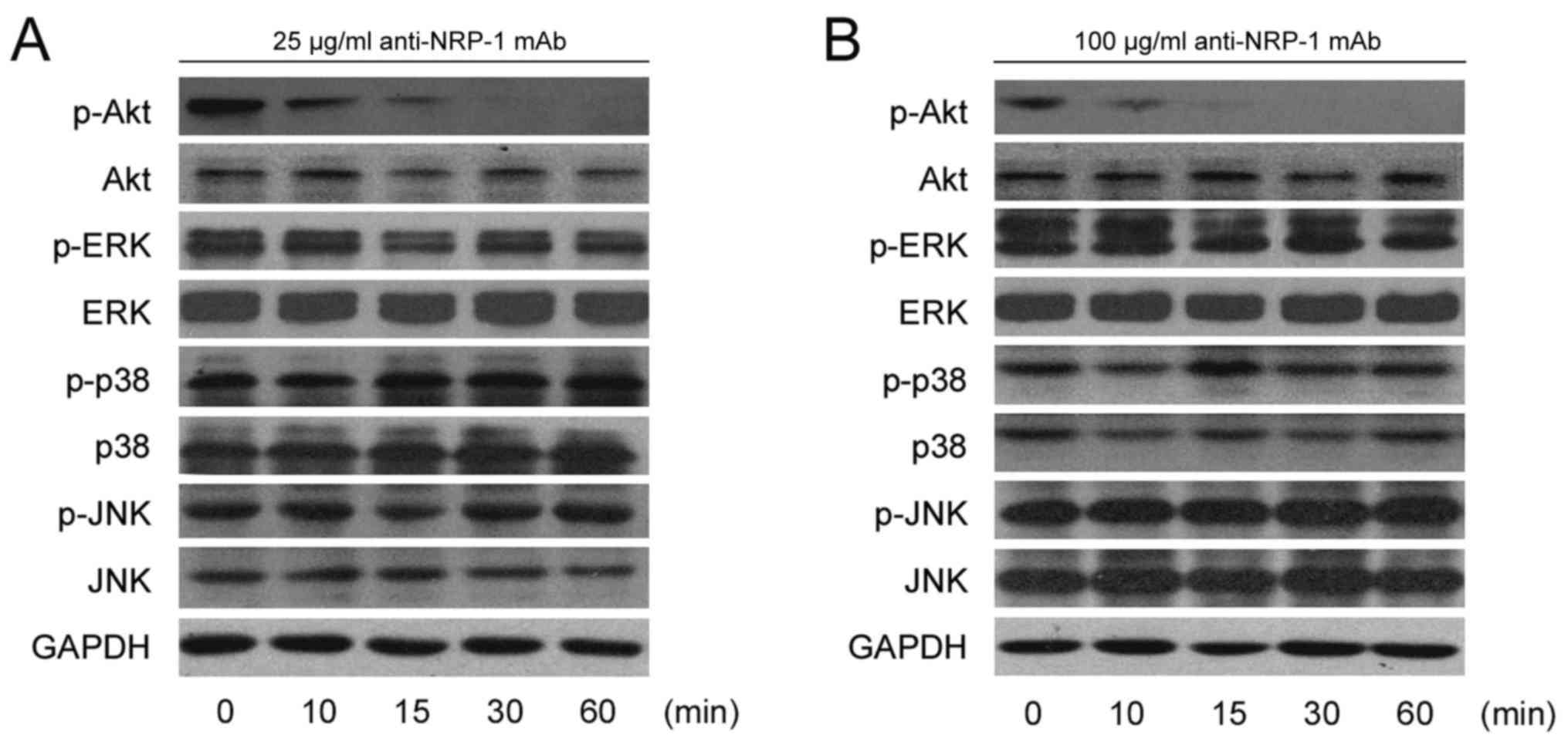 | Figure 4.Anti-NRP-1 mAb downregulates p-Akt in
human gastric cancer BGC-823 cells. Serum-starved BGC-823 cells
were incubated with (A) 25 µg/ml anti-NRP-1 mAb or (B) 100 µg/ml
anti-NRP-1 mAb for the indicated time periods. Cellular proteins
were extracted and 50 µg whole-cell protein extracts were subjected
to western blot analysis using primary antibodies against p-Akt,
Akt, p-ERK, ERK, p-JNK, JNK, p-P38, P38 and GAPDH. GAPDH was used
as a loading control. Representative immunoblots from three
independent experiments are shown. NRP-1, neuropilin 1; mAb,
monoclonal antibody; BGC-823, human gastric cancer cell line; p-,
phosphorylated; ERK, extracellular signal-regulated protein kinase;
JNK, c-Jun N-terminal kinase; P38, P38 mitogen-activated protein
kinase. |
Anti-NRP-1 mAb inhibits the growth of
human gastric cancer xenografts
Data on the growth characteristics of subcutaneous
xenograft tumors (Fig. 5A and B)
indicated that anti-NRP-1 mAb suppressed the growth of xenograft
tumors in nude mice. Following seven administrations of 1 and 5
mg/kg anti-NRP-1 mAb over 15 days, the final volumes of xenograft
tumors were reduced by 34.93 and 56.85%, respectively (Fig. 5A). In detail, for 5 mg/kg antibody,
tumor volume was significantly reduced on days 9, 11, 13 (all
P<0.05) and 15 (P<0.01), while 1 mg/ml antibody induced
significant reduction in tumor volume on day 15 (P<0.05),
relative to untreated controls. Similarly, treatment with 1 and 5
mg/kg anti-NRP-1 mAb decreased final tumor weights by 24.16 and
63.09% respectively; an effect deemed to be significant for 5 mg/kg
anti-NRP-1 mAb, relative to untreated controls (P<0.01; Fig. 5B). In addition, toxicity-dependent
weight loss was not observed in tumor-bearing mice treated with
anti-NRP-1 mAb (Fig. 5C).
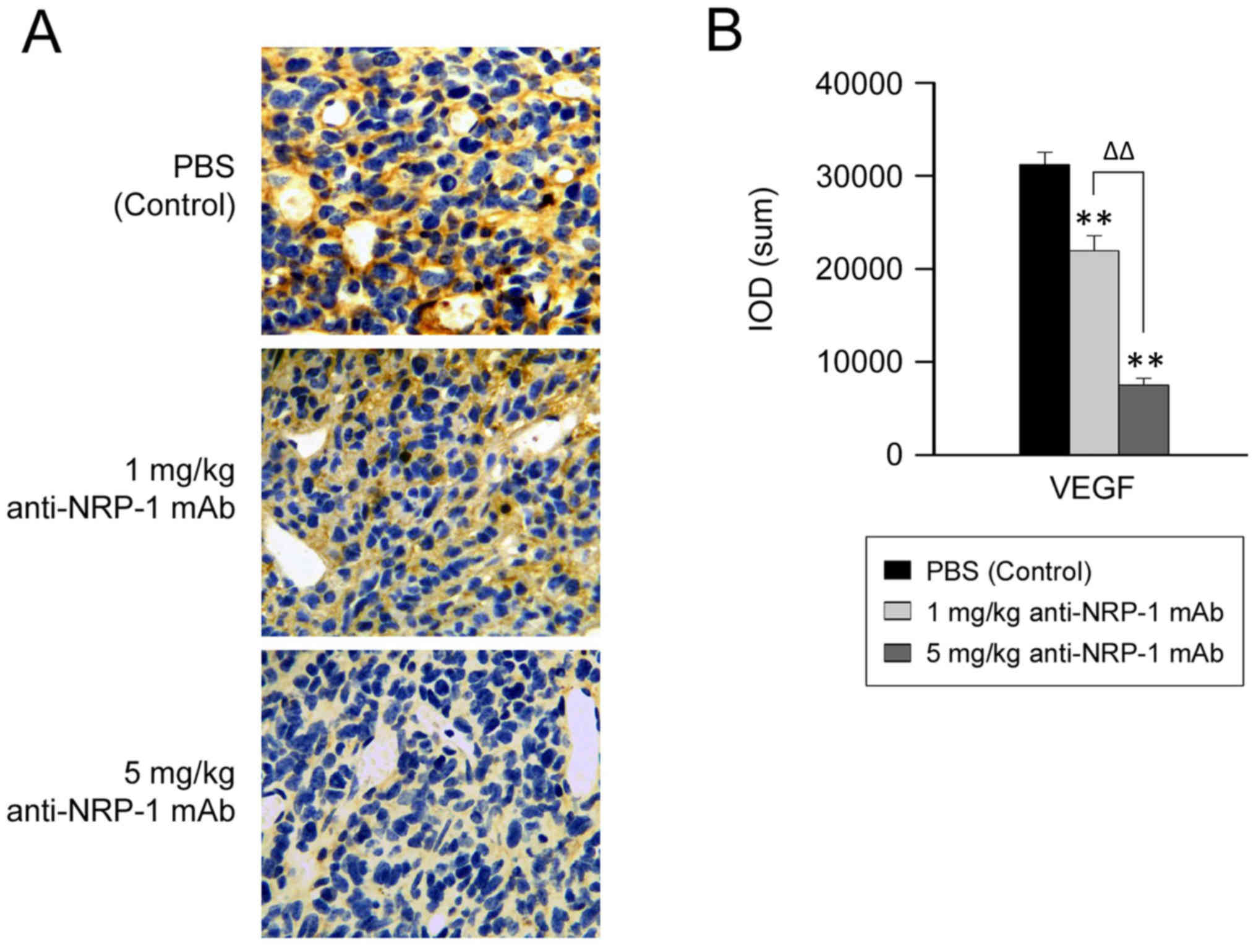
Anti-NRP-1 mAb downregulates VEGF
protein expression in human gastric cancer xenografts
As anti-NRP-1 mAb suppressed the growth of xenograft
tumors in nude mice, the potential underlying molecular events were
subsequently evaluated, by measuring the level of VEGF expression
in gastric cancer xenografts. Data from immunohistochemical
analysis (Fig. 6) demonstrated that
the IOD of VEGF was significantly decreased in the anti-NRP-1 mAb
treatment groups (1 and 5 mg/kg), relative to the negative control
group (P<0.01; Fig. 6B). In turn,
reduction in the IOD of VEGF was significantly greater in the 5
mg/kg anti-NRP-1 mAb treatment group, relative to that in the 1
mg/kg anti-NRP-1 mAb group (P<0.01; Fig. 6B). These data indicate that VEGF
expression may be downregulated by anti-NRP-1 mAb in a
dose-dependent manner.
Discussion
Preclinical data indicate that NRP-1 may have roles
in tumor cell proliferation and pathological angiogenesis, thus
making it a potential anticancer target (23,41).
However, for patients to clinically benefit from anti-NRP-1
therapies, it may be necessary to first determine the expression
patterns of NRP-1. Numerous studies have reported in situ
expression patterns of NRP-1 in human tissues, including gastric
(18), prostate (20), pancreatic (53), colorectal (15), breast (16,54) and
lung cancer (17,55). Therefore, the present study measured
the expression of NRP-1 in human gastric cancer cell lines
(BGC-823, SGC-7901 and MKN-74). Results demonstrated that all three
cell lines constitutively expressed NRP-1 at the mRNA and protein
level, with BGC-823 cells expressing relatively higher levels of
NRP-1. These data are consistent with a previous study by Akagi
et al (56), whereby the
expression of NRP-1 was detected in five of seven human gastric
cancer cell lines (TMK-1, AGS, NCI-N87, ST-2 and ST-7). It was
additionally observed in the current study that NRP-1 protein was
predominantly distributed in the cytomembrane and cytoplasm of
BGC-823 cells.
In a previous study by our group, the anti-NRP-1 mAb
(6.25–100 µg/ml) was demonstrated to inhibit the growth and
proliferation of human glioma cell lines (U251 and U87) and a rat
glioma cell line (C6) in a concentration- and time-dependent
manner. For instance, at a dose of 100 µg/ml anti-NRP-1 mAb, U251
cell growth was inhibited by 76.26% (43). Zeng et al (22) also demonstrated that the anti-NRP-1
mAb (200 and 400 µg/ml) significantly inhibited the proliferation
of MCF7 human breast cancer cells, observed as significant
reductions in the number and size of MCF7 cell colonies 7 days
after mAb administration. However, 25–100 µg/ml anti-NRP-1 mAb had
little to no effect on MCF7 cell growth and proliferation.
Similarly, the current study demonstrated that treatment with
<150 µg/ml anti-NRP-1 mAb had little to no effect on BGC-823
cell viability, while 200 or 400 µg/ml doses of anti-NRP-1 mAb were
able to significantly inhibit BGC-823 cell viability.
High-affinity mAbs that target the CUB domains
(anti-NRP-1A) or coagulation factors V/VIII domains (anti-NRP-1B)
of NRP-1 have been previously developed by Genetech, though it was
demonstrated that anti-NRP-1 mAb alone (anti-NRP-1A or −1B) or in
combination with anti-VEGF did not alter the proliferation of a
human NSCLC cell line SK-MES-1 in vitro. In addition, both
anti-NRP1 mAbs were unable to induce cytotoxicity in human ductal
carcinoma (BT-474) or SK-MES-1 cell lines (40). It has also been demonstrated that
anti-NRP-1A mAb had no effect on VEGF-induced endothelial cell (EC)
proliferation and anti-NRP-1B mAb only stimulated a slight
dose-responsive reduction, thus indicating that the inhibitory
effects of anti-NRP1 mAb on SK-MES-1 tumor growth are not through
the alteration of tumor blood vessels (41). Furthermore, knockdown of NRP-1 in ECs
by small interfering RNA only partially inhibited VEGF-induced
proliferation, suggesting that the primary role of NRP-1 in
VEGF-driven EC behavior is to mediate cell migration (57).
As anti-NRP-1 mAb exhibited no significant effect on
BGC-823 cell viability, the current study evaluated whether
anti-NRP-1 mAb regulated the migration and invasion of BGC-823
cells. Results of a standard Boyden chamber assay indicated that
anti-NRP-1 mAb may suppress BGC-823 cell migration and invasion. In
a previous study by our group, the migration and invasion of human
glioma U251 and U87 cells and rat glioma C6 cells were inhibited
after treatment with anti-NRP-1 mAb (43). Similarly, Pan et al (41) documented that treatment with
anti-NRP-1A and anti-NRP-1B significantly reduced EC migration,
with anti-NRP-1B exhibiting stronger inhibitory effects. Ochiumi
et al (8) also demonstrated
that cell migration was decreased following knockdown of NRP-1 in
WiDR cells (human colon adenocarcinoma cell line) by RNA
interference.
The PI3K/Akt and MAPK signaling pathways are
involved in a range of cellular functions, including cell survival,
growth, proliferation, migration and invasion (58–60). As
anti-NRP-1 mAb suppressed BGC-823 cell migration and invasion, the
current study investigated the potential molecular events involved.
The phosphorylation levels of key signaling molecules (Akt, ERK,
p38 and JNK) were measured following anti-NRP-1 mAb treatment. It
was observed that the anti-NRP-1 mAb inhibited the phosphorylation
of Akt in BGC-823 cells, though anti-NRP-1 mAb treatment did not
affect activation of MAPK signaling.
Results of the current study also indicated that
anti-NRP-1 mAb may suppress the growth of human gastric cancer
xenograft in nude mice in vivo. In addition, anti-NRP-1 mAb
treatment did not cause toxicity-dependent in tumor-bearing mice.
Consistent with present results, our previous study demonstrated
that the anti-NRP-1 mAb specifically targeted tumor cells in U87
xenografts, by reducing xenograft proliferation and growth rate
(43). Similarly, Pan et al
(41) used an NSCLC-SK-MES-1
xenograft model, specifically with high expression of NRP-1 in the
vascular and stromal tissue and intermediate expression in tumor
cells, to determine the effects of NRP-1 blockade on tumor growth
inhibition (TGI), initially provided by a VEGF blockade. In this
model, individual treatment with anti-VEGF mAb and anti-NRP-1B mAb
caused 52 and 37% TGI, respectively, while anti-NRP-1A mAb had no
significant effect on TGI. Ochiumi et al (8) has documented that cell proliferation
was not influenced by the inhibition of NRP-1 expression in WiDR
cells in vitro. However, it was observed in vivo that
the proliferation index of cells was significantly greater in
tissues exhibiting high levels of NRP-1 staining (8). The reasons for these inconsistencies
remain unknown, though it is possible that juxtacrine signaling
occurs, whereby VEGF bound to NRP-1 on tumor cells also binds to
VEGFR-2 on adjacent ECs, leading to the induction of tumor growth
(8,15,61). The
current study observed that anti-NRP-1 mAb significantly
downregulated VEGF expression in human gastric cancer xenografts,
and Xia et al (62)
previously documented that Akt1 is a key downstream regulator of
tumor growth, angiogenesis and VEGF expression. In addition, recent
studies have demonstrated that PI3K/Akt signaling regulates the
expression of hypoxia-inducible factor (HIF)-1α and VEGF in a
number of cancer cell types, including prostate, ovarian, and
pancreatic cancer cells, potentially by a cascade effect, whereby
activated Akt promotes HIF-1α upregulation, with HIF-1α then
inducing the transcriptional activation of VEGF (63–66).
Collectively, these results suggest that anti-NRP-1 mAb may inhibit
the activation of Akt, thus decreasing HIF-1 and VEGF expression
and inhibiting gastric cancer xenograft growth in vivo.
In conclusion, the present study demonstrated that
the migration and invasion of gastric cancer BGC-823 cells was
suppressed by treatment with anti-NRP-1 mAb in vitro. By
contrast, anti-NRP-1 mAb had little effect on BGC-823 cell
viability. While the underlying mechanisms regarding the effects of
anti-NRP-1 mAb remain unknown, it was observed that treatment with
anti-NRP-1 mAb inhibited the phosphorylation of Akt in BGC-823
cells. It was also demonstrated in vivo that anti-NRP-1 mAb
downregulated the expression of VEGF and suppressed the growth of
gastric cancer xenografts in mice. These results are consistent
with those of previous studies into the effects of anti-NRP-1 mAb
on different malignant tumors, thus indicating that NRP-1
inhibition with anti-NRP-1 mAb may have therapeutic effects in the
treatment of cancer.
Acknowledgements
Not applicable.
Funding
The current study was supported by the National
Natural Science Foundation of China (grant no. 81172970), the
Science and Technology Innovative Foundation of Xiamen (grant nos.
3502Z20134026 and 3502Z20144034) and the Medical Science and
Technology Innovation Fund of the Nanjing Military Region of the
People's Liberation Army (grant no. 14ZD33).
Availability of data and materials
The datasets used and/or analyzed during the current
study are available from the corresponding author on reasonable
request.
Authors' contributions
YQC and JHY designed the study, supervised the
experiments, reviewed the manuscript and approved the final version
to be published. YD performed the experiments, analyzed and
interpreted the data and drafted and revised the manuscript. JZ
participated in the design of the study, helped perform the
experiments and analyzed and interpreted the data. SYW instructed
in cellular and molecular techniques; YL, YJM, SHG and YX supplied
critical reagents, performed the experiments and participated in
the acquisition of data. All authors have read and approved the
final version of the manuscript.
Ethics approval and consent to
participate
All animal procedures were conducted under approved
guidelines of the Animal Care and Use Committee of Xiamen
University and ethical approval was obtained from the People's
Liberation Army 174th Hospital Medical Ethics Review board.
Consent for publication
Not applicable.
Competing interests
The authors declare that they have no competing
interests.
References
|
1
|
Fujisawa H, Takagi S and Hirata T:
Growth-associated expression of a membrane protein, neuropilin, in
Xenopus optic nerve fibers. Dev Neurosci. 17:343–349. 1995.
View Article : Google Scholar : PubMed/NCBI
|
|
2
|
Fujisawa H and Kitsukawa T: Receptors for
collapsin/semaphorins. Curr Opin Neurobiol. 8:587–592. 1998.
View Article : Google Scholar : PubMed/NCBI
|
|
3
|
Soker S: Neuropilin in the midst of cell
migration and retraction. Int J Biochem Cell Biol. 33:433–437.
2001. View Article : Google Scholar : PubMed/NCBI
|
|
4
|
He Z and Tessier-Lavigne M: Neuropilin is
a receptor for the axonal chemorepellent Semaphorin III. Cell.
90:739–751. 1997. View Article : Google Scholar : PubMed/NCBI
|
|
5
|
Gray MJ, Wey JS, Belcheva A, McCarty MF,
Trevino JG, Evans DB, Ellis LM and Gallick GE: Neuropilin-1
suppresses tumorigenic properties in a human pancreatic
adenocarcinoma cell line lacking neuropilin-1 coreceptors. Cancer
Res. 65:3664–3670. 2005. View Article : Google Scholar : PubMed/NCBI
|
|
6
|
Nakamura F, Tanaka M, Takahashi T, Kalb RG
and Strittmatter SM: Neuropilin-1 extracellular domains mediate
semaphorin D/III-induced growth cone collapse. Neuron.
21:1093–1100. 1998. View Article : Google Scholar : PubMed/NCBI
|
|
7
|
Pellet-Many C, Frankel P, Jia H and
Zachary I: Neuropilins: Structure, function and role in disease.
Biochem J. 411:211–226. 2008. View Article : Google Scholar : PubMed/NCBI
|
|
8
|
Ochiumi T, Kitadai Y, Tanaka S, Akagi M,
Yoshihara M and Chayama K: Neuropilin-1 is involved in regulation
of apoptosis and migration of human colon cancer. Int J Oncol.
29:105–116. 2006.PubMed/NCBI
|
|
9
|
Chaudhary B, Khaled YS, Ammori BJ and
Elkord E: Neuropilin 1: Function and therapeutic potential in
cancer. Cancer Immunol Immunother. 63:81–99. 2014. View Article : Google Scholar : PubMed/NCBI
|
|
10
|
Dzionek A, Fuchs A, Schmidt P, Cremer S,
Zysk M, Miltenyi S, Buck DW and Schmitz J: BDCA-2, BDCA-3, and
BDCA-4: Three markers for distinct subsets of dendritic cells in
human peripheral blood. J Immunol. 165:6037–6046. 2000. View Article : Google Scholar : PubMed/NCBI
|
|
11
|
Romeo PH, Lemarchandel V and Tordjman R:
Neuropilin-1 in the immune system. Adv Exp Med Biol. 515:49–54.
2002. View Article : Google Scholar : PubMed/NCBI
|
|
12
|
Tordjman R, Lepelletier Y, Lemarchandel V,
Cambot M, Gaulard P, Hermine O and Roméo PH: A neuronal receptor,
neuropilin-I, is essential for the initiation of the primary immune
response. Nat Immunol. 3:477–482. 2002. View Article : Google Scholar : PubMed/NCBI
|
|
13
|
Herzog Y, Kalcheim C, Kahane N, Reshef R
and Neufeld G: Differential expression of neuropilin-1 and
neuropilin-2 in arteries and veins. Mech Dev. 109:115–119. 2001.
View Article : Google Scholar : PubMed/NCBI
|
|
14
|
Battaglia A, Buzzonetti A, Monego G, Peri
L, Ferrandina G, Fanfani F, Scambia G and Fattorossi A:
Neuropilin-1 expression identifies a subset of regulatory T cells
in human lymph nodes that is modulated by preoperative
chemoradiation therapy in cervical cancer. Immunology. 123:129–138.
2008. View Article : Google Scholar : PubMed/NCBI
|
|
15
|
Parikh AA, Fan F, Liu WB, Ahmad SA,
Stoeltzing O, Reinmuth N, Bielenberg D, Bucana CD, Klagsbrun M and
Ellis LM: Neuropilin-1 in human colon cancer: Expression,
regulation, and role in induction of angiogenesis. Am J Pathol.
164:2139–2151. 2004. View Article : Google Scholar : PubMed/NCBI
|
|
16
|
Ghosh S, Sullivan CA, Zerkowski MP,
Molinaro AM, Rimm DL, Camp RL and Chung GG: High levels of vascular
endothelial growth factor and its receptors (VEGFR-1, VEGFR-2,
neuropilin-1) are associated with worse outcome in breast cancer.
Hum Pathol. 39:1835–1843. 2008. View Article : Google Scholar : PubMed/NCBI
|
|
17
|
Hong TM, Chen YL, Wu YY, Yuan A, Chao YC,
Chung YC, Wu MH, Yang SC, Pan SH, Shih JY, et al: Targeting
neuropilin 1 as an antitumor strategy in lung cancer. Clin Cancer
Res. 13:4759–4768. 2007. View Article : Google Scholar : PubMed/NCBI
|
|
18
|
Hansel DE, Wilentz RE, Yeo CJ, Schulick
RD, Montgomery E and Maitra A: Expression of neuropilin-1 in
high-grade dysplasia, invasive cancer, and metastases of the human
gastrointestinal tract. Am J Surg Pathol. 28:347–356. 2004.
View Article : Google Scholar : PubMed/NCBI
|
|
19
|
Evans IM, Yamaji M, Britton G, Pellet-Many
C, Lockie C, Zachary IC and Frankel P: Neuropilin-1 signaling
through p130Cas tyrosine phosphorylation is essential for growth
factor-dependent migration of glioma and endothelial cells. Mol
Cell Biol. 31:1174–1185. 2011. View Article : Google Scholar : PubMed/NCBI
|
|
20
|
Latil A, Bièche I, Pesche S, Valèri A,
Fournier G, Cussenot O and Lidereau R: VEGF overexpression in
clinically localized prostate tumors and neuropilin-1
overexpression in metastatic forms. Int J Cancer. 89:167–171. 2000.
View Article : Google Scholar : PubMed/NCBI
|
|
21
|
Prud'homme GJ and Glinka Y: Neuropilins
are multifunctional coreceptors involved in tumor initiation,
growth, metastasis and immunity. Oncotarget. 3:921–939. 2012.
View Article : Google Scholar : PubMed/NCBI
|
|
22
|
Zeng F, Luo F, Lv S, Zhang H, Cao C, Chen
X, Wang S, Li Z, Wang X, Dou X, et al: A monoclonal antibody
targeting neuropilin-1 inhibits adhesion of MCF7 breast cancer
cells to fibronectin by suppressing the FAK/p130cas signaling
pathway. Anticancer Drugs. 25:663–672. 2014.PubMed/NCBI
|
|
23
|
Jubb AM, Strickland LA, Liu SD, Mak J,
Schmidt M and Koeppen H: Neuropilin-1 expression in cancer and
development. J Pathol. 226:50–60. 2012. View Article : Google Scholar : PubMed/NCBI
|
|
24
|
Bergé M, Bonnin P, Sulpice E, Vilar J,
Allanic D, Silvestre JS, Lévy BI, Tucker GC, Tobelem G and
Merkulova-Rainon T: Small interfering RNAs induce
target-independent inhibition of tumor growth and vasculature
remodeling in a mouse model of hepatocellular carcinoma. Am J
Pathol. 177:3192–3201. 2010. View Article : Google Scholar : PubMed/NCBI
|
|
25
|
Raskopf E, Vogt A, Standop J, Sauerbruch T
and Schmitz V: Inhibition of neuropilin-1 by RNA-interference and
its angiostatic potential in the treatment of hepatocellular
carcinoma. Z Gastroenterol. 48:21–27. 2010. View Article : Google Scholar : PubMed/NCBI
|
|
26
|
Lu L, Zhang L, Xiao Z, Lu S, Yang R and
Han ZC: Neuropilin-1 in acute myeloid leukemia: Expression and role
in proliferation and migration of leukemia cells. Leuk Lymphoma.
49:331–338. 2008. View Article : Google Scholar : PubMed/NCBI
|
|
27
|
Barr MP, Byrne AM, Duffy AM, Condron CM,
Devocelle M, Harriott P, Bouchier-Hayes DJ and Harmey JH: A peptide
corresponding to the neuropilin-1-binding site on VEGF(165) induces
apoptosis of neuropilin-1-expressing breast tumour cells. Br J
Cancer. 92:328–333. 2005. View Article : Google Scholar : PubMed/NCBI
|
|
28
|
Tirand L, Frochot C, Vanderesse R, Thomas
N, Trinquet E, Pinel S, Viriot ML, Guillemin F and Barberi-Heyob M:
A peptide competing with VEGF165 binding on neuropilin-1 mediates
targeting of a chlorin-type photosensitizer and potentiates its
photodynamic activity in human endothelial cells. J Control
Release. 111:153–164. 2006. View Article : Google Scholar : PubMed/NCBI
|
|
29
|
Jia H, Cheng L, Tickner M, Bagherzadeh A,
Selwood D and Zachary I: Neuropilin-1 antagonism in human carcinoma
cells inhibits migration and enhances chemosensitivity. Br J
Cancer. 102:541–552. 2010. View Article : Google Scholar : PubMed/NCBI
|
|
30
|
Jarvis A, Allerston CK, Jia H, Herzog B,
Garza-Garcia A, Winfield N, Ellard K, Aqil R, Lynch R, Chapman C,
et al: Small molecule inhibitors of the neuropilin-1 vascular
endothelial growth factor A (VEGF-A) interaction. J Med Chem.
53:2215–2226. 2010. View Article : Google Scholar : PubMed/NCBI
|
|
31
|
Gagnon ML, Bielenberg DR, Gechtman Z, Miao
HQ, Takashima S, Soker S and Klagsbrun M: Identification of a
natural soluble neuropilin-1 that binds vascular endothelial growth
factor: In vivo expression and antitumor activity. Proc Natl Acad
Sci USA. 97:2573–2578. 2000. View Article : Google Scholar : PubMed/NCBI
|
|
32
|
Grandclement C and Borg C: Neuropilins: A
new target for cancer therapy. Cancers (Basel). 3:1899–1928. 2011.
View Article : Google Scholar : PubMed/NCBI
|
|
33
|
Starzec A, Ladam P, Vassy R, Badache S,
Bouchemal N, Navaza A, du Penhoat CH and Perret GY:
Structure-function analysis of the antiangiogenic ATWLPPR peptide
inhibiting VEGF(165) binding to neuropilin-1 and molecular dynamics
simulations of the ATWLPPR/neuropilin-1 complex. Peptides.
28:2397–2402. 2007. View Article : Google Scholar : PubMed/NCBI
|
|
34
|
Bondeva T, Rüster C, Franke S,
Hammerschmid E, Klagsbrun M, Cohen CD and Wolf G: Advanced
glycation end-products suppress neuropilin-1 expression in
podocytes. Kidney Int. 75:605–616. 2009. View Article : Google Scholar : PubMed/NCBI
|
|
35
|
Bondeva T and Wolf G: Advanced glycation
end products suppress neuropilin-1 expression in podocytes by a
reduction in Sp1-dependent transcriptional activity. Am J Nephrol.
30:336–345. 2009. View Article : Google Scholar : PubMed/NCBI
|
|
36
|
Bondeva T, Wojciech S and Wolf G: Advanced
glycation end products inhibit adhesion ability of differentiated
podocytes in a neuropilin-1-dependent manner. Am J Physiol Renal
Physiol. 301:F852–F870. 2011. View Article : Google Scholar : PubMed/NCBI
|
|
37
|
Teesalu T, Sugahara KN, Kotamraju VR and
Ruoslahti E: C-end rule peptides mediate neuropilin-1-dependent
cell, vascular, and tissue penetration. Proc Natl Acad Sci USA.
106:16157–16162. 2009. View Article : Google Scholar : PubMed/NCBI
|
|
38
|
Haspel N, Zanuy D, Nussinov R, Teesalu T,
Ruoslahti E and Aleman C: Binding of a C-end rule peptide to the
neuropilin-1 receptor: A molecular modeling approach. Biochemistry.
50:1755–1762. 2011. View Article : Google Scholar : PubMed/NCBI
|
|
39
|
Rizzolio S, Rabinowicz N, Rainero E,
Lanzetti L, Serini G, Norman JC, Neufeld G and Tamagnone L:
Neuropilin-1 dependent regulation of EGF-Receptor signaling. Cancer
Res. 72:5801–5811. 2012. View Article : Google Scholar : PubMed/NCBI
|
|
40
|
Liang WC, Dennis MS, Stawicki S, Chanthery
Y, Pan Q, Chen Y, Eigenbrot C, Yin J, Koch AW, Wu X, et al:
Function blocking antibodies to neuropilin-1 generated from a
designed human synthetic antibody phage library. J Mol Biol.
366:815–829. 2007. View Article : Google Scholar : PubMed/NCBI
|
|
41
|
Pan Q, Chanthery Y, Liang WC, Stawicki S,
Mak J, Rathore N, Tong RK, Kowalski J, Yee SF, Pacheco G, et al:
Blocking neuropilin-1 function has an additive effect with
anti-VEGF to inhibit tumor growth. Cancer Cell. 11:53–67. 2007.
View Article : Google Scholar : PubMed/NCBI
|
|
42
|
Li X, Luo F, Wang S, Ni E, Tang X, Lv H,
Chen X, Chen L and Yan J: Monoclonal Antibody Against NRP-1 b1b2.
Hybridoma (Larchmt). 30:369–373. 2011. View Article : Google Scholar : PubMed/NCBI
|
|
43
|
Chen L, Miao W, Tang X, Zhang H, Wang S,
Luo F and Yan J: Inhibitory Effect of Neuropilin-1 monoclonal
antibody (NRP-1 MAb) on glioma tumor in mice. J Biomed Nanotechnol.
9:551–558. 2013. View Article : Google Scholar : PubMed/NCBI
|
|
44
|
Dallas NA, Gray MJ, Xia L, Fan F, van
Buren G II, Gaur P, Samuel S, Lim SJ, Arumugam T, Ramachandran V,
et al: Neuropilin-2-mediated tumor growth and angiogenesis in
pancreatic adenocarcinoma. Clin Cancer Res. 14:8052–8060. 2008.
View Article : Google Scholar : PubMed/NCBI
|
|
45
|
Wang Y, Wang S, Ding Y, Ye Y, Xu Y, He H,
Li Q, Mi Y, Guo C, Lin Z, et al: A suppressor of cytokine signaling
1 antagonist enhances antigen-presenting capacity and tumor cell
antigen-specific cytotoxic T lymphocyte responses by human
monocyte-derived dendritic cells. Clin Vaccine Immunol.
20:1449–1456. 2013. View Article : Google Scholar : PubMed/NCBI
|
|
46
|
Wu N, Wang Y, Wang S, Chen Y and Yan J:
Recombinant human leptin induces growth inhibition and apoptosis in
human gastric cancer MGC-803 cells. Clin Exp Med. 13:305–314. 2013.
View Article : Google Scholar : PubMed/NCBI
|
|
47
|
Li YJ, Sun LC, He Y, Liu XH, Liu M, Wang
QM and Jin XM: The anti-tumor properties of two tumstatin peptide
fragments in human gastric carcinoma. Acta Pharmacologica Sinica.
30:1307–1315. 2009. View Article : Google Scholar : PubMed/NCBI
|
|
48
|
Cai Y, Wang R, Zhao YF, Jia J, Sun ZJ and
Chen XM: Expression of Neuropilin-2 in salivary adenoid cystic
carcinoma: Its implication in tumor progression and angiogenesis.
Pathol Res Pract. 206:793–799. 2010. View Article : Google Scholar : PubMed/NCBI
|
|
49
|
Sulpice E, Plouët J, Bergé M, Allanic D,
Tobelem G and Merkulova-Rainon T: Neuropilin-1 and neuropilin-2 act
as coreceptors, potentiating proangiogenic activity. Blood.
111:2036–2045. 2008. View Article : Google Scholar : PubMed/NCBI
|
|
50
|
O'Reilly MS, Boehm T, Shing Y, Fukai N,
Vasios G, Lane WS, Flynn E, Birkhead JR, Olsen BR and Folkman J:
Endostatin: An endogenous inhibitor of angiogenesis and tumor
growth. Cell. 88:277–285. 1997. View Article : Google Scholar : PubMed/NCBI
|
|
51
|
Zhang JL, Chen GW, Liu YC, Wang PY, Wang
X, Wan YL, Zhu J, Gao HQ, Yin J, Wang W and Tian ML: Secreted
protein acidic and rich in cysteine (SPARC) suppresses angiogenesis
by down-regulating the expression of VEGF and MMP-7 in gastric
cancer. PLoS One. 7:e446182012. View Article : Google Scholar : PubMed/NCBI
|
|
52
|
Xavier LL, Viola GG, Ferraz AC, Da Cunha
C, Deonizio JM, Netto CA and Achaval M: A simple and fast
densitometric method for the analysis of tyrosine hydroxylase
immunoreactivity in the substantia nigra pars compacta and in the
ventral tegmental area. Brain Res Brain Res Protoc. 16:58–64. 2005.
View Article : Google Scholar : PubMed/NCBI
|
|
53
|
Fukahi K, Fukasawa M, Neufeld G, Itakura J
and Korc M: Aberrant expression of neuropilin-1 and −2 in human
pancreatic cancer cells. Clin Cancer Res. 10:581–590. 2004.
View Article : Google Scholar : PubMed/NCBI
|
|
54
|
Stephenson JM, Banerjee S, Saxena NK,
Cherian R and Banerjee SK: Neuropilin-1 is differentially expressed
in myoepithelial cells and vascular smooth muscle cells in
preneoplastic and neoplastic human breast: A possible marker for
the progression of breast cancer. Int J Cancer. 101:409–414. 2002.
View Article : Google Scholar : PubMed/NCBI
|
|
55
|
Lantuéjoul S, Constantin B, Drabkin H,
Brambilla C, Roche J and Brambilla E: Expression of VEGF,
semaphorin SEMA3F, and their common receptors neuropilins NP1 and
NP2 in preinvasive bronchial lesions, lung tumours, and cell lines.
J Pathol. 200:336–347. 2003. View Article : Google Scholar : PubMed/NCBI
|
|
56
|
Akagi M, Kawaguchi M, Liu W, McCarty MF,
Takeda A, Fan F, Stoeltzing O, Parikh AA, Jung YD, Bucana CD, et
al: Induction of neuropilin-1 and vascular endothelial growth
factor by epidermal growth factor in human gastric cancer cells. Br
J Cancer. 88:796–802. 2003. View Article : Google Scholar : PubMed/NCBI
|
|
57
|
Murga M, Fernandez-Capetillo O and Tosato
G: Neuropilin-1 regulates attachment in human endothelial cells
independently of vascular endothelial growth factor receptor-2.
Blood. 105:1992–1999. 2005. View Article : Google Scholar : PubMed/NCBI
|
|
58
|
Manning BD and Cantley LC: AKT/PKB
signaling: Navigating downstream. Cell. 129:1261–1274. 2007.
View Article : Google Scholar : PubMed/NCBI
|
|
59
|
Engelman JA: Targeting PI3K signalling in
cancer: Opportunities, challenges and limitations. Nat Rev Cancer.
9:550–562. 2009. View Article : Google Scholar : PubMed/NCBI
|
|
60
|
Burotto M, Chiou VL, Lee J-M and Kohn EC:
The MAPK pathway across different malignancies: A new perspective.
Cancer. 120:3446–3456. 2014. View Article : Google Scholar : PubMed/NCBI
|
|
61
|
Miao HQ, Lee P, Lin H, Soker S and
Klagsbrun M: Neuropilin-1 expression by tumor cells promotes tumor
angiogenesis and progression. FASEB J. 14:2532–2539. 2000.
View Article : Google Scholar : PubMed/NCBI
|
|
62
|
Xia C, Meng Q, Ca Z, Shi X and Jiang B-H:
Regulation of angiogenesis and tumor growth by p110 alpha and AKT1
via VEGF expression. J Cell Physiol. 209:56–66. 2006. View Article : Google Scholar : PubMed/NCBI
|
|
63
|
Mazure NM, Chen EY, Laderoute KR and
Giaccia AJ: Induction of vascular endothelial growth factor by
hypoxia is modulated by a phosphatidylinositol 3-kinase Akt
signaling pathway in Ha-ras-transformed cells through a hypoxia
inducible factor-1 transcriptional element. Blood. 90:3322–3331.
1997.PubMed/NCBI
|
|
64
|
Zhong H, Chiles K, Feldser D, Laughner E,
Hanrahan C, Georgescu MM, Simons JW and Semenza GL: Modulation of
hypoxia-inducible factor 1 alpha expression by the epidermal growth
factor/phosphatidylinositol 3-kinase/PTEN/AKT/FRAP pathway in human
prostate cancer cells: Implications for tumor angiogenesis and
therapeutics. Cancer Res. 60:1541–1545. 2000.PubMed/NCBI
|
|
65
|
Jiang BH, Jiang G, Zheng JZ, Lu Z, Hunter
T and Vogt PK: Phosphatidylinositol 3-kinase signaling controls
levels of hypoxia-inducible factor. Cell Growth Differ. 12:363–369.
2001.PubMed/NCBI
|
|
66
|
Skinner HD, Zheng JZ, Fang J, Agani F and
Jiang BH: Vascular endothelial growth factor transcriptional
activation is mediated by hypoxia-inducible factor 1 alpha, HDM2,
and p70S6K1 in response to phosphatidylinositol 3-kinase/AKT
signaling. J Biol Chem. 279:45643–45651. 2004. View Article : Google Scholar : PubMed/NCBI
|