Introduction
Essential tremor (ET) is a chronic progressive
neurological syndrome characterised by involuntary tremors of the
hands or arms, and progressively of the head, jaw and voice
(1-4).
ET can present with heterogeneous clinical phenotypes, and some
patients manifest extensive and complex deficits (5-10).
ET is often considered a hereditary benign condition, with an
isolated tremor and no further neurological signs (11); however, it is clearly defined in
the new classification of tremors that ET is a syndrome that may
have multiple aetiologies (5). The
new diagnostic criteria for ET require the occurrence of isolated
action tremor syndrome of the bilateral upper limbs with a minimum
duration of 3 years, and/or tremors in the head, voice and lower
limbs and mild neurological signs, such as dystonia, ataxia and/or
Parkinsonism. Soft neurological deficits, such as mild memory
impairment and impaired tandem gait, are also accepted; however,
such patients are now diagnosed with ET-plus syndrome (5).
Numerous studies have been conducted to clarify the
pathological, neuroimaging, physiological and clinical features of
ET; however, the pathophysiological mechanism is not clearly
understood (6-48).
The cerebellum is one of the brain structures that has been
extensively investigated at the macroscopic and microscopic levels
(6-8).
A number of studies have reported different levels of structural
and functional alterations in the cerebellum of ETs. For example,
neuroimaging studies have revealed a reduction in the volume of the
cerebellar vermis with marked atrophy, atrophy of the cerebellar
cortex, white matter changes in multiple cerebellar areas, and
overactivity of the deep cerebellar nuclei and the cerebellar
cortex and their connections (11-29).
Meanwhile, additional studies have failed to identify any
significant differences in the cerebellum between patients with ET
and normal controls (NCs) (11,15,16,28-40).
Neuropathological studies have also reported heterogeneous
findings, with a decrease in the linear density of Purkinje cells,
alterations in dendritic arborisation, axonal torpedoes,
heterotopic Purkinje cells, hypertrophy of the basket cells and
decreased climbing fibre-Purkinje cell synaptic density (29,44-50).
The present study investigated the morphological and
morphometric changes in Purkinje cells in ET and ET-plus brains
compared with age-matched control brains.
Materials and methods
Patients with ET and NC samples
In the present study, the morphology of the
dendritic arborisation of Purkinje cells from different parts of
the cerebellum was analysed in 12 patients with ET (7 males and 5
females) and 15 individuals (8 males and 7 females) with no history
of neurological conditions, who died accidentally and were used as
NC samples (Table I). The mean age
was 61.3±6.4 years (range, 50-74 years) and 65.6±6.0 years (range,
50-74 years) for patients with ET and NCs, respectively. Three
parts were excised from the cerebellum: The first from the
flocculonodular lobe or lobule X, the second from the anterior lobe
(lobule IV) and the third from the posterior lobe (lobule crus I).
These parts were selected as representative parts of the three
functional divisions of the cerebellum. All the brain tissues were
obtained from the Laboratory of Forensic Medicine and Toxicology of
the Aristotle University of Thessaloniki between January 2009 and
December 2016. For each brain, written informed consent was
obtained from the relatives of the deceased. The research was
performed in full accordance with the Greek Democracy legislation
(v. 2472/1997, 2819/2000, 2915/2001, 3235/2004, 3471/2006) and the
Committee for Research Deontology Principles of the Aristotle
University of Thessaloniki (ethical approval no. 12/2/4431/2019)
(51). The average time from death
to autopsy for all individuals was 8±2.9 h. All patients in the ET
group were clinically diagnosed with ET. Their history and
neurological examination were retrospectively reviewed, and they
were divided into three groups: ET with head and arm tremor (ET-h;
n=5); ET with arms tremor only (ET-a; n=4); and ET-plus (n=3). An
independent neuropathologist who was blinded to the medical history
examined the brain. After the autopsy, the brains were immersed at
18˚C in a 10% solution of formaldehyde for 25 days (52). Subsequently, small parts of the
anterior, posterior and flocculonodular lobes were excised and used
for Golgi staining at 120 µm-thick sections, as described in our
previous work (52), and studied
with an Axiostar plus Carl Zeiss light microscope at a standard
(magnification, x400). Golgi method stains the whole dendritic
field and makes the tracing of cells possible in a 3-dimensional
manner.
 | Table IDemographic features of patients and
NCs. |
Table I
Demographic features of patients and
NCs.
| A, Patients with
ET |
|---|
| Case | Age, years | Sex | Diagnosis |
|---|
| 1 | 57 | M | ET-h |
| 2 | 68 | M | ET-a |
| 3 | 50 | M | ET-a |
| 4 | 62 | F | ET-h |
| 5 | 63 | M | ET-plus |
| 6 | 61 | F | ET-a |
| 7 | 74 | F | ET-plus |
| 8 | 55 | M | ET-h |
| 9 | 63 | M | ET-plus |
| 10 | 62 | F | ET-h |
| 11 | 59 | F | ET-a |
| 12 | 54 | M | ET-a |
| Average/total | 61.3±6.4 | 7M/5F | |
| B, NCs |
| Case | Age, years | Sex | Diagnosis |
| 1 | 67 | M | NA |
| 2 | 59 | F | NA |
| 3 | 69 | F | NA |
| 4 | 72 | F | NA |
| 5 | 70 | M | NA |
| 6 | 62 | M | NA |
| 7 | 68 | M | NA |
| 8 | 62 | F | NA |
| 9 | 63 | M | NA |
| 10 | 74 | F | NA |
| 11 | 71 | M | NA |
| 12 | 69 | F | NA |
| 13 | 66 | F | NA |
| 14 | 50 | M | NA |
| 15 | 63 | M | NA |
| Average/total | 65.6±6.0 | 8M/7F | |
Cell selection criteria
Based on the criteria suggested by Jacobs et
al (53), 25 Purkinje cells
were selected from each brain. To avoid experimental bias, all
selected cells were randomly pooled and were assigned serial
numbers, and every third cell was then chosen.
Neuronal tracing and dendritic
quantification
Neuronal tracing and dendritic measurements were
performed as described in our previous study (52) using a motorised XYZ microscope
stage system (MLS203/MZS500-E; ThorLabs, Inc.), with the movement
on the Z-axis being controlled by the MZS500-E-Z-Axis Piezo Stage
and Controller Kit, with the aim of the APC software provided by
Thorlabs, Inc. with a JogStep of 1 µm a Travel Range of 250 µm. The
video captures were analysed in digital image sequences of 200
serial pictures. They were then imported into neuromantic
application with the help of which the cells were traced and
quantified along x-, y- and z-coordinates (52). Neuronal tracing was performed using
a semi-automatic form by two different investigators. All the
measurements were used for statistical analysis. The quantitative
evaluation of the dendritic trees was based on the method suggested
by Uylings et al (54).
Dendritic measures and Sholl's
analysis
Neuromantic automatically provides all the
measurements for the total length of dendritic trees, total number
of dendritic segments and terminal branches, total dendritic area
and total dendritic volume, which were analysed further in the
statistical analysis. The tracings were additionally analysed using
the Fiji (version 2017; Fiji) and Simple Neurite Tracer plugin on
ImageJ (v.1.8; National Institutes of Health) based on Sholl's
method of concentric spheres centred on the cell's soma at
intervals of 10 µm (55).
Spine counts
Spine counts were performed for 500 images. Images
were captured with an AxioCam HR (Zeiss GmbH), at a magnification
of x1,000, with an Axiostar plus photomicroscope. All the visible
spines were measured on the three segments of the dendritic field:
The first segment, 20-30 µm in length at a distance of 50 µm from
the cell soma; the second segment, 20-30 µm in length within 150
µm; and the third segment, 20-30 µm within 250 µm from the cell
body.
Purkinje cell density
The linear density of Purkinje cells was measured in
Nissl-stained specimens on 30 randomly selected images
(magnification, x20), using ImageJ (v.1.8; National Institutes of
Health) and the cell counter plugin (56).
Statistical analysis
RStudio (version, 1.4.1717) with R (version, 4.1.1)
was used for the statistical analysis and plotting of graphs
(57). All data was stored in
comma-separated value files and presented for statistical analysis
as data frames. One-way ANOVA followed by Tukey's post hoc test was
used to determine whether significant differences existed across
the independent parameters of Purkinje cells among the four groups.
ANOVA mixed-effects model followed by Tukey's post hoc test was
used for the statistics of Sholl's analysis, based on an R Script.
Pearson's correlation test was performed to identify any
correlation between the autolysis time and Purkinje cell dendritic
complexity variables. P<0.05 was considered to indicate a
statistically significant difference.
Results
Total dendritic length
The total dendritic length of Purkinje cells from
the cerebellar vermis was significantly lower in the ET-h group
compared with the ET-a, ET-plus and NC groups (Table II). No significant difference was
identified between the ET-a, ET-plus and NC groups (Fig. 1A). However, the total dendritic
length of Purkinje cells in the cerebellar hemispheres was
significantly decreased in the ET-a group compared with ET-plus and
NC groups (Fig. 1B). Similarly,
the difference was statistically significant between the ET-h,
ET-plus and NC groups, and between the ET-plus and NC groups. No
significant differences were found between the ET-a and ET-h groups
(Fig. 1B).
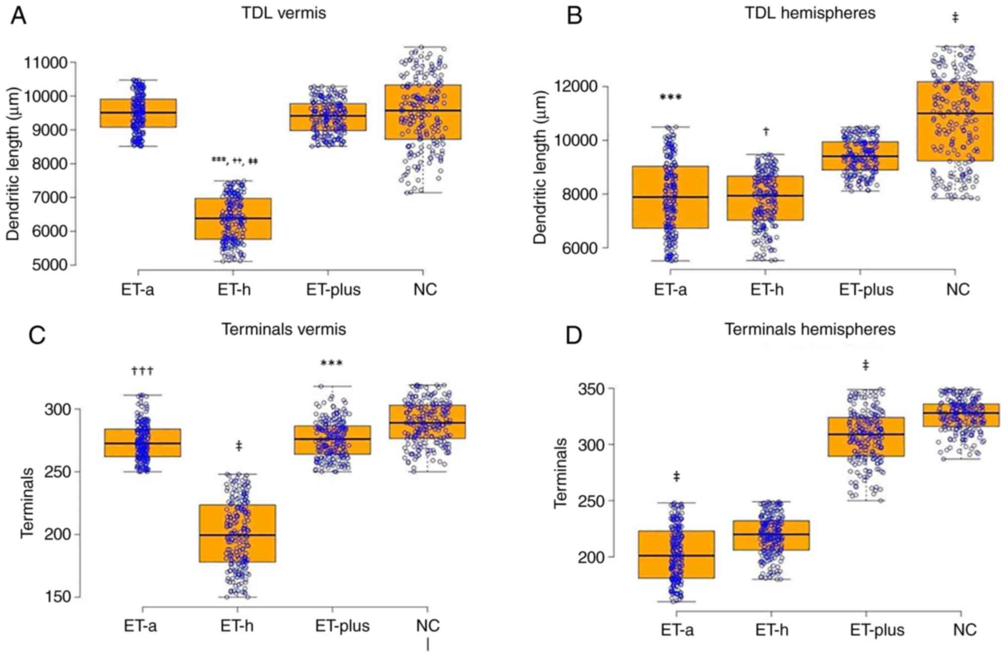 | Figure 1TDL of Purkinje cells from (A) the
cerebellar vermis and (B) the cerebellar hemispheres. Total number
of terminal branches of the Purkinje cells from (C) the cerebellar
vermis and (D) the hemispheres. Results are presented as boxplot
charts and the statistical significance in ANOVA test was expressed
as symbols (A) ***P<0.001 ET-h vs. ET-a;
††P<0.003 ET-h vs. ET-plus; ‡‡P<0.00001
ET-h vs. NC; (B) ***P<0.001 ET-a vs. ET-plus, NC;
‡P<0.0001 NC vs. ET-plus, ET-h;
†P<0.002 ET-h vs. ET-plus; (C)
‡P<0.0001 ET-h vs. ET-a, ET-plus, NC;
†††P<0.005 ET-a vs. NC; ***P<0.001
ET-plus vs. NC; (D) ‡P<0.0001 ET-a vs. ET-plus, ET-h,
NC, ET-plus vs. NC. TDL, total dendritic length; ET, essential
tremor; ET-a, ET with arm tremor only; ET-h, ET with head and arm
tremor; NC, normal control. |
| Table IIANOVA analysis of Purkinje cell
properties. |
Table II
ANOVA analysis of Purkinje cell
properties.
| A, Vermis |
|---|
| Parameter | ET-a | ET-h | ET-plus | NC | F-value |
|---|
| Total dendritic
length, µm |
9,487.73±551.06 |
6,361.28±661.17a |
9,388.75±486.88b |
9,507±1053.13c | 922.1 |
| Number of
terminals | 273.84±14.36 |
199.86±27.04d |
276.12±14.64c |
288.94±16.66c,e,f | 915.43 |
| Branch length,
µm | 562.9±33.79 | 513.25±35.51 |
570.56±39.05a,c |
587.35±37.68a,c | 151.89 |
| Branch order | 26.38±1.19 | 20.24±2.48 | 26.61±1.37 | 27.04±0.88 | 816.06 |
| Spines | 9.73±0.67 |
7.87±0.71a |
9.84±0.68g |
9.37±0.93f,g | 290.99 |
| B, Hemispheres |
| Parameter | ET-a | ET-h | ET-plus | NC | F-value |
| Total dendritic
length, µm |
7,889.4±1,387.22g,h |
7,794.89±1,017.91b,c |
9,407.22±642.27a,b |
10,757.3±1,666.24a,c,f | 257.33 |
| Number of
terminals | 202.72±23.98 |
219.01±17.73d |
306.86±23.04d |
325.69±14.39d,h | 1,871.33 |
| Branch length,
µm | 531.66±31.72 |
538.44±31.54a |
594.69±36.65d,g |
597.62±36.54d,g | 214.29 |
| Branch order | 22.96±1.66 |
24.64±1.58d |
28.41±1.75a,c |
30.61±1.62a,c,e | 890.35 |
| Spines | 7.75±0.67 |
7.4±0.49d |
9.76±0.66a |
9.98±0.68a | 902.82 |
Terminal branches
The total number of terminal branches of Purkinje
cells from the cerebellar vermis was significantly lower in the
ET-h group compared with the ET-a, ET-plus and NC groups. No
significant difference was found between the ET-a and ET-plus
groups, but there was a statistically significant difference
between the ET-a and NC groups, and between the ET-plus and NC
groups (Figs. 1C and 2 and Table
II). Purkinje cells from the cerebellar hemispheres from the
ET-a group had significantly fewer terminal branches than the
ET-plus, NC and the ET-h groups (Fig.
1D). In addition, the ET-plus group displayed a significantly
lower number of terminal branches compared with the NC group
(Fig. 1D).
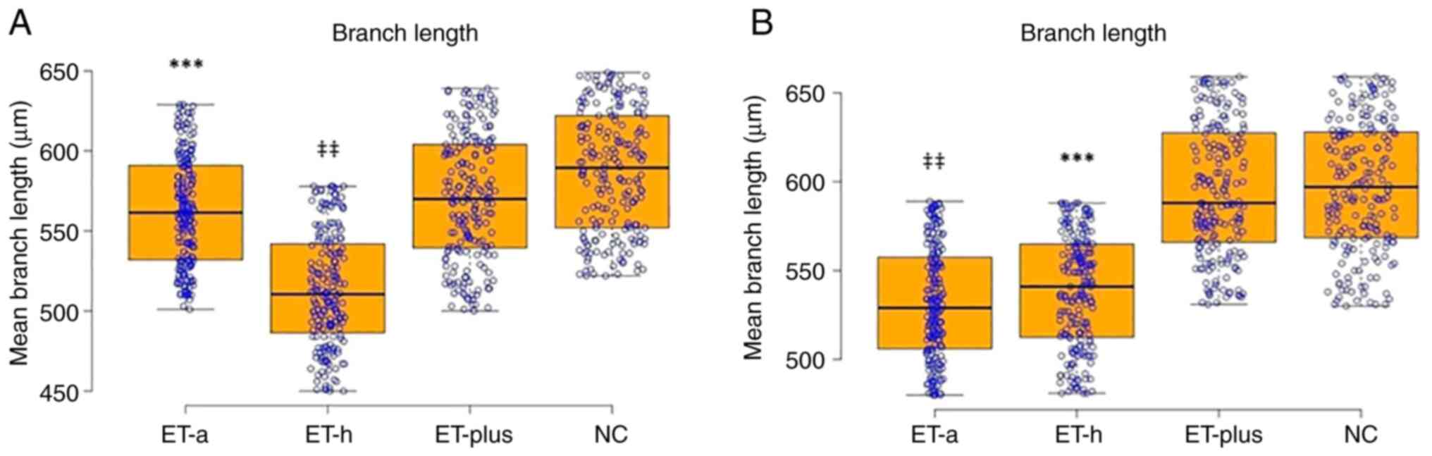
Branch length and branch order
The mean branch length (Fig. 3A and Table II) and the maximum branch order of
the cerebellar vermis were both significantly decreased in the ET-h
group compared with the ET-plus and NC groups. The ET-a group also
indicated a significantly lesser branch length and maximum branch
order than the ET-plus and NC groups. The ET-plus group
demonstrated a decreased branch length and branch order compared
with the NC group. Purkinje cells from the cerebellar hemispheres
exhibited a decreased branch length in the ET-a group compared with
the other groups, and in the ET-h group compared with the ET-plus
and NC groups. No statistically significant difference was observed
between the ET-plus and NC groups (Fig. 3B). The maximum branch order was
similarly decreased in the ET-h group compared with the other
groups, in the ET-a group compared with the ET-plus and NC groups,
and in the ET-plus group compared with the NC group.
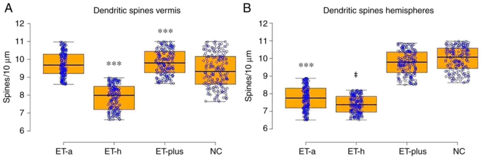
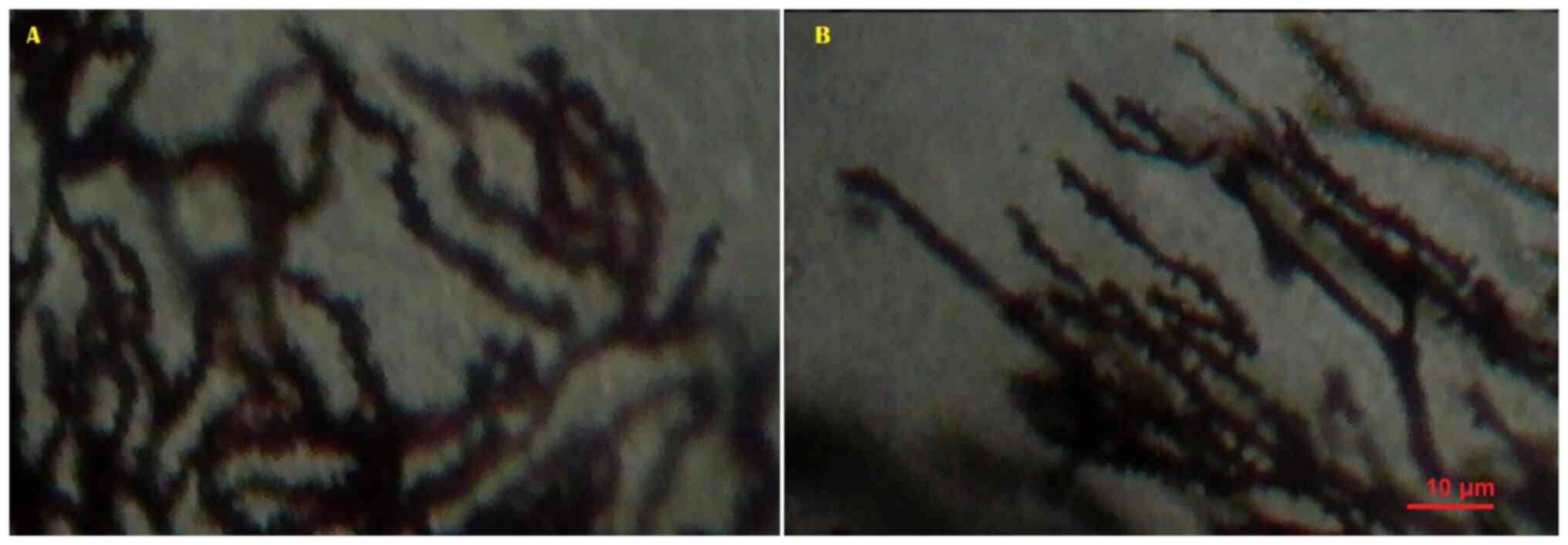
Dendritic spines
The dendritic spine density was reduced in the ET-h
group compared with the ET-a, ET-plus and NC groups in Purkinje
cells from the cerebellar vermis; however, no significant
difference was found between the ET-a and ET-plus groups. The
latter displayed notably more dendritic spines compared with
Purkinje cells from the NC group (Figs. 4A and 5 and Table
II). Meanwhile, Purkinje cells from the cerebellar hemispheres
demonstrated a significant difference between the ET-h and ET-a
groups, between the ET-plus and NC groups, between the ET-a and
ET-plus, and between the ET-a and NC groups (Figs. 4B, 5 and Table
II). Moreover, Pearson's correlation test did not reveal a
significant correlation between autolysis time and dendritic and
spinal measurements.
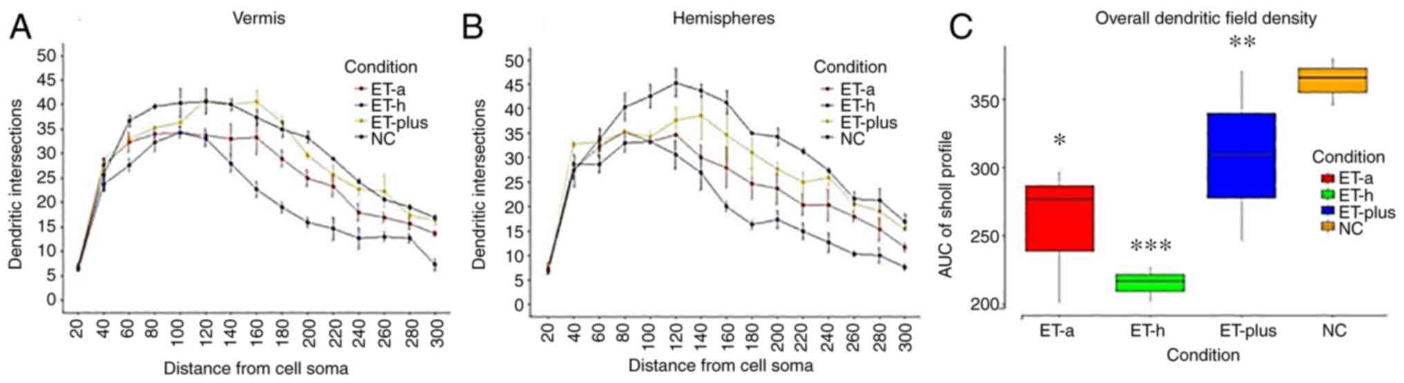
Sholl's analysis
Sholl's concentric circle analysis revealed
significant restriction of the dendritic field of Purkinje cells
from the cerebellar vermis and cerebellar hemispheres in the ET-h
and ET-a groups compared with the ET-plus and NC groups for
distances >120 µm from the cell soma (Fig. 6A and B). The areas under the curve also
confirmed the significance of the differences in the overall
dendritic field density (Fig.
6C).
Discussion
Multiple studies have reported neuropathological
changes in ET, with morphological and morphometric alterations of
Purkinje cells and Lewy bodies in the locus coeruleus; however,
whether these changes are the characteristic features of the
condition is unclear (15,16,11-49).
One possible explanation may be that different types of essential
tremor exhibit different pathological mechanisms and
neuropathological backgrounds.
Two previous studies demonstrated a significant
reduction in the linear Purkinje cell density in ET, and greater
distances between single Purkinje cell bodies (28,29);
however, another study failed to confirm these findings (58). Further studies reported increased
numbers of heterotopic Purkinje cells in ET, up to three times
compared with controls (59), and
certain morphological changes, with a substantial loss of dendritic
spines, total dendritic length and restriction of the dendritic
fields (44).
The most common neuropathological feature of ET is
axonal changes, known as axonal torpedoes; they are focal swellings
of Purkinje cell axons containing an accumulation of
hyperphosphorylated neurofilaments and disrupted organelles, and
can also be found in spinocerebellar ataxias (46,47).
Additional changes, which could potentially explain the
pathophysiology of ET, are also referred to as the basket cell
morphology (47) and climbing
fibre-Purkinje cells' synaptic density (49).
Patients with ET may have tremors of the hands,
head, voice and rarely the jaw. According to the 2016
classification of ET, patients who develop soft neurological
symptoms, such as mild rigidity, balance impairment or mild memory
impairment are classified as experiencing ET-plus (5). However, whether ET-plus or ET are
different entities remains controversial.
In the present study, significant morphological
changes were found in Purkinje cells, including a decreased
dendritic length and field density, an overall loss of terminal
branches, and a decreased density of dendritic spines in ET
patients compared with controls. Furthermore, ET patients were
divided into three groups based on the revised criteria for ET. The
results indicated that the ET-h group exhibited significant changes
in both the cerebellar vermis and hemispheres, while the ET-a group
displayed significant differences only in Purkinje cells from the
cerebellar hemispheres compared with controls. Meanwhile, Purkinje
cells from patients with ET-plus demonstrated only minor changes.
The significant loss of dendritic spines and terminal branches,
which are the most plastic components of the dendritic field
(52), could lead to a substantial
decrease in the synaptic contacts of Purkinje cells. This could be
of significant importance in the pathophysiology of ET.
The flocculonodular lobe of the cerebellum
corresponds to the vestibulocerebellum and is functionally related
to head movements, while the anterior and posterior lobes of the
hemisphere are related to limb movements and movement scheduling
(60,61). Different Purkinje cell pathologies
were identified in the different groups of the study, which could
explain the different symptoms in each patient group. Patients with
ET-a did not exhibit significant pathology in the Purkinje cells of
the cerebellar vermis. However, patients with ET-a exhibited
pathology in the cerebellar hemispheres, which are functionally
related to Purkinje cells. Patients with ET-h exhibited pathology
in the Purkinje cells of the cerebellar hemispheres and vermis.
Patients with ET-plus did not display any significant pathology in
the Purkinje cells of the cerebellum, which may suggest that this
condition has a different pathophysiological background; however,
whether these changes are primary and degenerative, or are instead
compensatory, remains unclear.
The heterogeneous findings among the different
groups of ET could correspond to clinical heterogeneity. Therefore,
it can be assumed that ET-plus syndrome is different from ET
syndrome in terms of not only pathology and physiopathology, but
also clinical aspects. Although the cause of ET remains unknown,
the morphological changes found in Purkinje cells could be the
structural background of ET symptomatology. Differences in the
Purkinje cell pathology between ET-h and ET-a also reflected the
differences in the clinical presentation of ET.
Acknowledgements
Not applicable.
Funding
Funding: No funding was received.
Availability of data and materials
The datasets used and/or analyzed during the current
study are available from the corresponding author on reasonable
request.
Authors' contributions
IM made substantial contributions to conception and
design of the study. DK, FP, SC and EK contributed to data
acquisition and analysis. IM, DK, SNN, VC, AC, CT, IMB and SJB
contributed to data interpretation and preparation of the
manuscript. IM, VC, CT, IMB and SJB supervised the study and
critically reviewed the manuscript. IM, DK, FP, SC, EK, AC, VC, CT
and SJB confirm the authenticity of all the raw data. All authors
have read and approved the final manuscript.
Ethics approval and consent to
participate
The research was performed in full accordance with
the Greek Democracy legislation (v. 2472/1997, 2819/2000,
2915/2001, 3235/2004, 3471/2006) and the Committee for Research
Deontology Principles of the Aristotle University of Thessaloniki.
The ethical approval number of this study was 12/2/4431/2019. For
each brain, written informed consent was obtained from the
relatives of the deceased.
Patient consent for publication
Not applicable.
Competing interests
The authors declare that they have no competing
interests.
References
|
1
|
Putzke JD, Whaley NR, Baba Y, Wszolek ZK
and Uitti RJ: Essential tremor: Predictors of disease progression
in a clinical cohort. J Neurol Neurosurg Psychiatry. 77:1235–1237.
2006.PubMed/NCBI View Article : Google Scholar
|
|
2
|
Louis ED: The primary type of tremor in
essential tremor is kinetic rather than postural: Cross-sectional
observation of tremor phenomenology in 369 cases. Eur J Neurol.
20:725–727. 2013.PubMed/NCBI View Article : Google Scholar
|
|
3
|
Cohen O, Pullman S, Jurewicz E, Watner E
and Louis ED: Rest tremor in patients with essential tremor:
Prevalence, clinical correlates, and electrophysiologic
characteristics. Arch Neurol. 60:405–410. 2003.PubMed/NCBI View Article : Google Scholar
|
|
4
|
Sternberg EJ, Alcalay RN, Levy OA and
Louis ED: Postural and intention tremors: A detailed clinical study
of essential tremor vs. Parkinson's disease. Front Neurol.
4(51)2013.PubMed/NCBI View Article : Google Scholar
|
|
5
|
Gitchel GT, Wetzel PA and Baron MS: Slowed
saccades and increased square wave jerks in essential tremor.
Tremor Other Hyperkinet Mov (NY).
3(tre-03-178-4116-2)2013.PubMed/NCBI View
Article : Google Scholar
|
|
6
|
Louis ED, Galecki M and Rao AK: Four
essential tremor cases with moderately impaired gait: How impaired
can gait be in this disease? Tremor Other Hyperkinet Mov (NY).
3(tre-03-200-4597-1)2013.PubMed/NCBI View
Article : Google Scholar
|
|
7
|
Benito-Leon J, Louis ED and Bermejo-Pareja
F: Neurological Disorders in Central Spain (NEDICES) Study Group.
Population-based case-control study of cognitive function in
essential tremor. Neurology. 66:69–74. 2006.PubMed/NCBI View Article : Google Scholar
|
|
8
|
Louis ED, Bromley SM, Jurewicz EC and
Watner D: Olfactory dysfunction in essential tremor: A deficit
unrelated to disease duration or severity. Neurology. 59:1631–1633.
2002.PubMed/NCBI View Article : Google Scholar
|
|
9
|
Ondo WG, Sutton L, Vuong KD, Lai D and
Jankovic J: Hearing impairment in essential tremor. Neurology.
61:1093–1097. 2003.PubMed/NCBI View Article : Google Scholar
|
|
10
|
Shin H, Lee DK, Lee JM, Huh YE, Youn J,
Louis ED and Cho JW: Atrophy of the cerebellar vermis in essential
tremor: Segmental volumetric MRI analysis. Cerebellum. 15:174–181.
2016.PubMed/NCBI View Article : Google Scholar
|
|
11
|
Quattrone A, Cerasa A, Messina D,
Nicoletti G, Hagberg GE, Lemieux L, Novellino F, Lanza P, Arabia G
and Salsone M: Essential head tremor is associated with cerebellar
vermis atrophy: A volumetric and voxel-based morphometry MR imaging
study. AJNR Am J Neuroradiol. 29:1692–1697. 2008.PubMed/NCBI View Article : Google Scholar
|
|
12
|
Klaming R, Kayano J, Bartsch H and Annese
J: Multimodal longitudinal imaging and close monitoring of
single-case studies may demonstrate the relationship between
structural and behavioral markers of Essential Tremor. This poster
(Program#/Poster#: 553.16/D49) was presented at the Annual Meeting
of the Society for Neuroscience Nov 14-16 2011 in Washington, DC,
USA.
|
|
13
|
Sharifi S, Nederveen AJ, Booij J and van
Rootselaar AF: Neuroimaging essentials in essential tremor: A
systematic review. Neuroimage Clin. 5:217–231. 2014.PubMed/NCBI View Article : Google Scholar
|
|
14
|
Benito-Leon J, Alvarez-Linera J,
Hernandez-Tamames JA, Alonso-Navarro H, Jimenez-Jimenez FJ and
Louis ED: Brain structural changes in essential tremor: Voxel-based
morphometry at 3-Tesla J Neurol. Sci. 287:138–142. 2009.PubMed/NCBI View Article : Google Scholar
|
|
15
|
Lin CH, Chen CM, Lu MK, Tsai CH, Chiou JC,
Liao JR and Duann JR: VBM reveals brain volume differences between
Parkinson's disease and essential tremor patients. Front Hum
Neurosci. 7(247)2013.PubMed/NCBI View Article : Google Scholar
|
|
16
|
Klein JC, Lorenz B, Kang JS, Baudrexel S,
Seifried C, van de Loo S, Steinmetz H, Deichmann R and Hilker R:
Diffusion tensor imaging of white matter involvement in essential
tremor. Hum Brain Mapp. 32:896–904. 2011.PubMed/NCBI View Article : Google Scholar
|
|
17
|
Nicoletti G, Manners D, Novellino F,
Condino F, Malucelli E, Barbiroli B, Tonon C, Arabia G, Salsone M,
Giofre L, et al: Diffusion tensor MRI changes in cerebellar
structures of patients with familial essential tremor. Neurology.
74:988–994. 2010.PubMed/NCBI View Article : Google Scholar
|
|
18
|
Saini J, Bagepally BS, Bhatt MD, Chandran
V, Bharath RD, Prasad C, Yadav R and Pal PK: Diffusion tensor
imaging: Tract based spatial statistics study in essential tremor
Parkinsonism Relat. Disord. 18:477–482. 2012.PubMed/NCBI View Article : Google Scholar
|
|
19
|
Shin DH, Han BS, Kim HS and Lee PH:
Diffusion tensor imaging in patients with essential tremor. AJNR Am
J Neuroradiol. 29:151–153. 2008.PubMed/NCBI View Article : Google Scholar
|
|
20
|
Jia L, Jia-Lin S, Qin D, Qing L and Yan Z:
Diffusion tensor imaging study in essential tremor. J Neuroimaging.
21:370–374. 2011.PubMed/NCBI View Article : Google Scholar
|
|
21
|
Martinelli P, Rizzo G, Manners D, Tonon C,
Pizza F, Testa C, Scaglione C, Barbiroli B and Lodi R:
Diffusion-weighted imaging study of patients with essential tremor.
Mov Disord. 22:1182–1185. 2007.PubMed/NCBI View Article : Google Scholar
|
|
22
|
Pinto AD, Lang AE and Chen R: The
cerebellothalamocortical pathway in essential tremor. Neurology.
60:1985–1987. 2003.PubMed/NCBI View Article : Google Scholar
|
|
23
|
Deuschl G and Elble RJ: The
pathophysiology of essential tremor. Neurology. 54 (11 Suppl
4):S14–S20. 2000.PubMed/NCBI
|
|
24
|
Wills AJ, Jenkins IH, Thompson PD, Findley
LJ and Brooks DJ: A positron emission tomography study of cerebral
activation associated with essential and writing tremor. Arch
Neurol. 52:299–305. 1995.PubMed/NCBI View Article : Google Scholar
|
|
25
|
Paris-Robidas S, Brochu E, Sintes M, Emond
V, Bousquet M, Vandal M, Pilote M, Tremblay C, Paolo TD, Rajput AH,
et al: Defective dentate nucleus GABA receptors in essential
tremor. Brain. 135:105–116. 2012.PubMed/NCBI View Article : Google Scholar
|
|
26
|
Rajput AH, Adler CH, Shill HA and Rajput
A: Essential tremor is not a neurodegenerative disease.
Neurodegener Dis Manag. 2:259–268. 2012.
|
|
27
|
Louis ED and Vonsattel JP: The emerging
neuropathology of essential tremor. Mov Disord. 23:174–182.
2008.PubMed/NCBI View Article : Google Scholar
|
|
28
|
Axelrad JE, Louis ED, Honig LS, Flores I,
Ross GW, Pahwa R, Lyons KE, Faust PL and Vonsattel JP: Reduced
Purkinje cell number in essential tremor: A postmortem study. Arch
Neurol. 65:101–107. 2008.PubMed/NCBI View Article : Google Scholar
|
|
29
|
Louis ED, Faust PL, Vonsattel JP, Honig
LS, Rajput A, Robinson CA, Rajput A, Pahwa R, Lyons KE, Ross GW, et
al: Neuropathological changes in essential tremor: 33 cases
compared with 21 controls. Brain. 130:3297–3307. 2007.PubMed/NCBI View Article : Google Scholar
|
|
30
|
Cameron E, Dyke JP, Hernandez N, Louis ED
and Dydak U: Cerebral gray matter volume losses in essential
tremor: A case control study using high resolution tissue
probability maps. Parkinsonism Relat Disord. 51:85–90.
2018.PubMed/NCBI View Article : Google Scholar
|
|
31
|
Daniels C, Peller M, Wolff S, Alfke K,
Witt K, Gaser C, Jansen O, Siebner HR and Deuschl G: Voxel-based
morphometry shows no decreases in cerebellar gray matter volume in
essential tremor. Neurology. 67:1452–1456. 2006.PubMed/NCBI View Article : Google Scholar
|
|
32
|
Fang W, Lv F, Luo T, Cheng O, Liao W,
Sheng K, Wang X, Wu F, Hu Y, Luo J, et al: Abnormal regional
homogeneity in patients with essential tremor revealed by
resting-state functional MRI. PLoS One. 8(e69199)2013.PubMed/NCBI View Article : Google Scholar
|
|
33
|
Nicoletti V, Cecchi P, Frosini D, Pesaresi
I, Fabbri S, Diciotti S, Bonuccelli U, Cosottini M and Ceravolo R:
Morphometric and functional MRI changes in essential tremor with
and without resting tremor. J Neurol. 262:719–728. 2014.PubMed/NCBI View Article : Google Scholar
|
|
34
|
Buijink AW, Broersma M, van der Stouwe AM,
Sharifi S, Tijssen MA, Speelman JD, Maurits NM and van Rootselaar
AF: Cerebellar atrophy in cortical myoclonic tremor and not in
hereditary essential tremor-a voxel-based morphometry study.
Cerebellum. 15:696–704. 2016.PubMed/NCBI View Article : Google Scholar
|
|
35
|
Fang W, Chen H, Wang H, Zhang H, Puneet M,
Liu M, Lv F, Luo T, Cheng O, Wang X and Lu X: Essential tremor is
associated with disruption of functional connectivity in the
ventral intermediate nucleus-motor cortex-cerebellum circuit. Hum
Brain Mapp. 37:165–178. 2016.PubMed/NCBI View Article : Google Scholar
|
|
36
|
Archer DB, Coombes SA, Chu WT, Chung JW,
Burciu RG, Okun MS, Shukla AW and Vaillancourt DE: A widespread
visually-sensitive functional network relates to symptoms in
essential tremor. Brain. 141:472–485. 2018.PubMed/NCBI View Article : Google Scholar
|
|
37
|
Gallea C, Popa T, Garcia-Lorenzo D,
Valabregue R, Legrand AP, Marais L, Degos B, Hubsch C,
Fernandez-Vidal S, Bardinet E, et al: Intrinsic signature of
essential tremor in the cerebello-frontal network. Brain.
138:2920–2933. 2015.PubMed/NCBI View Article : Google Scholar
|
|
38
|
Cao H, Wang R, Luo X, Li X, Hallett M,
Thompson-Westra J, Yang J, Qu Q and Yang X: A voxel-based magnetic
resonance imaging morphometric study of cerebral and cerebellar
gray matter in patients under 65 years with essential tremor. Med
Sci Monit. 24:3127–3135. 2018.PubMed/NCBI View Article : Google Scholar
|
|
39
|
Han Q, Hou Y and Shang H: A voxel-wise
meta-analysis of gray matter abnormalities in essential tremor.
Front Neurol. 26(495)2018.PubMed/NCBI View Article : Google Scholar
|
|
40
|
Luo R, Pan P, Xu Y and Chen L: No reliable
gray matter changes in essential tremor. Neurol Sci. 40:2051–2063.
2019.PubMed/NCBI View Article : Google Scholar
|
|
41
|
Radua J and Mataix-Cols D: Voxel-wise
meta-analysis of grey matter changes in obsessive-compulsive
disorder. Br J Psychiatry. 195:393–402. 2009.PubMed/NCBI View Article : Google Scholar
|
|
42
|
Louis ED, Lee M, Babij R, Ma K, Cortes C,
Vonsattel JP and Faust PL: Reduced Purkinje cell dendritic
arborization and loss of dendritic spines in essential tremor.
Brain. 137:3142–3148. 2014.PubMed/NCBI View Article : Google Scholar
|
|
43
|
Louis ED, Vonsattel JP, Honig LS, Ross GW,
Lyons KE and Pahwa R: . Neuropathologic findings in essential
tremor. Neurology. 66:1756–1759. 2006.PubMed/NCBI View Article : Google Scholar
|
|
44
|
Louis ED, Yi H, Erickson-Davis C,
Vonsattel JPG and Faust PL: Structural study of Purkinje cell
axonal torpedoes in essential tremor. Neurosci Lett. 450:287–291.
2009.PubMed/NCBI View Article : Google Scholar
|
|
45
|
Mann DM, Stamp JE, Yates PO and Bannister
CM: The fine structure of the axonal torpedo in Purkinje cells of
the human cerebellum. Neurol Res. 1:369–378. 1980.PubMed/NCBI View Article : Google Scholar
|
|
46
|
Babij R, Lee M, Cortes E, Vonsattel JP,
Faust PL and Louis ED: Purkinje cell axonal anatomy: Quantifying
morphometric changes in essential tremor versus control brains.
Brain. 136:3051–3061. 2013.PubMed/NCBI View Article : Google Scholar
|
|
47
|
Erickson-Davis CR, Faust PL, Vonsattel JP,
Gupta S, Honig LS and Louis ED: ‘Hairy baskets’ associated with
degenerative Purkinje cell changes in essential tremor. J
Neuropathol Exp Neurol. 69:262–271. 2010.PubMed/NCBI View Article : Google Scholar
|
|
48
|
Lin CY, Louis ED, Faust PL, Koeppen AH,
Vonsattel JP and Kuo SH: Abnormal climbing fibre-Purkinje cell
synaptic connections in the essential tremor cerebellum. Brain.
137:3149–3159. 2014.PubMed/NCBI View Article : Google Scholar
|
|
49
|
Their S, Lorenz D, Nothnagel M, Poremba C,
Papengut F, Appenzeller S, Paschen S, Hofschulte F, Hussl AC,
Hering S, et al: Polymorphisms in the glial glutamate transporter
SLC1A2 are associated with essential tremor. Neurology. 79:243–248.
2012.PubMed/NCBI View Article : Google Scholar
|
|
50
|
Yu SW, Chen CM, Chen YC, Chang CW, Chang
HS, Lyu RK, Ro LS and Wu YR: SLC1A2 variant is associated with
essential tremor in Taiwanese population. PLoS One.
8(e71919)2013.PubMed/NCBI View Article : Google Scholar
|
|
51
|
Research Committee: Research Deontology
Principles. 2nd edition, Aristotle University of Thessaloniki,
Thessaloniki, Greece, pp22-25, 2010.
|
|
52
|
Mavroudis IA, Petrides F, Manani M,
Chatzinikolaou F, Ciobică AS, Pădurariu M, Kazis D, Njau SN, Costa
VG and Baloyannis SJ: Purkinje cells pathology in schizophrenia. A
morphometric approach. Rom J Morphol Embryol. 58:419–424.
2017.PubMed/NCBI
|
|
53
|
Jacobs B, Driscoll L and Schall M:
Life-span dendritic and spine changes in areas 10 and 18 of human
cortex: A quantitative Golgi study. J Comp Neurol. 386:661–680.
1997.PubMed/NCBI
|
|
54
|
Uylings HBM, Van Eden CG, Parnavelas JG
and Kalsbeek A: The prenatal and postnatal development of rat
cerebral cortex. In: The Cerebral Cortex of the Rat. Kolb B and
Tees RC (eds). MIT Press, Cambridge, MA, pp35-76, 1990.
|
|
55
|
Sholl DA: The organization of the visual
cortex in the cat. J Physiol. 124:23–4P. 1954.PubMed/NCBI
|
|
56
|
Rueden CT, Schindelin J, Hiner MC, DeZonia
BE, Walter AE, Arena ET and Eliceir KW: ImageJ2: ImageJ for the
next generation of scientific image data. BMC Bioinformatics.
18(529)2017.PubMed/NCBI View Article : Google Scholar
|
|
57
|
R Core Team (2021). R: A language and
environment for statistical computing. R Foundation for Statistical
Computing, Vienna, Austria. URL http://www.R-project.org/.
|
|
58
|
Kuo SH, Erickson-Davis C, Gillman A, Faust
PL, Vonsattel JP and Louis ED: Increased number of heterotopic
Purkinje cells in essential tremor. J Neurol Neurosurg Psychiatry.
82:1038–1040. 2010.PubMed/NCBI View Article : Google Scholar
|
|
59
|
Symanski C, Shill HA, Dugger B, Hentz JG,
Adler CH, Jacobson SA, Driver-Dunckley E and Beach TG: Essential
tremor is not associated with cerebellar Purkinje cell loss. Mov
Disord. 29:496–500. 2014.PubMed/NCBI View Article : Google Scholar
|
|
60
|
Lance J and McLeod J (eds): The cerebellum
and its disorders. Chapter 8. In: A Physiological Approach to
Clinical Neurology. 3rd edition. Butterworth-Heinemann, Oxford,
pp191-218, 1981.
|
|
61
|
Rilling JK: The Evolution of the
Cerebellum in Anthropoid Primates. Chapter 10. In: Evolution of
Nervous Systems. Kaas J (ed). Volume 4. 2nd edition. Academic
Press, Oxford, pp149-156, 2007.
|