Introduction
Methotrexate (MTX) is widely used to treat
rheumatoid arthritis (RA) (1).
However, it has been linked with MTX-associated lymphoproliferative
disorder (MTX-LPD), a malignant lymphoma (2). According to the World Health
Organization's (WHO) histological classification (Revision 4),
MTX-LPD is currently classified as other iatrogenic
immunodeficiency-associated LPD (OIIA-LPD), a subgroup of
immunodeficiency-associated LPDs (IA-LPDs). Harigai (2) characterized MTX-LPD to be prevalent
at an age of 60-70 years, associated with more than 10 years of MTX
use, predominant in patients having B-cell non-Hodgkin's lymphoma,
and in those having a positive laboratory test for Epstein-Barr
virus (EBV). Spontaneous remission with the discontinuation of MTX
alone is another feature (3,4), and
this tendency is particularly strong in EBV-positive cases
(5). MTX-OIIA-LPD is characterized
by extranodal involvement at various sites, including the skin,
lungs, muscles, and gastrointestinal tract (5). However, reports of MTX-OIIA-LPD
involvement in the central nervous system (CNS) are rare, with only
15 cases reported in the literature (3,4,6-16),
and none in the field of oral surgery. In the present study, we
report the case of a patient with RA who developed lightheadedness
and floating dizziness during treatment for medication-related
osteonecrosis of the jaw (MRONJ). Furthermore, close examination
revealed neoplastic lesions in the left cerebellum and the right
frontal lobe, which proved to be diffuse B-cell lymphoma (DLBCL).
Thus, a diagnosis of MTX-OIIA-LPD of the CNS was made. Also, we
review the literature relevant to this case.
Case report
The patient was a 76-year-old woman, who presented
to the referring dentist with a chief complaint of bone exposure
and pain in the right mandibular molar. The patient was then
referred to our department for further examination and treatment.
Her medical history included MTX, iguratimod, and tocilizumab use,
which were initiated in 2016 to treat RA. Alendronate sodium
hydrate was also used in 2016 for the treatment of osteoporosis.
However, a switch to denosumab was made in 2019 to control the
progression of bone destruction caused by osteoporosis and RA. The
patient had a stroke in 2000 but was independent in her activities
of daily living. In 2021, the patient underwent endoscopic
submucosal dissection for colorectal cancer at another hospital,
and the pathological diagnosis was adenocarcinoma pT1b. No
metastasis or recurrence was noted before her initial visit to our
department.
At the initial examination, the patient experienced
pain in the mandibular right molar and 65 (indicates right side
mandibular) mobility, 6 mild swelling in the distal buccal gingiva,
but no inflammation in the 6 proximal buccal and lingual gingiva
and 54 buccal and lingual gingiva. Furthermore, 6 of the proximal
to 5 buccal gingiva and 5 of the distal to 6 lingual gingiva showed
bone exposure and retraction (Fig.
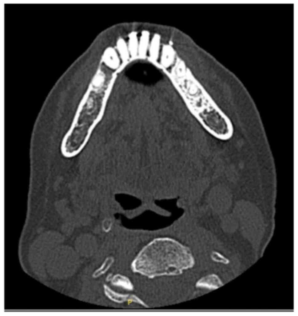
1). Panoramic radiography and computed tomography (CT) showed
no obvious bone resorption or osteosclerosis in the 654 area
(Figs. 2 and 3), and magnetic resonance imaging (MRI)
showed high signal in the T2 short τ inversion recovery image
(Fig. 4). Based on these clinical
and imaging findings, the patient was diagnosed with MRONJ on the
right side of the mandible. Amoxicillin was administered for
anti-inflammatory purposes, and the patient's pain improved. At the
follow-up visit, the patient was found to have severe
lightheadedness and floating dizziness. Therefore, a CT scan was
performed, which suggested a cerebellar infarction (Fig. 5). Subsequent MRI showed scattered
neoplastic lesions in the left cerebellum and the right frontal
lobe on gadolinium contrast T1-weighted volumetric interpolated
breath-hold examination (Fig. 6).
Neurosurgery was performed to remove the tumor from the cerebellum
because of severe dizziness and nausea and anticipated tumor
compression into the brainstem. As a metastatic brain tumor was
suspected as the brain lesion,
2-[18F]-fluoro-2-deoxy-D-glucose positron emission
tomography/computed tomography (FDG-PET/CT) was performed
postoperatively for the purpose of a systemic search. No obvious
lymphadenopathy or other lesions suggestive of malignancy were
found, except for a strong accumulation of FDG in the right adrenal
gland (Fig. 7). The pathological
results showed DLBCL with CD20, CD30, and EBV positivity (Fig. 8). Peripheral blood EBV-PCR testing
was performed within our hospital and results were detected at 70
copies/µg DNA but was not high; thus, retesting was deemed
clinically unnecessary and was not performed during or after
treatment. Because the patient had a history of MTX therapy and the
central lesion was identified as DLBCL, the patient was diagnosed
with MTX-OIIA-LPD. MTX treatment was immediately discontinued. Due
to the patient's age and poor performance status, high-dose MTX was
deemed difficult to tolerate. Whole-brain irradiation would have
been an option if only central lesions were present; however,
because of adrenal involvement, R-CHASE (cyclophosphamide,
cytarabine, etoposide, dexamethasone, rituximab) was administered
as systemic chemotherapy, including cytarabine, which has central
transferring activity. During chemotherapy, oral care was performed
regularly to prevent acute inflammation of the MRONJ in the
mandibular right molar and keep the exposed bone area clean.
However, the extent of bone exposure expanded to 7654. During the
outpatient follow-up after chemotherapy, 654 had spontaneously
fallen off; therefore, we sculpted the bone exposure area while
waiting for the patient's physical condition to recover. The
multiple brain lesions and MTX-OIIA-LPD lesions in the right
adrenal gland completely disappeared eight months after onset.
Subsequently, the patient's physical condition improved without
recurrence, and the bone-exposed area became epithelialized.
Discussion
MTX-LPD is a lymphoproliferative disorder that
occurs in patients receiving MTX and was first reported by Ellman
et al (17) in 1991. Since
then, similar reports have been published, and the term MTX-LPD has
been proposed. In 2001, the WHO histological classification of
hematopoietic and lymphoid tissue tumors (3rd edition) included the
classification of MTX-LPD (1).
However, in 2008, because LPD occurred with biological agents, such
as anti-tumor necrosis factor-α agents and tacrolimus, in addition
to MTX, the WHO Tissue Classification (4th edition) classified it
as OIIA-LPD, a subgroup of IA-LPDs (18). According to WHO, the
characteristics of MTX-OIIA-LPD are: (1) presence of RA (85% of patients with
MTX-OIIA-LPD have RA), (2)
similarity to lymphomas occurring in non-immunocompromised
individuals, (3) EBV involvement
(occurs in approximately half of the cases, and (4) most common type of lymphoma (35%)
being DLBCL. On average, the disease occurs ~3 years (0.5-5.5
years) after MTX administration. Furthermore, 60% of patients
achieve at least partial remission after discontinuation of MTX
(1). In our case, all the
characteristics were met.
Although MTX-OIIA-LPD is prone to extranodal
involvement (19), there are few
reports of MTX-OIIA-LPD in the CNS. We have identified 15 such
reported cases in the literature (3,4,6-16)
(Table I). The mean age of the
patients in these cases was 67.7 years (range: 50-86 years, except
for the patient in one case where the details were unknown), the
primary disease was RA, and the mean duration of MTX use was 5.6
years (range: 2-13 years; reports stating more than 10 years were
considered as 10 years). The histopathologic diagnoses were DLBCL
in eight patients, T-cell lymphoma in two,
pleomorphic/lymphoplasmacytic lymphoproliferative disease in one,
and intravascular large B-cell lymphoma in one; in three cases, the
primary disease was not described (20). Reportedly, EBV-DNA in the
peripheral blood was positive in patients with MTX-OIIA-LPD who did
not require chemotherapy after discontinuation of MTX and went into
remission, whereas EBV-DNA was negative in patients who required
chemotherapy.
 | Table IMTX-LPD cases in the central nervous
system. |
Table I
MTX-LPD cases in the central nervous
system.
| No. | Author | Years | Age, years | Sex | Primary illness | Central nervous
system symptom | LPD site | MTX duration,
years | Pathology | EBER | Regression after
cessation of MTX | Additional
treatment | (Refs.) |
|---|
| 1 |
Kleinschmidt-DeMasters et al | 2008 | 78 | F | RA | Neurological
symptoms | Cerebrum | ≥10 | P/L LPD | + | + | - | (3) |
| 2 | Fukushima et
al | 2013 | 64 | F | RA | Impairment of
cognitive function | Cerebrum | 4 | PTCL-NOS | + | + transient | High-dose MTX with
leucovorin rescue | (4) |
| 3 | Migita et
al | 2013 | 53 | F | RA | Vertigo, vomiting,
and double vision | Medulla | 1 | DLBCL | + | + | Resection of
mass | (6) |
| 4 | Liu et al | 2015 | 58 | M | RA | Depressive disorder,
dizziness, vomiting staggering gait | Cerebrum | 2 | DLBCL | - | NA | Radiotherapy | (7) |
| 5 | Shimada et
al | 2015 | 60sa | F | RA | Convulsion, impaired
consciousness | Cerebrum | 7 | NA | + | + | - | (8) |
| 6 | Kikuchi et
al | 2016 | 50 | F | RA | - | Hypertrophy of
frontal dura mater | 3 | Intravascular large B
cell lymphoma | - | - | R-CHOP + intrathecal
chemotherapy (prednisolone+ MTX + cytarabine) | (9) |
| 7 | Matsuda et
al | 2018 | 76 | F | RA | Staggering gait | Cerebrum | 2 | DLBCL | + | + | - | (10) |
| 8 | Miyaza et
al | 2019 | 68 | F | RA | Hemiparesis | Cerebrum | 5 | DLBCL | + | + | - | (11) |
| 9 | Kawazoe et
al | 2020 | 75 | M | RA | Gait ataxia | Cerebrum pons | 9 | DLBCL | Brain- +
Stomach | Brain- +
Stomach | High-dose MTX
rituximab | (12) |
| 10 | Uneda et
al | 2020 | 68 | F | RA | Motor aphasia,
right hemiparesis | Cerebrum | ≥10 | DLBCL | + | + | - | (13) |
| 11 | Kawano et
al | 2021 | 75 | F | RA | Unsteady gait,
nausea and vomiting | Cerebellum,
cerebrum | 3 | T-cell
lymphoma | - | + | - | (14) |
| 12 | Ueno et
al | 2022 | 64 | M | RA | Ataxia | Cerebellum,
cerebrum | 3 | DLBCL | - | + | - | (15) |
| | | | 86 | F | RA | Weakness | Cerebrum,
thalamus | 3 | NA | + | + | - | |
| | | | 66 | F | RA | Gait
disturbance | Cerebrum | 13 | NA | + | + | - | |
| 13 | Mizushima et
al | 2022 | 59 | F | RA | Staggering gait,
memory deficit | Cerebrum | 9 | DLBCL | - | - | R-MPV
Radiotherapy | (16) |
| 14 | Kato et
al | 2023 | 75 | F | RA | Dizziness | Cerebellum,
cerebrum | 6 | DLBCL | + | + | R-CHASE | Present case |
Among the 15 cases of MTX-OIIA-LPD that developed in
the CNS, 11 involved patients were EBV-positive. Among them, in 10
cases, the lesions resolved with interruption of MTX, whereas in
one case, the tumor shrank after MTX withdrawal, but chemotherapy
was administered because the tumor worsened. Considering that it
takes ~4 weeks from MTX discontinuation to remission (19) and that remission is not achieved in
all cases after MTX withdrawal, we decided that we could not wait
for disease shrinkage by MTX discontinuation and decided to
administer chemotherapy.
Several patients with RA also have osteoporosis
(2), and a number of patients are
likely to have been administered with MTX and bone resorption
inhibitors concomitantly. However, There are few reports of cases
with MRONJ and MTX-OIIA-LPD (21).
Furthermore, to the best of our knowledge, there are no reports of
concomitant MRONJ and CNS MTX-OIIA-LPD in the literature, as in our
own case. Unless there is evidence of metastatic lesions to the
jawbone in MRONJ, surgical biopsy is generally not recommended
because of the possibility of exposing new fresh bone surfaces
(22,23); thus, the diagnosis of MRONJ is made
based on clinical symptoms (24)
without biopsy. Furukawa et al (21) reported a case of MRONJ and
MTX-OIIA-LPD in a patient who had been taking bisphosphonates (BP)
and MTX. They reported that clinicians should keep in mind that
intraoral MTX-OIIA-LPD is sometimes complicated by MRONJ, and that
pathological examination is necessary for a final diagnosis.
There have been few reports of MTX-OIIA-LPD in the
oral and maxillofacial region (21), and dentists are thought to be less
aware of MTX-OIIA-LPD than MRONJ. We were able to identify only one
case of MTX-OIIA-LPD and MRONJ occurring simultaneously in the oral
and maxillofacial region (21).
Moreover, our case is the first to report extraoral MTX-OIIA-LPD
and MRONJ. Although simultaneous occurrence of MTX-OIIA-LPD and
MRONJ is rare, when a patient with MRONJ is taking MTX, biopsy of
the oral lesions should be performed considering oral and extraoral
MTX-OIIA-LPD. In addition, systemic search should be performed in
collaboration with related departments, such as rheumatology and
hematology, to promptly diagnose MTX-OIIA-LPD and initiate prompt
treatment. One of the side effects of MTX is dizziness. In our
case, the patient already had CNS and progressive symptoms at the
disease onset, different from MTX side effects, and imaging tests
were performed, leading to the discovery of a neoplastic lesion in
the brain. Neurosurgery was performed to remove the tumor from the
cerebellum immediately because of severe dizziness and nausea and
anticipated tumor compression into the brainstem and a pathological
diagnosis was done. Thus, although MTX-OIIA-LPD of the CNS is rare,
we were able to diagnose it early.
In conclusion, oral and maxillofacial surgeons
should perform biopsies of oral lesions in patients with RA who are
taking BP and MTX and have MRONJ. Further, collaboration with
related diagnostic departments, such as rheumatology and
hematology, is essential to enable early diagnosis and treatment of
extraoral MTX-LPD. Moreover, although central MTX-LPD is rare, as
in our case, it is also necessary to collaborate with neurosurgery
and other related departments depending on the symptoms.
Acknowledgements
The authors would like to thank Dr Hiroki Yamada
(Department of Neurosurgery, Graduate School of Medicine, Kyoto
University, Kyoto, Japan) who contributed to the diagnosis and
treatment of the patient.
Funding
Funding: No funding was received.
Availability of data and materials
The datasets used and/or analyzed during the current
study are available from the corresponding author on reasonable
request.
Authors' contributions
TK acquired the patient's clinical data, performed
the literature review and edited the manuscript. TK, FI, TW, SY and
SF contributed substantially to the conception and design of the
study. CM and KN acquired data, provided clinical advice and
revised the manuscript. TK played a major role in writing the
manuscript. TK and CM confirm the authenticity of all the raw data.
All the authors read and approved the final version of the
manuscript.
Ethics approval and consent to
participate
Not applicable.
Patient consent for publication
Written informed consent was obtained from the
patient for the publication of this case report and accompanying
images.
Competing interests
The authors declare that they have no competing
interests.
References
|
1
|
Wang W, Zhou H and Liu L: Side effects of
methotrexate therapy for rheumatoid arthritis: A systematic review.
Eur J Med Chem. 158:502–516. 2018.PubMed/NCBI View Article : Google Scholar
|
|
2
|
Harigai M: Lymphoproliferative disorders
in patients with rheumatoid arthritis in the era of widespread use
of methotrexate: A review of the literature and current
perspective. Mod Rheumatol. 28:1–8. 2018.PubMed/NCBI View Article : Google Scholar
|
|
3
|
Kleinschmidt-DeMasters BK, Damek DM,
Lillehei KO, Dogan A and Giannini C: Epstein Barr virus-associated
primary CNS lymphomas in elderly patients on immunosuppressive
medications. J Neuropathol Exp Neurol. 67:1103–1111.
2008.PubMed/NCBI View Article : Google Scholar
|
|
4
|
Fukushima M, Katayama Y, Yokose N, Kura Y,
Sawada U, Kotani A and Yoshino A: Primary central nervous system
malignant lymphoma in a patient with rheumatoid arthritis receiving
low-dose methotrexate treatment. Br J Neurosurg. 27:824–826.
2013.PubMed/NCBI View Article : Google Scholar
|
|
5
|
Hoshida Y, Xu JX, Fujita S, Nakamichi I,
Ikeda J, Tomita Y, Nakatsuka S, Tamaru J, Iizuka A, Takeuchi T and
Aozasa K: Lymphoproliferative disorders in rheumatoid arthritis:
Clinicopathological analysis of 76 cases in relation to
methotrexate medication. J Rheumatol. 34:322–331. 2007.PubMed/NCBI
|
|
6
|
Migita K, Miyashita T, Mijin T, Sakito S,
Kurohama H, Ito M, Toda K, Tsustumi K, Baba H, Izumi Y, et al:
Epstein-Barr virus and methotrexate-related CNS lymphoma in a
patient with rheumatoid arthritis. Mod Rheumatol. 23:832–836.
2013.PubMed/NCBI View Article : Google Scholar
|
|
7
|
Liu W, Xue J, Yu S, Chen Q, Li X and Yu R:
Primary central nervous system lymphoma mimicking recurrent
depressive disorder: A case report. Oncol Lett. 9:1819–1821.
2015.PubMed/NCBI View Article : Google Scholar
|
|
8
|
Shimada H, Dobashi H, Morimoto H, Kameda
T, Susaki K, Izumikawa M, Takeuchi Y, Nakashima S, Imataki O and
Bandoh S: Primary central nervous system lymphoma in a rheumatoid
arthritis patient treated with methotrexate: A case report. BMC Res
Notes. 8(88)2015.PubMed/NCBI View Article : Google Scholar
|
|
9
|
Kikuchi J, Kaneko Y, Kasahara H, Emoto K,
Kubo A, Okamoto S and Takeuchi T: Methotrexate-associated
intravascular large B-cell lymphoma in a patient with rheumatoid
arthritis. Intern Med. 55:1661–1665. 2016.PubMed/NCBI View Article : Google Scholar
|
|
10
|
Matsuda I and Hirota S:
Methotrexate-associated lymphoproliferative disorder masquerading
as multiple cerebral metastases. Br J Haematol.
180(628)2018.PubMed/NCBI View Article : Google Scholar
|
|
11
|
Miyaza S, Matsuda R, Nakamura M, Nakagawa
I, Motoyama Y and Nakase H: Intracranial methotrexate-associated
lymphoproliferative disorder in rheumatoid arthritis. World
Neurosurg. 130:138–141. 2019.PubMed/NCBI View Article : Google Scholar
|
|
12
|
Kawazoe M, Kaneko K and Nanki T:
Methotrexate-associated lymphoproliferative disorders in the
central nervous system and stomach: A case report. Medicine
(Baltimore). 99(e19850)2020.PubMed/NCBI View Article : Google Scholar
|
|
13
|
Uneda A, Hirashita K, Kanda T, Yunoki M,
Yoshino K, Kurozumi K and Date I: rimary central nervous system
methotrexate-associated lymphoproliferative disorder in a patient
with rheumatoid arthritis: Case report and review of literature.
NMC Case Rep J. 7:121–127. 2020.PubMed/NCBI View Article : Google Scholar
|
|
14
|
Kawano H, Kitamura T, Higuchi K and Nozaki
K: Primary central nervous system T-cell lymphoma as
methotrexate-associated lymphoproliferative disorders: Case report.
NMC Case Rep J. 8:253–259. 2021.PubMed/NCBI View Article : Google Scholar
|
|
15
|
Ueno H, Ohno N, Abe T, Kimoto K, Matsuoka
C, Giga M, Naito H, Kono T, Takasu M, Kidani N, et al: Prognosis
prediction using magnetic resonance spectroscopy and oligoclonal
bands in central nervous system methotrexate-associated
lymphoproliferative disorder. Intern Med. 61:3733–3738.
2022.PubMed/NCBI View Article : Google Scholar
|
|
16
|
Mizushima M, Ishi Y, Ikeda H, Echizenya I,
Otsuka T, Mitsuhashi T, Yamaguchi S and Fujimura M: Successful
treatment of intracranial methotrexate-associated
lymphoproliferative disorder without Epstein-Barr virus infection
using rituximab, methotrexate, procarbazine, and vincristine: A
case report. NMC Case Rep J. 9:237–242. 2022.PubMed/NCBI View Article : Google Scholar
|
|
17
|
Ellman MH, Hurwitz H, Thomas C and Kozloff
M: Lymphoma developing in a patient with rheumatoid arthritis
taking low dose weekly methotrexate. J Rheumatol. 18:1741–1743.
1991.PubMed/NCBI
|
|
18
|
Gaulard P, Swerdlow SH, Harris NL,
Sundstrom C and Jaffe ES: Other iatrogenic
immunodeficiency-associated lymphoproliferative disorders. In:
Swerdlow SH, Campo E, Harris NL, Jaffe ES, Pileri SA, Stein H and
Thiele J (eds). WHO classification of tumours of haematopoietic and
lymphoid tissues. Revised 4th edition. IARC, Lyon, France,
pp462-464, 2017.
|
|
19
|
Rizzi R, Curci P, Delia M, Rinaldi E,
Chiefa A, Specchia G and Liso V: Spontaneous remission of
‘methotrexate-associated lymphoproliferative disorders’ after
discontinuation of immunosuppressive treatment for autoimmune
disease. Review of the literature. Med Oncol. 26:1–9.
2009.PubMed/NCBI View Article : Google Scholar
|
|
20
|
Katsuyama T, Sada KE, Yan M, Zeggar S,
Hiramatsu S, Miyawaki Y, Ohashi K, Morishita M, Watanabe H,
Katsuyama E, et al: Prognostic factors of methotrexate-associated
lymphoproliferative disorders associated with rheumatoid arthritis
and plausible application of biological agents. Mod Rheumatol.
27:773–777. 2017.PubMed/NCBI View Article : Google Scholar
|
|
21
|
Furukawa S, Oobu K, Moriyama M, Kawano S,
Sako S, Hayashida JN, Matsubara R, Ogata KI, Kiyoshima T and
Nakamura S: Oral methotrexate-related lymphoproliferative disease
presenting with severe osteonecrosis of the jaw: A case report and
literature review. Intern Med. 57:575–581. 2018.PubMed/NCBI View Article : Google Scholar
|
|
22
|
Ruggiero SL, Dodson TB, Assael LA,
Landesberg R, Marx RE and Mehrotra B: American Association of Oral
and Maxillofacial Surgeons. American association of oral and
maxillofacial surgeons position paper on bisphosphonate-related
osteonecrosis of the Jaws-2009 update. J Oral Maxillofac Surg. 67
(Suppl):S2–S12. 2009.PubMed/NCBI View Article : Google Scholar
|
|
23
|
Ficarra G and Beninati F:
Bisphosphonate-related osteonecrosis of the Jaws: An update on
clinical, pathological and management aspects. Head Neck Pathol.
1:132–140. 2007.PubMed/NCBI View Article : Google Scholar
|
|
24
|
Ruggiero SL, Dodson TB, Aghaloo T, Carlson
ER, Ward BB and Kademani D: American association of oral and
maxillofacial surgeons' position paper on medication-related
osteonecrosis of the Jaws-2022 update. J Oral Maxillofac Surg.
80:920–943. 2022.PubMed/NCBI View Article : Google Scholar
|