Introduction
Renal cell carcinoma (RCC) is a metastatic,
heterogeneous disease that is resistant to conventional treatment
modalities (1,2). Resistance to therapy continues to be
a major challenge to the effective treatment of patients with
metastatic RCC (2,3). It is estimated that ~25% of patients
have advanced stage RCC, which includes locally invasive or
metastatic RCC. In addition, it was reported that one-third of
patients who underwent partial lesion resection experienced
recurrence (4). The median
survival rate of patients with metastatic cancer is ~13 months
(5). Therefore, a more effective
treatment for RCC is urgently required. In previous years,
gene-targeted therapy has become promising for the treatment of
RCC. Over the last decade, the treatment of RCC has evolved to
include drugs that target the vascular endothelial growth factor
(VEGF) and mammalian target of rapamycin (mTOR) pathways, resulting
in substantially improved outcomes for the patient population
(6). However, despite prolonged
survival rates, the majority of patients develop resistance to
VEGF- and mTOR-targeted therapies. It is important to identify
novel anticancer agents to combat this life-threatening disease
(6).
Junctional adhesion molecule 3 (Jam3) is a vascular
adhesion molecule, which regulates adhesion and interactions among
or between cells and the extracellular matrix (5). It has been reported that Jam3
enhances the expression and activation of adhesion molecules in
vascular endothelial cells (7,8).
The methylation status of Jam3 is a biomarker for the diagnosis of
preneoplastic and neoplastic lesions of the cervix and promotes
cervical cancer progression (9).
Therefore, the present study examined whether Jam3 enhanced and
influenced migration and apoptosis in RCC. E-cadherins are
calcium-dependent cell adhesion proteins, and their enhancement can
reduce cancer migration (10).
Integrins are cell adhesion receptors, which are important in the
interactions between cells and the extracellular matrix, and in
cell-cell interactions. Integrin β1 has also been shown to
contribute to cancer migration (11). Matrix metalloproteinase (MMP)-2 is
known as an interstitial collagenase and fibroblast collagenase
encoded by the MMP-2 gene in humans. Tumor cells promote migration
by degrading collagen through the secretion of MMP-2. MMP-2 may
alter tumor viability and invasion by regulating lymphangiogenesis
in addition to angiogenesis (12).
However, no study published to date has examined the
role of Jam3 in renal carcinoma cells. The present study
investigated whether the expression of Jam3 is increased in renal
carcinoma cells and determined the important migration protein of
RCC in this process. The results may provide information to assist
in the development of novel therapeutic targets and strategies for
the treatment of RCC.
Materials and methods
Cell culture
The HK-2 cell line (adenovirus 50-immortalized line
of human renal tubular epithelial cells), Caki-1 and 786-0 cell
lines (Simian virus 26-immortalized line of human ACHN) were
obtained from American Type Culture Collection (Manassas, VA, USA)
and cultured in Dulbecco's modified Eagle's medium (DMEM) with 10%
fetal bovine serum (FBS; Gibco; Thermo Fisher Scientific, Inc.,
Waltham, MA, USA). The cells were cultured at 37°C in a 5%
CO2 atmosphere, and growth status was measured using an
inverted microscope. On reaching 80-90% confluence, the cells were
subcultured and digested with trypsin. The cell activities were
examined by a routine MTT assay. The detailed protocol has been
described previously (13-16).
Western blot analysis
The HK-2, Caki-1 and 786-0 cells were homogenized
with M2 buffer containing 20 mM Tris-HCl (pH 7.6), 0.5% NP-40, 250
mM NaCl, 3 mM EDTA, 2 mM DTT, 0.5 mM phenylmethylsulfonylfluoride,
20 mM β-glycerophosphate, 1 mM sodium vanadate, and 1 µg/ml
leupeptin. Protein concentrations were determined using the BCA
assay (Bioteke Corporation, Beijing, China). Equal quantities (40
µg) of protein homogenates were analyzed using 12% sodium dodecyl
sulfate polyacrylamide gel electrophoresis and transferred onto a
polyvinylidene fluoride membrane (EMD Millipore, Billerica, MA,
USA), which was then blocked with Tris-buffered saline (TBS) and
0.1% Tween-20 (TBS-T) containing 5% bovine serum albumin (Sangon
Biotech Co., Ltd., Shanghai, China) for 2 h at 37°C. The membrane
was subsequently washed three times with TBS-T. The membranes were
probed with primary antibody overnight at 4°C followed by
incubation with secondary antibody for 1 h at room temperature. The
signals were detected via enhanced chemiluminescence (GE Healthcare
Life Sciences, Little Chalfont, UK) according to the manufacturer's
protocol. The band density was quantified using ImageJ software 5.0
(National Institutes of Health, Bethesda, MD, USA) and normalized
relative to glyceraldehyde 3-phosphate dehydrogenase (GAPDH). The
primary antibodies used were as follows: Monoclonal anti-Jam3
(WH0083700M1; 1:2,000 dilution; human; Sigma-Aldrich; Merck KGaA,
Darmstadt, Germany), E-cadherin (5296; 1:1,000 dilution; human;
Cell Signaling Technology, Inc., Danvers, MA, USA), N-cadherin
(14215; 1:1,000 dilution; human; Cell Signaling Technology, Inc.),
integrin β1 (ab24693; 1:2,000 dilution; human; Abcam, Cambridge,
UK), MMP-2 (ab86607; 1:2,000 dilution; human; Abcam), B cell
lymphoma 2 (Bcl-2; ab692; 1:2,000 dilution; human; Abcam)
Bcl-2-associated X protein (Bax; ab77566; 1:2,000 dilution; human;
Abcam), and GAPDH (sc293335; 1:2,000 dilution; human; Santa Cruz
Biotechnology, Inc., Santa Cruz, CA, USA). A horseradish
peroxidase-labeled secondary antibody (7076; 1:1,000 dilution;
human; Cell Signaling Technology, Inc.) was used as the secondary
antibody.
Reverse transcription-quantitative
polymerase chain reaction (RT-qPCR) analysis
The HK-2, Caki-1 and 786-0 cells were cultured in
six-well plates for 24 h. RNA was obtained from these cells using
lysis buffer (Bioteke Corporation) and reversed transcribed using a
Prime ScriptRT reagent kit (Takara Bio, Inc., Shiga, Japan)
according to the manufacturer's protocol. The mRNA expression level
of Jam3 was determined via qPCR analysis. The reaction was
processed using Real Master mix (SYBR-Green; Tiangen Biotech Co.,
Ltd., Beijing, China) in a total volume of 10 µl containing 2 µl
cDNA, 5 µl SYBR solution, 0.15 µl each of forward and reverse
primers, and up to 2.7 µl ddH2O. The primer sequences
for Jam3 were as follows: Forward 5′-CGT AGT TAG GGT TGG GAT TC-3′
and reverse 5′-GAA ATC CGA CGA CTA TCC GA-3′; and the primer
sequences for β-actin were as follows: Forward 5′-GTG GAC ATC CGC
AAA GAC-3′ and reverse 5′-GAA AGG GTG TAA CGC AAC T-3′. The PCR
amplification was performed as follows: 95°C for 10 min, followed
by 40 cycles at 95°C for 15 sec, and 58-60°C for 1 min, in a total
volume of 10 µl based on the 7900HT Fast Real-Time PCR system
(Applied Biosystems; Thermo Fisher Scientific, Inc.). The
expression of Jam3 was analyzed using the 2−ΔΔCq method
(17).
Cell transfection
The Caki-1 and 786-0 cells were cultured in
serum-free DMEM without antibiotics. Jam3 small interfering (si)RNA
(sc-43872) and negative siRNAs (sc-37007) were obtained from Santa
Cruz Biotechnology, Inc. The cells were transfected using
Lipofectamine 2000 (Invitrogen; Thermo Fisher Scientific, Inc.)
according to the manufacturer's protocol. Following 6 h of
transfection, fresh medium was added to the transfected cells and
cultured for up to 24 h. The cells were collected to verify gene
silencing by assessing the protein expression of Jam3 using western
blot analysis.
Flow cytometry
Following transfection with or without Jam3 siRNA
for 6 h, fresh medium was added to the transfected cells and
cultured for up to 24 h, Subsequently, Annexin V/propidium iodide
staining was detected by flow cytometry using the APC Annexin V kit
(BD Biosciences, Franklin Lakes, NJ, USA) according to the
manufacturer's protocol. The flow cyto-metric analysis was
performed using SLR II flow cytometer software. Data were analyzed
using FACSDIVA software 6.0 (BD Biosciences).
Wound-healing assay
The Caki-1 and 786-0 cells were seeded in six-well
plates at a density of 2×10⁵ cells per well. The cells
were then transfected with negative or Jam3 siRNA for 24 h and
grown to 80-90% confluence. The cell layers were scratched with a
20-µl pipette tip, washed three times with phosphate-buffered
saline and cultured in medium without FBS. Images of the wound area
were captured using a fluorescence microscope every 24 h. The cells
on either side of the wound migrated into the cell-free area. The
widths of three different wound surfaces of each group were
recorded and measured using ImageJ software. The experiment was
repeated at least three times.
Cell migration assay
The Caki-1 and 786-0 cells were transfected with
non-specific or Jam3 siRNA for 24 h, and the cells were digested,
counted and suspended in serum-free DMEM. Following this, 100 µl
(2×10⁵ cells) were seeded in the upper chamber of a
Transwell unit with an 8.0-µm polycarbonate membrane (EMD
Millipore) inserted in a 24-well plate, and 500 µl of culture
medium with 10% FBS was added to the lower chamber. The cells were
cultured for 24 h at 37°C, following which the cells on the top
surface of the Transwell chamber were removed using a cotton swab.
The cells adhering to the lower surface were fixed with 4%
paraformaldehyde for 15 min and stained with crystal violet. The
migration was assessed using an inverted phase contrast microscope
in five randomly selected fields.
Statistical analysis
Data in the present study were analyzed using
GraphPad Prism 5.0 software (GraphPad Software, Inc., La Jolla, CA,
USA) and are presented as the mean ± standard deviation.
Statistical differences between multiple groups were compared using
one-way analysis of variance followed by Tukey's post hoc test or
Student's t-test (parametric)/Mann-Whitney (non-parametric) test.
If an overall test was significant, Tukey's test was used for
specific comparisons between individual groups. P<0.05 was
considered to indicate a statistically significant difference.
Results
mRNA and protein levels of Jam3 in renal
cancer cells
To assess the differential expression of Jam3 in
renal cancer cells and normal renal cells, the present study
determined the mRNA and protein expression of Jam3 in the HK-2
human renal tubular epithelial cell line and the Caki-1 and 786-0
renal cancer cell lines. It was found that the protein expression
of Jam3 in Caki-1 and 786-0 cells was higher than that in HK-2
cells (P<0.05), as shown by the western blot analysis (Fig. 1A). The PCR results showed that the
mRNA expression of Jam3 was also higher in the Caki-1 and 786-0
cells compared with that in the HK-2 cells (P<0.05; Fig. 1B). These results suggested that
the mRNA and protein levels of Jam3 were higher in renal carcinoma
cells. Therefore, its effects were examined in subsequent
experiments.
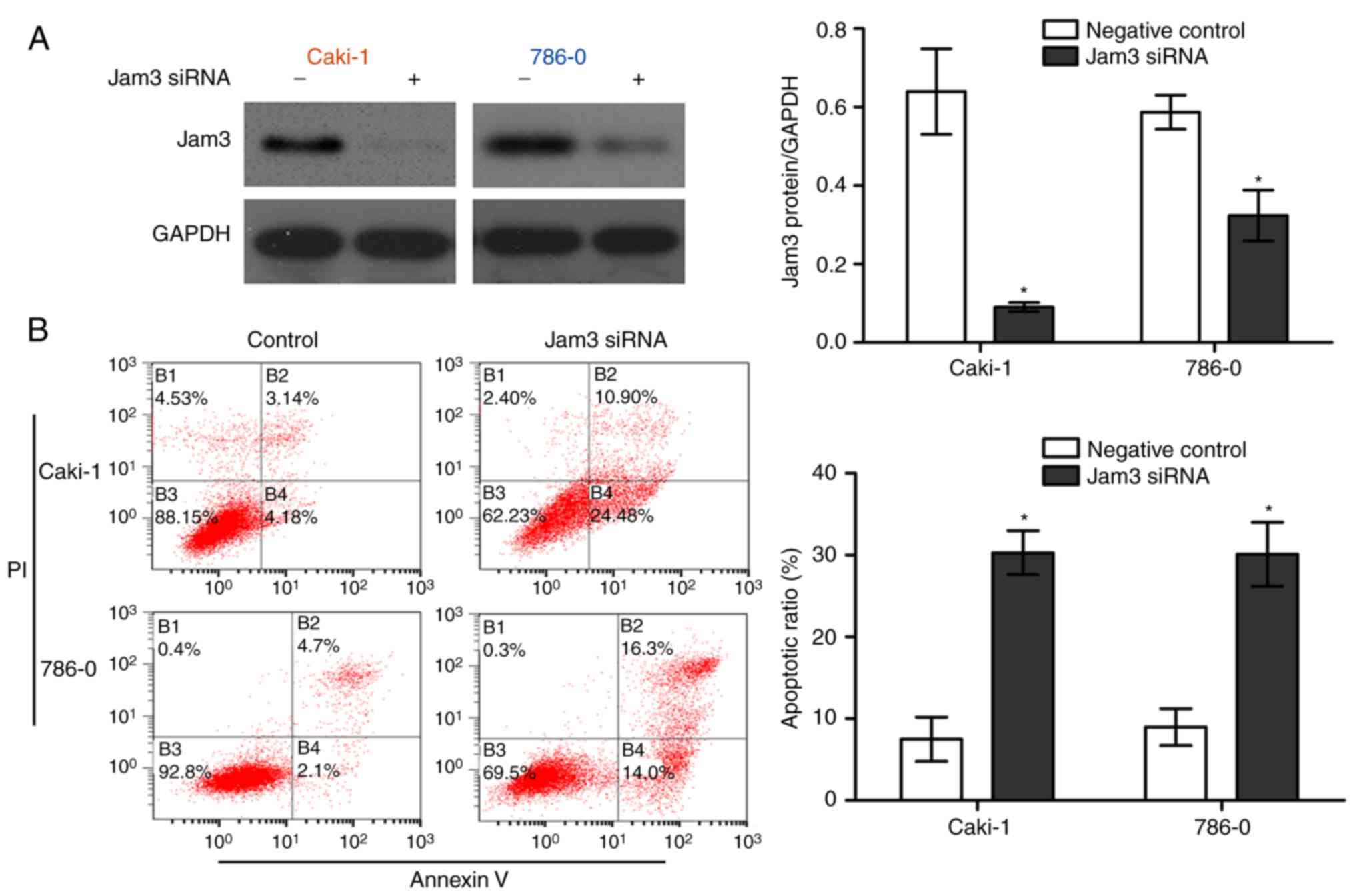
Jam3 degeneration induces apoptosis of
renal carcinoma cells
To investigate the function of Jam3 on the
proliferation and apoptosis of Caki-1 and 786-0 cells, siRNAs were
used. An siRNA targeting the Jam3 mRNA sequence was transfected
into Caki-1 and 786-0 cells, and the protein levels of Jam3 were
assessed by western blot analysis. The results showed that Jam3
siRNA significantly silenced the protein expression of Jam3
(P<0.05), compared with that in the control group (Fig. 2A). The present study also examined
whether Jam3 affected the apoptosis of Caki-1 and 786-0 cells using
flow cytometry with FITC-Annexin V. The flow cytometry results
showed that the percentage of apoptotic cells was higher in cells
in which the Jam3 gene was silenced (Fig. 2B).
Jam3 enhances the migratory ability of
renal carcinoma cells
To further support a function of Jam3 in renal
cancer cell migration, Jam3 was exogenously knocked down in Caki-1
and 786-0 cells using siRNA. The wound-healing assay revealed that,
24 h post-wounding, the wound width was larger in the Caki-1 and
786-0 cells in which the Jam3 gene was silenced, compared with that
in the non-specific siRNA group (P<0.05; Fig. 3A and B). Notably, cell migration
was measured via Transwell assays. Jam3 siRNA knockdown
significantly enhanced the migration of the Caki-1 and 786-0 cells.
Fewer cells migrated in the Jam3 siRNA group (P<0.05; Fig. 3C), compared with the non-specific
siRNA group. These results suggested that Jam3 exhibited a marked
effect on renal carcinoma cells, promoting the migration of Caki-1
and 786-0 cells and inhibiting Caki-1 and 786-0 cell migration when
downregulated.
Effects of Jam3 on E-cadherin,
N-cadherin, integrin β1, MMP-2, Bcl-2 and Bax in renal carcinoma
cells
E-cadherin and N-cadherin are link proteins between
cells and are important for the migration of cancer cells (18). Therefore, the present study
measured the protein levels of E-cadherin and N-cadherin in Jam3
siRNA-transfected Caki-1 and 786-0 cells compared with the
non-specific siRNA-transfected cells. It was found that the
expression of E-cadherin was increased and the expression of
N-cadherin was decreased following transfection with Jam3 siRNA,
compared with the negative siRNA (Fig. 4A). Integrin β1 can induce cancer
cell migration (19), and it was
found that the protein level of integrin β1 was also reduced in the
Jam3 siRNA group, compared with that in the control group
(P<0.05; Fig. 4A). MMP-2 can
degrade type IV collagen and contributes to cancer cell invasion
(20). The knockdown of Jam3 in
Caki-1 and 786-0 cells reduced the protein levels of MMP-2
(Fig. 4A). These data indicated
that Jam3 knockdown caused an increase in E-cadherin and Bax, and a
decrease in N-cadherin, integrin β1, MMP-2 and Bcl-2. All data for
the Jam3 siRNA group were statistically significant when compared
with the non-specific siRNA group (P<0.05, Fig. 4B).
 | Figure 4Differential expression of
E-cadherin, N-cadherin, integrin β1 and MMP-2 in Caki-1 and 786-0
cells transfected with non-specific siRNA and Jam3 siRNA. (A)
Cellular protein was collected from Caki-1 and 786-0 cells
transfected with non-specific siRNA or Jam3 siRNA. Western blot
analysis was performed to assess the levels of E-cadherin,
N-cadherin, integrin β1, MMP-2, Bax and Bcl-2. GAPDH was used as a
loading control. (B) Average grey values represented as a histogram
(*P<0.05). Jam3, junctional adhesion molecule 3;
MMP-2, matrix metalloproteinase 2; Bcl-2, B-cell lymphoma 2; Bax,
Bcl-2-associated X protein; GAPDH, glyceraldehyde 3-phosphate
dehydrogenase; siRNA, small interfering RNA. |
Discussion
As a common type of renal cancer, RCC has a high
annual mortality rate (21-24). RCC accounts for ~3% of adult
malignant tumors and 85% of primary renal tumors. In China, renal
cancer was expected to account for 1.5% of all new cancer cases in
2015, with a mortality rate of ~35% (25). Therefore, the prognosis of RCC is
poor (21-23,26,27). Surgical resection of RCC is only
suitable for a limited number of patients with early stage tumors
(21,28-30). Several studies (13,31) have examined novel oncotargets for
RCC. The present study identified Jam3 as an important gene in RCC,
which has been reported as a junctional adhesion molecule. In the
present study, the protein and mRNA levels of Jam3 were higher in
renal carcinoma cells (Caki-1 and 786-0), compared with those in
renal tubular epithelial cells (HK-2). It was reported previously
that Jam3 was overexpressed in gastric adenocarcinoma tumors and
was involved with the progression of gastric tumor cells (32). Junctional adhesion molecule-C
(JAM-C) is a tight junction-associated transmembrane protein
expressed on mammalian endothelial cells. It is involved in
leukocyte diapedesis and interacts with the leukocyte integrin M2
(14). Jam3 is involved in
leukocyte transendothelial migration and can form homophilic
(JAM-C/JAM-C) and heterophilic interactions with the leukocyte
integrin αMβ2. In a previous study, the
effect of early administration of monoclonal antibodies directed
against JAM-C in cerulein-induced acute pancreatitis was assessed
(33). It was suggested that Jam3
expressed by endothelial cells contributes to the pathophysiology
of acute pancreatitis and may be considered a target for clinical
applications (33). The
methylation status of Jam3 has been used as a biomarker for the
diagnosis of preneoplastic and neoplastic lesions of the cervix
(10). The present study found
that renal carcinoma cell apoptosis was enhanced following
transfection with Jam3 siRNA. Therefore, it was hypothesized that
Jam3 inhibits renal carcinoma cell apoptosis. Previous results have
shown that the expression of Jam3 can be regulated during B-cell
maturation. Furthermore, the expression of JAM-C has been used to
divide CD27+ B cells into two subtypes:
JAM-C+ cells, as the major proportion of circulating
CD27+ cells in the peripheral blood, and
JAM-C− cells, identified as a phenotype of germinal
center B cells of secondary lymphoid organs. JAM-C-cells exhibit
high expression of BCL6, a nuclear proto-oncogene with a pivotal
role in germinal center formation (33), whereas JAM-C+ cells
express a low level of BCL6, which is a signature of
extrafollicular cells (34).
According to the data obtained in the present study,
Jam3 knockdown in Caki-1 and 786-0 cells inhibited cancer cell
migration and the protein expression of N-cadherin, and increased
the expression of E-cadherin. The PDZ domain-binding motif located
in the cytoplasmic tail of JAMs mediates interactions with
intracellular scaffolding proteins, including zonula occludens 1,
thereby providing a link to the cytoskeleton (35). Integrin β1 is considered to be
important in cell migration by redistributing integrins from the
retracting edges to the migrating front (36). Li et al and Dai et
al previously demonstrated that endocytic integrin β1
accumulated at the recycling endosome under starvation conditions,
and was recycled following acute stimulation (37,38). In human aortic smooth muscle
cells, KP-10 significantly suppressed angio-tensin II-induced
migration and proliferation, but increased apoptosis and the
activities of MMP-2 and MMP-9 via the upregulation of extracellular
signal-regulated kinase 1 and 2, p38, Bax, and caspase-3. MMP-2
increases cancer cell migration by degrading extracellular collagen
tissue (39).
In conclusion, the present study showed that Jam3
was expressed at a high level in renal carcinoma cells. The
downregulation of Jam3 significantly enhanced apoptosis and
suppressed migration of renal carcinoma cells. It is known that the
link proteins E-cadherin and N-cadherin, and integrin β1 and MMP-2
are associated with tumor migration ability. According to the
present study, transfection of Jam3 siRNA in renal carcinoma cells
promoted the protein level of E-cadherin, and downregulated the
protein levels of N-cadherin, integrin β1 and MMP-2. Therefore,
Jam3 may affect renal carcinoma cell migration and apoptosis by
regulating E-cadherin, N-cadherin, integrin β1 and MMP-2. However,
whether Jam3 is also expressed at a high level in clinical samples
remains to be elucidated. The present study did not examine in
vivo Jam3 levels in patients with RCC; however, this question
is to be addressed in future investigations. Jam3 may be a
potential target for clinical applications, therefore, it is would
be useful to examine this novel RCC medicine in future
investigations.
Funding
This study was supported by the National Nature
Science Foundation of China (grant no. 81200528) and Prevention and
Control Strategies for Major Diseases, Diagnosis and Treatment
Practices, and Evaluation of Effects (grant no. 2015BAI12B05; Type:
12th five-year public relations).
Availability of data and materials
All data generated or analyzed during this study are
included in this published article.
Authors' contributions
XL and JS conceived the study; AY and WZ performed
the experiments; FZ, JiaL and JingL analyzed the data; JS wrote the
manuscript. All authors have read and approved the final version of
the manuscript.
Ethics approval and consent to
participate
Not applicable.
Patient consent for publication
Not applicable.
Competing interests
The authors declare that they have no competing
interests.
Acknowledgments
Not applicable.
References
|
1
|
Hirbod-Mobarakeh A, Gordan HA, Zahiri Z,
Mirshahvalad M, Hosseinverdi S, Rini BI and Rezaei N: Specific
immunotherapy in renal cancer: A systematic review. Ther Adv Urol.
9:45–58. 2017. View Article : Google Scholar : PubMed/NCBI
|
|
2
|
Turajlic S, Larkin J and Swanton C:
SnapShot: Renal cell carcinoma. Cell. 163:1556–1556.e1. 2015.
View Article : Google Scholar : PubMed/NCBI
|
|
3
|
Ling S, Nheu L and Komesaroff PA: Cell
adhesion molecules as pharmaceutical target in atherosclerosis.
Mini Rev Med Chem. 12:175–183. 2012. View Article : Google Scholar
|
|
4
|
Mazza C, Escudier B and Albiges L:
Nivolumab in renal cell carcinoma: Latest evidence and clinical
potential. Ther Adv Med Oncol. 9:171–181. 2017. View Article : Google Scholar : PubMed/NCBI
|
|
5
|
Thakor P, Song W, Subramanian RB, Thakkar
VR, Vesey DA and Gobe GC: Maslinic acid inhibits proliferation of
renal cell carcinoma cell lines and suppresses angiogenesis of
endothelial cells. J Kidney Cancer VHL. 4:16–24. 2017. View Article : Google Scholar : PubMed/NCBI
|
|
6
|
Heng DY: The next 10 years: Challenges for
the future and overcoming resistance to targeted therapies for
renal cell carcinoma. Can Urol Assoc J. 10(11-12 Suppl 7):
S256–S258. 2016. View Article : Google Scholar
|
|
7
|
Lim SH, Hwang IG, Ji JH, Oh SY, Yi JH, Lim
DH, Lim HY, Lee SJ and Park SH: Intrinsic resistance to sunitinib
in patients with metastatic renal cell carcinoma. Asia Pac J Clin
Oncol. 13:61–67. 2017. View Article : Google Scholar
|
|
8
|
Santoso S, Sachs UJ, Kroll H, Linder M,
Ruf A, Preissner KT and Chavakis T: The junctional adhesion
molecule 3 (JAM-3) on human platelets is a counterreceptor for the
leukocyte integrin Mac-1. J Exp Med. 196:679–691. 2002. View Article : Google Scholar : PubMed/NCBI
|
|
9
|
Galkina E and Ley K: Vascular adhesion
molecules inatherosclerosis. Arterioscler Thromb Vasc Biol.
27:2292–2301. 2007. View Article : Google Scholar : PubMed/NCBI
|
|
10
|
Yin A, Zhang Q, Kong X, Jia L, Yang Z,
Meng L, Li L, Wang X, Qiao Y, Lu N, et al: JAM3 methylation status
as a biomarker for diagnosis of preneoplastic and neoplastic
lesions of the cervix. Oncotarget. 6:44373–44387. 2015. View Article : Google Scholar : PubMed/NCBI
|
|
11
|
Gurzu S, Silveanu C, Fetyko A, Butiurca V,
Kovacs Z and Jung I: Systematic review of the old and new concepts
in the epithelial-mesenchymal transition of colorectal cancer.
World J Gastroenterolog. 22:6764–6775. 2016. View Article : Google Scholar
|
|
12
|
Song Y, Wang L, Yang F, Wu X, Duan Q and
Gong Z: Increased expressions of integrin subunit β1, β2 and β3 in
patients with acute infection. Int J Med Sci. 12:639–643. 2015.
View Article : Google Scholar :
|
|
13
|
Zheng B, Zhu H, Gu D, Pan X, Qian L, Xue
B, Yang D, Zhou J and Shan Y: MiRNA-30a-mediated autophagy
inhibition sensitizes renal cell carcinoma cells to sorafenib.
Biochem Biophys Res Commun. 459:234–239. 2015. View Article : Google Scholar : PubMed/NCBI
|
|
14
|
Zheng B, Mao JH, Qian L, Zhu H, Gu DH, Pan
XD, Yi F and Ji DM: Pre-clinical evaluation of AZD-2014, a
novelmTORC1/2 dual inhibitor, against renal cell carcinoma. Cancer
Lett. 357:468–475. 2015. View Article : Google Scholar
|
|
15
|
Zheng B, Mao JH, Li XQ, Qian L, Zhu H, Gu
DH and Pan XD: Overexpression of DNA-PKcs in renal cell
carcinomaregulates mTORC2 activation, HIF-2alpha expression and
cell proliferation. Sci Rep. 6:294152016. View Article : Google Scholar
|
|
16
|
Wu D, Ding J, Wang L, Pan H, Zhou Z, Zhou
J and Qu P: microRNA-125b inhibits cell migration and invasion
bytargeting matrix metallopeptidase 13 in bladder cancer. Oncol
Lett. 5:829–834. 2013. View Article : Google Scholar : PubMed/NCBI
|
|
17
|
Livak KJ and Schmittgen TD: Analysis of
relative gene expression data using real-time quantitative PCR and
the 2−ΔΔCT method. Methods. 25:402–408. 2001.
View Article : Google Scholar
|
|
18
|
Mook OR, Frederiks WM and Van Noorden CJ:
The role of gelatinases in colorectal cancer progression and
metastasis. Biochim Biophysica Acta. 1705:69–89. 2004.
|
|
19
|
Su NW, Wu SH, Chi CW, Liu CJ, Tsai TH and
Chen YJ: Metronomic cordycepin therapy prolongs survival of oral
cancer-bearing mice and inhibits epithelial-mesenchymal transition.
Molecules. 22:E6292017. View Article : Google Scholar : PubMed/NCBI
|
|
20
|
Matsubara M and Bissell MJ: Inhibitors of
Rho kinase (ROCK) signaling revert the malignant phenotype of
breast cancer cells in 3D context. Oncotarge. 7:31602–31622. 2016.
View Article : Google Scholar
|
|
21
|
Moroz A, Delella FK, Lacorte LM, Deffune E
and Felisbino SL: Fibronectin induces MMP2 expression in human
prostate cancer cells. Biochem Biophys Res Commun. 430:1319–1321.
2013. View Article : Google Scholar
|
|
22
|
Motzer RJ, Hutson TE, Cella D, Reeves J,
Hawkins R, Guo J, Nathan P, Staehler M, de Souza P, Merchan JR, et
al: Pazopanib versus sunitinibin metastatic renal-cell carcinoma. N
Engl J Med. 369:722–731. 2013. View Article : Google Scholar : PubMed/NCBI
|
|
23
|
Cohen HT and McGovern FJ: Renal-cell
carcinoma. N Engl J Med. 353:2477–2490. 2005. View Article : Google Scholar : PubMed/NCBI
|
|
24
|
Motzer RJ, Bander NH and Nanus DM:
Renal-cell carcinoma. N Engl J Med. 335:865–875. 1996. View Article : Google Scholar : PubMed/NCBI
|
|
25
|
Siegel R, Ma J, Zou Z and Jemal A: Cancer
statistics, 2014. CA Cancer J Clin. 64:9–29. 2014. View Article : Google Scholar : PubMed/NCBI
|
|
26
|
Chen W, Zheng R, Baade PD, Zhang S, Zeng
H, Bray F, Jemal A, Yu XQ and He J: Cancer statistics in China,
2015. CA Cancer J Clin. 66:115–132. 2016. View Article : Google Scholar : PubMed/NCBI
|
|
27
|
Fiori E, De Cesare A, Galati G, Bononi M,
D'Andrea N, Barbarosos A, Izzo L and Bolognese A: Prognostic
significanceof primary-tumor extension, stage and grade of
nucleardifferentiation in patients with renal cell carcinoma. J Exp
Clin Cancer Res. 21:229–232. 2002.PubMed/NCBI
|
|
28
|
Uygur MC, Usubütün A, Ozen H, Ayhan A and
Kendi S: Prognosticfactors and the role of nephrectomy in
metastatic renal cellcarcinoma. J Exp Clin Cancer Res. 18:397–401.
1999.PubMed/NCBI
|
|
29
|
Amato RJ: Chemotherapy for renal cell
carcinoma. Semin Oncol. 27:177–186. 2000.PubMed/NCBI
|
|
30
|
Ljungberg B, Cowan NC, Hanbury DC, Hora M,
Kuczyk MA, Merseburger AS, Patard JJ, Mulders PF and Sinescu IC;
European Association of Urology Guideline Group: EAU guidelines on
renal cell carcinoma: The 2010 update. Eur Urol. 58:398–406. 2010.
View Article : Google Scholar : PubMed/NCBI
|
|
31
|
Dutcher JP and Nanus D: Long-term survival
of patients with sarcomatoid renal cell cancer treated with
chemotherapy. Med Oncol. 28:1530–1533. 2011. View Article : Google Scholar
|
|
32
|
Hajjari M, Behmanesh M, Sadeghizadeh M and
Zeinoddini M: Junctional adhesion molecules 2 and 3 may potentially
be involved in progression of gastric adenocarcinoma tumors. Med
Oncol. 30:3802013. View Article : Google Scholar : PubMed/NCBI
|
|
33
|
Vonlaufen A, Aurrand-Lions M, Pastor CM,
Lamagna C, Hadengue A, Imhof BA and Frossard JL: The role of
junctional adhesion molecule C (JAM-C) in acute pancreatitis. J
Pathol. 209:540–548. 2006. View Article : Google Scholar : PubMed/NCBI
|
|
34
|
Phan RT and Dalla-Favera R: The BCL6
proto-oncogene suppresses p53 expression in germinal-centre B
cells. Nature. 432:635–639. 2004. View Article : Google Scholar : PubMed/NCBI
|
|
35
|
Ebnet K, Aurrand-Lions M, Kuhn A, Kiefer
F, Butz S, Zander K, Meyer zu Brickwedde MK, Suzuki A, Imhof BA and
Vestweber D: The junctional adhesion molecule (JAM) family members
JAM-2 and JAM-3 associate with the cell polarity protein PAR-3: A
possible role for JAMs in endothelial cell polarity. J Cell Sci.
116:3879–3891. 2003. View Article : Google Scholar : PubMed/NCBI
|
|
36
|
Ebnet K, Suzuki A, Ohno S and Vestweber D:
Junctional adhesion molecules (JAMs): More molecules with dual
functions. J Cell Sci. 117:19–29. 2004. View Article : Google Scholar
|
|
37
|
Li J, Ballif BA, Powelka AM, Dai J, Gygi
SP and Hsu VW: Phosphorylation of ACAP1 by Akt regulates the
stimulation-dependent recycling of integrin beta1 to control cell
migration. Dev Cell. 9:663–673. 2005. View Article : Google Scholar : PubMed/NCBI
|
|
38
|
Dai J, Li J, Bos E, Porcionatto M, Premont
RT, Bourgoin S, Peters PJ and Hsu VW: ACAP1 promotes endocytic
recycling by recognizing recycling sorting signals. Dev Cell.
7:771–776. 2004. View Article : Google Scholar : PubMed/NCBI
|
|
39
|
Sato K, Shirai R, Hontani M, Shinooka R,
Hasegawa A, Kichise T, Yamashita T, Yoshizawa H, Watanabe R,
Matsuyama TA, et al: Potent vasoconstrictor kisspeptin-10 induces
atherosclerotic plaque progression and instability: Reversal by its
receptor GPR54 antagonist. J Am Heart Assoc. 6:e0057902017.
View Article : Google Scholar : PubMed/NCBI
|