Introduction
Mesenchymal stem cells (MSCs) were first identified
in the bone marrow, but can also be obtained from various tissues,
such as fat, synovial membrane, muscle, skin, trabecular bone,
articular cartilage, umbilical cord and placenta, and they may
differentiate into osteoblasts, chondroblasts, myoblasts,
adipoblasts, myocardium and skin (1,2).
Due to their multiple differentiation potential, bone marrow MSCs
(BMSCs) have played a key role in cell-based therapy and tissue
engineering in recent years. However, limitations associated with
cell isolation, aging and limited proliferative capacity have
restricted the potential clinical applicability of BMSCs (3,4).
Gingiva-derived MSCs (GMSCs) are a novel type of pluripotent MSCs
that exhibit self-renewal, multipotent differentiation potential
and immunomodulatory capacities (4-6).
As novel postnatal stem cells, GMSCs have been attracting
increasing attention due to their easy isolation, high
proliferative capacity, homogeneity, stable phenotype and, notably,
the fact that they maintain a normal karyotype and telomerase
activity during prolonged culture (4). Therefore, GMSCs are considered to be
an optimal candidate cell resource for tissue engineering and
cell-based therapies.
Concentrated growth factors (CGF), initially
identified by Sacco (unpublished data), represent a novel
generation of platelet concentrate products (3,7).
CGF are produced by centrifuging venous blood using a specialized
centrifugation procedure. The alternated and controlled speed
centrifugation permits the isolation of a larger and denser fibrin
matrix (8), which forms richer
layers of growth factors and produces an enriched fibrin clot
(9). This fibrin clot exhibits
high cohesion due to the agglutination of fibrinogen, factor XIII
and thrombin factor XIIIa, and may provide protection from plasmin
degradation, resulting in higher fibrin tensile strength and
stability (10). A number of
previous studies have demonstrated that CGF can promote, improve
and enhance tissue repair and regeneration (7,11,12). However, those studies were largely
clinical and the underlying mechanism has not yet been explored in
depth. The aim of the present study was to investigate the effect
of different concentrations of CGF on GMSC proliferation and
osteogenic differentiation.
Materials and methods
Isolation and culture of GMSCs
In the present study, samples were harvested from
the normal gingival tissue of patients undergoing crown lengthening
surgery. The study protocol was approved by the Ethics Committee of
the Affiliated Stomatological Hospital of Southwest Medical
University and the patients provided written informed consent prior
to tissue collection. The gingival tissues were washed three times
with PBS containing 400 μg/ml streptomycin and 400 U/ml
penicillin, the epithelial layer was separated from connective
tissue and the connective tissue was minced into 0.5-mm3
pieces. The tissue explants were then placed into Dulbecco's
modified Eagle's medium (DMEM; HyClone; GE Healthcare Life
Sciences) supplemented with 15% fetal bovine serum (Hangzhou
Sijiqing Biological Engineering Materials Co., Ltd.) and incubated
at 37°C in a humidified atmosphere of 5% CO2. Cells were
subcultured to 80% confluence with 0.25% trypsin/EDTA solution
Flow cytometry for surface marker
analysis
GMSCs were prepared as single-cell suspensions via
trypsinization and resuspended in blocking buffer containing Hank's
balanced salt solution supplemented with 1% BSA for 30 min.
Approximately 1×10⁶ cells/ml were incubated with primary
antibodies against CD90 (mouse anti-human monoclonal antibody,
H30901-09G, Tianjin Sungene Biotech Co., Ltd.), CD105 (mouse
anti-human monoclonal antibody, H31051-09H, Tianjin Sungene Biotech
Co., Ltd.), CD73 (mouse anti-human monoclonal antibody,
85-11-0739-41; eBioscience) and CD45 (mouse anti-human monoclonal
antibody, H20451-09G, Tianjin Sungene Biotech Co., Ltd.) for 30 min
at 4°C in the dark. After washing three times with PBS, the cells
were fixed in fluorescence-activated cell sorting fix solution and
then analyzed using a Beckman Coulter flow cytometer and FACScan
Cytomics (Becton, Dickinson and Company).
Immunofluorescence analysis
Following fixation with 4% paraformaldehyde, the
GMSCs were permeabilized with methanol and blocked with 5% BSA in
PBS for 20 min. Subsequently, the GMSCs were immunolabeled with
antibodies against CD90 (rabbit anti-human monoclonal antibody,
1:100, EPR313, Abcam) S100A4 (rabbit anti-human monoclonal
antibody, 1:200, EPR2761(2),
Abcam), vimentin (rabbit anti-human monoclonal antibody, 1:100,
EPR3776, Abcam) and cytokeratin (mouse anti-human monoclonal
antibody, 1:100, AM10031PU-S, OriGene Technologies, Inc.).
Following incubation, the cells were washed with PBS, incubated
with fluorescein isothiocyanate-conjugated secondary antibody and
counterstained with DAPI. The samples were observed under a
fluorescence microscope (Olympus Corporation).
Immunohistochemical analysis
Briefly, endogenous peroxidase activity within the
sections was quenched by incubating the sections with 3%
H2O2 for 10 min following dewaxing and
hydration. GMSCs were incubated with primary antibodies against
vimentin (rabbit anti-human monoclonal antibody, 1:100, EPR3776,
Abcam), CD90 (rabbit anti-human monoclonal antibody, 1:100, EPR313,
Abcam), CD73 (rabbit anti-human monoclonal antibody, 1:200,
EPR6114, Abcam) and cytokeratin (mouse anti-human monoclonal
antibody, 1:100, AM10031PU-S, OriGene Technologies, Inc.) for 30
min at room temperature, followed by incubation with a secondary
antibody. In negative controls, the primary antibody was replaced
with PBS. The sections were then counterstained with
hematoxylin.
Cell viability analysis
Cell viability was evaluated using the MTT assay
(13). GMSCs were incubated with
MTT in culture medium at 37°C for 4 h. The medium was then
aspirated from the well, and 150 μl dimethyl sulfoxide
(Hebei Bio-High Technology Development Co.) was added to each well.
The plates were agitated on a plate shaker for 20 min, and 150 ml
of this solution was transferred to a 96-well plate (Costar;
Corning, Inc.) using opaque-walled transparent-bottomed plates. The
optical density was read at 570-650 nm on a plate reader, and data
are expressed as absorbance.
Cell activity at different concentrations
of CGF
Passage 3 GMSCs were prepared into a cell suspension
and inoculated in 96-well plates at a density of 1×10⁵
cells/well. After 24 h of culture, DMEM was replaced with 5, 10, 20
and 40% CGF at 24, 48, 72 and 92 h in the same conditioning
culture. All treated cells were assessed via CCK-8 assay.
Osteogenic differentiation
Passage 3 GMSCs were seeded in a 24-well plate
(Costar; Corning, Inc.) at a density of 2×10⁴ cells/well
and incubated overnight. The following day, cells were divided into
three groups as follows: i) The control group, cultured with DMEM;
ii) the pure mineralization group, cultured with osteogenesis
induction medium; and iii) the experimental group, cultured with
osteogenesis induction medium + 10% CGF. On days 7, 14 and 21,
Alizarin Red S staining (Sigma-Aldrich; Merck KGaA) was performed
to observe the formation of mineralized nodules.
Alkaline phosphatase (ALP) enzyme
activity assay
ALP activity was assessed to determine ALP
expression of GMSCs cultured under different conditions. GMSCs were
cultured in the three culture media detailed above and the ALP
activity of GMSCs was detected using an ALP assay kit (Thermo
Fisher Scientific, Inc.). The results were measured at 405 nm in a
spectrophotometer using a microplate reader (Sunrise; Tecan Group,
Ltd.).
Reverse transcription-quantitative
polymerase chain reaction (RT-qPCR) analysis
Cells in all three groups were harvested and RNA was
extracted using TRIzol reagent (Qiagen GmbH) according to the
manufacturer's protocol. RT to cDNA was then performed using a
Sensiscript RT kit (Thermo Fisher Scientific, Inc.), followed by
qPCR. The thermocycling conditions were as follows: 95°C for 10
min; 40 cycles at 95°C for 20 sec, 56°C for 30 sec and extension at
72°C for 31 sec. The relative quantities of mRNA were calculated
using the 2-∆∆Cq method (14) and normalized to the housekeeping
gene GAPDH. The PCR primer sequences are listed in Table I.
 | Table IPrimers used for qPCR analysis. |
Table I
Primers used for qPCR analysis.
| Gene | Forward
(5′-3′) | Reverse
(3′-5′) |
|---|
| DSPP |
GGCGATGCAGGTCACAATGA |
GTGCCTGTGTTACCTCAGC |
| DMP1 |
TCAGGAAGAGGTGGTGAGTGAGTC |
ACTGGATTCGCTGTCTGCTTGC |
| BMP2 |
TGACGAGGTCCTGAGCGAGTTC |
TGAGTGCCTGCGATACAGGTCTAG |
| RUNX2 |
AACAGCAGCAGCAGCAGCAG |
GCACCGAGCACAGGAAGTTGG |
| GAPDH |
GGTGAAGGTCGGTGTGAACG |
CTCGCTCCTGGAAGATGGTG |
Western blot analysis
Cells were lysed with buffer containing 50 mM
Tris-HCl (pH 7.5), 5 mM EDTA, 150 mM NaCl, 0.5% Triton X-100, 10 mM
sodium fluoride, 20 mM 2-ME, 250 μM sodium orthovanadate and
1 mM phenylmethane sulfonyl fluoride, and incubated at 4°C for 1 h.
The cell lysates were ultrasonicated and centrifuged at 12,000 × g
for 10 min. Protein concentrations were determined with the
bicinchoninic acid assay. Protein samples were separated by 8%
SDS-PAGE and electroblotted onto nitrocellulose membranes (EMD
Millipore). Following blocking with TBS and 5% non-fat dry milk for
2 h, the membrane was incubated overnight at 4°C with antibodies
against dentin sialophosphoprotein (DSPP, rabbit anti-human
polyclonal antibody, 1:1,000, bs-10316R, Bioss), bone morphogenetic
protein (BMP)2 (rabbit anti-human polyclonal antibody, 1:1,000,
bs-1012R, Bioss), dentin matrix acidic phosphoprotein 1 (DMP1,
rabbit anti-human polyclonal antibody, 1:1,000, bs-12359R, Bioss)
and runt-related transcription factor (RUNX)2 (rabbit anti-human
polyclonal antibody, 1:1,000, bs-1134R, Bioss), followed by
incubation with a horseradish peroxidase-conjugated secondary
antibody for 45 min at room temperature. After each incubation, the
membrane was thoroughly washed with TBS-Tween-20. Subsequently, a
coloring reaction was performed with ECL and the ratio was
quantified using a GelDoc XR System (Bio-Rad Laboratories,
Inc.).
Statistical analysis
All data were analyzed with SPSS 19.0 (IBM Corp.)
and are expressed as mean ± standard deviation. Statistical
analysis was performed using one-way ANOVA followed by a SNK-q post
hoc test. P<0.05 was considered to indicate a statistically
significant difference.
Results
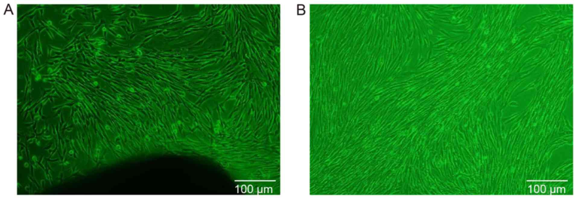
Cytoskeletal morphology of GMSCs
The first adherent cells appeared 3-14 days after
initiation of the primary culture, at which time the cells
displayed a long fusiform or polygonal shape (Fig. 1A). When the primary cells reached
80% confluence, cell subculture was conducted. Following cell
subculture, the cells exhibited consistent morphology, including a
fusiform shape with a plump cell body, a clear nucleus and a
fibroblast-like phenotype. When the cells were densely clustered,
they were arranged in a circinate or radial pattern (Fig. 1B).
Characterization of GMSCs
Flow cytometric analysis revealed that GMSCs were
uniformly positive for CD73, CD105 and CD90, but did not express
the hematopoietic stem cell marker CD45. The positive cell count
was 99.83% for CD73, 99.74% for CD90, 82.35% for CD105 and 2.05%
for CD45. These findings suggested that the cultured cells derived
from gingival tissue had the biological characteristics of MSCs
(Fig. 2A).
To further identify the GMSCs, immunohistochemical
and immunofluorescence staining were conducted. Immunohistochemical
peroxidase stained the cytoplasm brown and the nuclei
blueish-purple for CD73, CD90 and vimentin, whereas on cytokeratin
staining both the cytoplasm and nucleus remained blueish-purple
(Fig. 2B). Notably, similar
results were obtained by DAPI staining. The cytoplasm of CD90- and
vimentin-stained cells exhibited red fluorescence, and the
cytoplasm of S100A4-stained cells exhibited green fluorescence. The
nuclei of all cells exhibited blue fluorescence, except the cells
stained positive for cytokeratin, which exhibited no fluorescence
in the cytoplasm. These results indicated that the cultured cells
were stem cells of mesenchymal origin, and were determined to be
GMSCs (Fig. 2C).
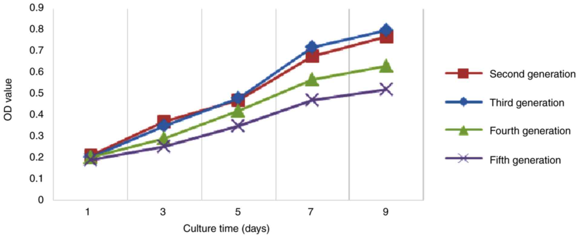
Proliferative activity of GMSCs
In order to determine the optimal proliferative
capacity of GMSCs, the MTT assay was performed to plot the growth
curve of each generation of GMSCs. The results of the MTT assay
indicated that the GMSCs exhibited logarithmic growth on days 3-4
following inoculation, and the cell number tended to stabilize on
days 7 and 8. The cell growth curve had a typical 'S' shape, and it
displayed three growth stages: Latency, logarithmic growth period
and plateau. The line chart demonstrated that second- and
third-generation cells exhibited a strong proliferation capacity,
whereas fourth- and fifth-generation cells exhibited a weaker
capacity (Fig. 3). Therefore,
third-generation cells were used in the following experiments.
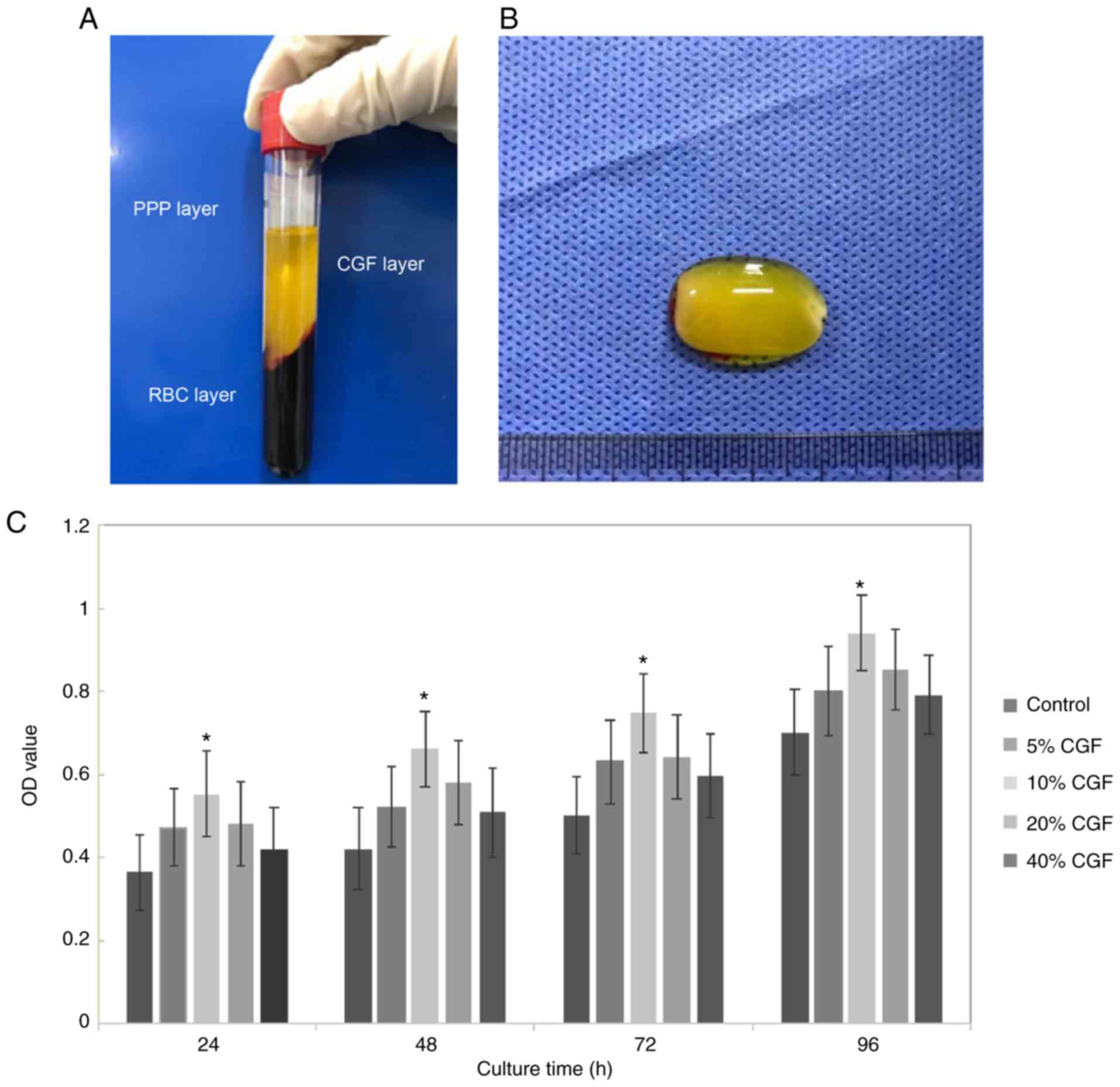
CGF promote the proliferation of
GMSCs
CGF can promote, improve and enhance tissue repair
and regeneration. Following centrifugation with a specialized
centrifugation procedure, CGF generation is characterized by three
phases: i) A superior phase represented by the serum; ii) an
interim phase represented by a very large and dense polymerized
fibrin clot with aggregated platelets and CGF; and iii) a dense,
viscous lower red portion consisting of coagulated red blood cells
(Fig. 4A). As shown in Fig. 4B, CGF gels appeared with a light
yellow gelatinous, translucent, soft, elastic, smooth surface.
The CCK-8 assay results indicated that the optical
density value increased gradually during the experimental period.
Compared with the control group, 10% CGF significantly promoted the
proliferation of GMSCs (P<0.05; Fig. 4C). However, at concentrations
>10%, the proliferative activity gradually decreased, although
it remained higher compared with the control group (Fig. 4C).
CGF promote osteogenic differentiation of
GMSCs
ALP is an exoenzyme of osteoblasts and its
expression is an obvious marker of osteoblast differentiation.
Through analyzing the activity of ALP in the three groups, it was
demonstrated that the ALP activity in the experimental group was
highest at 7, 14 and 24 days. Furthermore, significant differences
were observed on pairwise analysis between groups (P<0.01 vs.
control group; P<0.05 vs. pure mineralization group; Fig. 5A). These findings suggest that CGF
can induce osteogenic differentiation of GMSCs.
To further verify that CGF can induce osteogenic
differentiation of GMSCs, Alizarin Red S staining was performed at
7, 14 and 24 days, and the results indicated that the experimental
and the pure mineralization groups exhibited mineralized nodules.
With the extension of experimental time, the staining area and
density were gradually increased, and a large number of nodules
were observed. The mineralized nodules were the most prevalent and
the osteogenic induction was most prominent on the 21st day.
Through comparative analysis, it was demonstrated that the
mineralized nodules in the experimental group with the addition of
CGF appeared earlier and with a higher prevalence and density
(Fig. 5B). This further supports
the previous finding that CGF can induce osteogenic differentiation
of GMSCs.
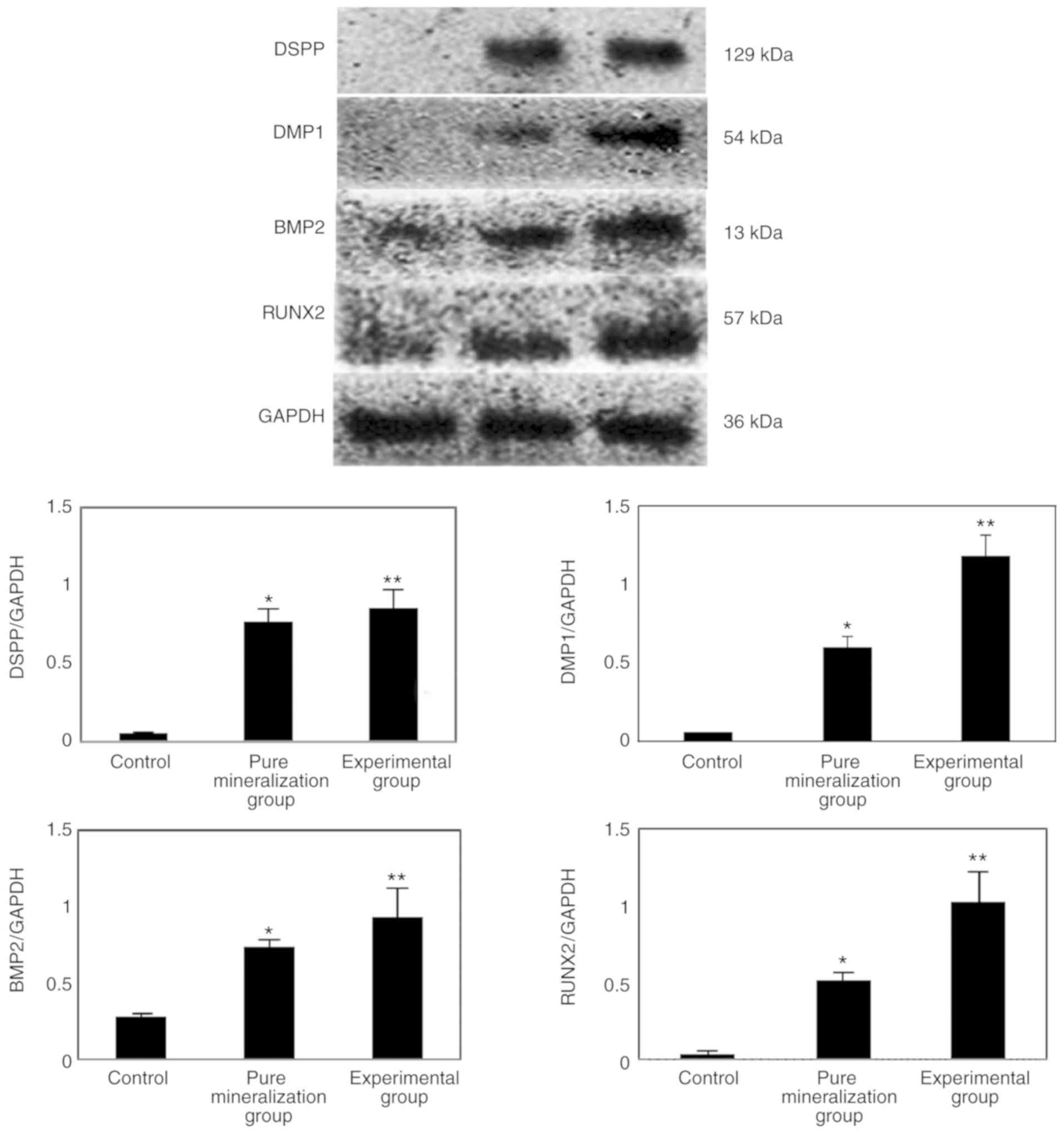
CGF promote the mRNA expression of DMP1,
DSPP, BMP2 and RUNX2
In order to elucidate whether DMP1, DSPP, BMP2 and
RUNX2 are involved in the CGF-induced differentiation of GMSCs,
RT-qPCR was used to detect their mRNA expression. The results
indicated that the mRNA expression of these four genes was
significantly higher in the experimental group compared with that
in the control group (P<0.01; Fig.
6). Therefore, it was hypothesized that DMP1, DSPP, BMP2 and
RUNX2 are involved in the CGF-induced mineralization of GMSCs.
CGF promote the protein expression of
DMP1, DSPP, BMP2 and RUNX2
In order to further elucidate the mechanism of
action of CGF, the protein expression of DMP1, DSPP, BMP2 and RUNX2
was measured by western blotting, and the results indicated that
the protein expression was significantly increased compared with
the control group (P<0.01; Fig. 7A
and B). This finding further confirmed that CGF can promote the
osteogenic differentiation of GMSCs.
Discussion
MSCs play an important role in tissue engineering
and immunotherapy due to their multidirectional differentiation
potential and ability to regulate immune responses. At present, a
variety of MSCs have been identified in the oral cavity, including
dental pulp stem cells, dental follicle stem cells, stem cells from
the apical papilla, stem cells from human exfoliated deciduous
teeth, periodontal vascular stem cells and GMSCs (15). It was previously reported that
GMSCs display characteristics similar to those of stem cells and
have a multidirectional differentiation potential. Therefore, the
gingiva may be the source of novel seed cells (16,17). Previous studies have also
demonstrated that GMSCs have the ability to differentiate into
bone, cartilage and adipose tissue, as well as endothelial-like
cells and smooth muscle-like cells, under the influence of
different cytokines (18-21).
Compared with BMSCs, GMSCs are easy to isolate and
homogenize, they proliferate quickly and exhibit a stable
morphology following multiple generations (4). In the present study, samples were
collected from the normal gingival tissue of patients undergoing
crown lengthening surgery. Through flow cytometry,
immunohistochemical staining and immunofluorescence staining, the
expanded culture cells were identified. The results of flow
cytometry demonstrated that the positive rates of CD73, CD90, CD105
and CD45 were 99.83, 99.74, 82.35 and 2.05%, respectively,
suggesting that the cells cultured in the present study displayed
the biological characteristics of MSCs. To verify this result,
immunohistochemical and immunofluorescence staining was performed.
Immunohistochemical staining revealed positive expression of
vimentin, CD73 and CD90, and negative expression of cytokeratin.
Immunofluorescence staining revealed positive expression of CD90,
S100A4 and vimentin, and negative expression of CK. Taken together,
these findings indicated that the cultured cells in the present
study were GMSCs.
CGF represent a novel generation of platelet
concentrate (22). Due to the
advanced extraction technology and specialized equipment, the
preparation process is simple and does not require any synthetic or
catalytic substances. CGF derived from the patient's own venous
blood exclude potential cross-infection, toxicity and
immunogenicity; thus, the application is highly safe (23). As CGF are derived from autologous
venous blood, the growth factor levels are higher. Previous studies
have demonstrated that CGF can promote bone formation and
differentiation (24,25). It has also been reported that CGF
can improve and enhance tissue repair and regeneration (7,11,12).
In order to elucidate whether CGF also affect the
proliferation and differentiation of GMSCs, a preliminary analysis
of cell activity was subsequently conducted by adding CGF extract
in vitro. During the experiment, five different CGF
concentration gradients were used. The CCK-8 results demonstrated
that CGF enhanced the proliferation of GMSCs within the range of
10% concentration. However, when the concentration was >10%, a
decrease in proliferative activity was observed, although it
remained higher compared with the control group. Based on the
analysis detailed above, it was demonstrated that CGF can promote
the proliferation of GMSCs cells at an optimal concentration of
10%. The effect of different concentrations of CGF on the
proliferation capacity of GMSCs was also analyzed through a
literature review. The proliferative effect of CGF is not only
attributed to the various growth factors, but also to the
three-dimensional fibrin network structure of platelets, white
blood cells and growth factors (9). Platelets and various growth factors
bond with fibrin molecules, which facilitate the adhesion of cell
components and create an appropriate microenvironment for cell
migration. However, the inability of high CGF concentration to
promote gingival stem cell proliferation may be associated with the
pH value in the cell environment. It has previously been proposed
that pH value is affected by the change in platelet count, which
may negatively affect cell proliferation (26).
ALP, as a marker of osteoblast differentiation,
first appears in the early stages of cell mineralization induction
and may be used as an early osteogenesis marker (27,28). As the induction time increases and
the osteogenic differentiation of cells progresses, its activity
gradually increases. Therefore, the osteogenic function of the
GMSCs can be evaluated. In order to explore the ability of CGF to
induce osteogenic differentiation of GMSCs, ALP activity was
detected. The results indicated that the ALP activity of the
experimental group was highest at 7, 14 and 21 days, suggesting
that CGF can promote the osteogenesis of GMSCs. Changing calcium
salt levels is a marker of bone cell proliferation and
differentiation, and of the osteogenic potential of bone tissue.
Therefore, the ability of osteoblasts to differentiate may be
identified by determining the calcium salt deposition in the cell
matrix of each group by Alizarin Red S staining. Through comparison
of the experimental data among the three groups, it was
demonstrated that the mineralized nodules in the experimental group
with added CGF appeared earlier, and the number and density of the
nodules were higher compared with the other two groups.
Mineralization images revealed that the mineralized nodules were
the most prevalent and the osteogenic induction was the most marked
on day 21. This further verified that CGF can promote the
osteoblastic differentiation of GMSCs.
DSPP is an extracellular matrix protein closely
associated with tooth development, which has been demonstrated to
play a central role in the formation and growth of hydroxyapatite
crystals in the extracellular matrix of hard tissues, such as bones
and teeth (29). DMP1 is a type
of hyperphosphorylated hyperacidic non-collagen, which is mainly
expressed in mineralized tissues, including bones and teeth. Due to
its acidic domain and negative charge, it strongly binds with
calcium ions and promotes the formation of hydroxyapatite (30-32). RUNX2 is associated with
transcription during bone and tooth development, participates in
dental crown formation and promotes odontoblast differentiation
(33,34). BMP2 promotes the self-renewal of
stem cells, promotes the differentiation of mesenchymal cells into
bone and cartilage, and participates in the development of various
organs (35,36). In order to investigate whether
these four genes are involved in the induction of osteogenic
differentiation of GMSCs by CGF, RT-qPCR and western blot analyses
were conducted to quantify RNA and protein expression of the
experimental, pure mineralization and control groups on the 21st
day after mineralization induction. The RT-qPCR results indicated
that DSPP, DMP1, BMP2 and RUNX2 were significantly upregulated
compared with the control group. This finding indicates that CGF
likely promotes osteoblastic differentiation of GMSCs cells by
regulating the expression of DSPP, DMP1, BMP2 and RUNX2. The
western blotting results were consistent with the RT-qPCR results,
further supporting the hypothesis that CGF upregulates DSPP, DMP1,
BMP2 and RUNX2 to promote the proliferation and osteogenic
differentiation of GMSCs.
In conclusion, the findings of the present study
revealed that CGF can significantly promote the proliferation and
osteogenic differentiation of GMSCs. Therefore, CGF appears to be
promising for certain applications in tissue engineering for tooth
regeneration and repair. However, the mechanisms underlying the
regulation of the DSPP, DMP1, BMP2 and RUNX2 signaling pathways by
CGF remain to be fully elucidated.
Funding
The present study was supported by grants from the
Medical Science Project of Sichuan Province (no. S17073) and the
University-level Scientific Research Project of Southwest Medical
University (no. 2017-ZRQN-083) and the General Program of Sichuan
Provincial Science and Technology Department Applied Basic Research
(no. 2018JY040).
Availability of data and materials
The datasets generated and/or analyzed during the
present study are available from the corresponding author on
reasonable request.
Authors' contributions
XL conceived of the study, provided materials and
samples, and participated in data collection, analysis and
interpretation of the results. MN provided administrative support.
XC, YC and MZ provided materials and samples. YC and YH contributed
to data collection. XC and PS contributed to interpretation of the
results. All authors have read and approved the final version of
this manuscript for publication.
Ethics approval and consent to
participate
The study was approved by the Ethics Committee of
the Affiliated Stomatological Hospital of Southwest Medical
University and all the patients included in the study signed an
informed consent prior to tissue collection. All procedures were
performed in accordance with the World Medical Association's
Declaration of Helsinki.
Patient consent for publication
Not applicable.
Competing interests
All the authors declare that they have no competing
interests to disclose.
Acknowledgments
Not applicable.
References
|
1
|
Xu S, De Veirman K, De Becker A,
Vanderkerken K and Van Riet I: Mesenchymal stem cells in multiple
myeloma: A thera-peutical tool or target? Leukemia. 32:1500–1514.
2018. View Article : Google Scholar : PubMed/NCBI
|
|
2
|
Dominici M, Le Blanc K, Mueller I,
Slaper-Cortenbach I, Marini F, Krause D, Deans R, Keating A,
Prockop D and Horwitz E: Minimal criteria for defining multipotent
mesenchymal stromal cells. The International Society for Cellular
Therapy position statement. Cytotherapy. 8:315–317. 2006.
View Article : Google Scholar : PubMed/NCBI
|
|
3
|
Stenderup K, Justesen J, Clausen C and
Kassem M: Aging is associated with decreased maximal life span and
accelerated senescence of bone marrow stromal cells. Bone.
33:919–926. 2003. View Article : Google Scholar : PubMed/NCBI
|
|
4
|
Tomar GB, Srivastava RK, Gupta N,
Barhanpurkar AP, Pote ST, Jhaveri HM, Mishra GC and Wani MR: Human
gingiva-derived mesenchymal stem cells are superior to bone
marrow-derived mesenchymal stem cells for cell therapy in
regenerative medicine. Biochem Biophys Res Commun. 393:377–383.
2010. View Article : Google Scholar : PubMed/NCBI
|
|
5
|
Mitrano TI, Grob MS, Carrión F,
Nova-Lamperti E, Luz PA, Fierro FS, Quintero A, Chaparro A and Sanz
A: Culture and characterization of mesenchymal stem cells from
human gingival tissue. J Periodontol. 81:917–925. 2010. View Article : Google Scholar : PubMed/NCBI
|
|
6
|
Zhang Q, Shi S, Liu Y, Uyanne J, Shi Y,
Shi S and Le AD: Mesenchymal stem cells derived from human gingiva
are capable of immunomodulatory functions and ameliorate
inflammation-related tissue destruction in experimental colitis. J
Immunol. 183:7787–7798. 2009. View Article : Google Scholar : PubMed/NCBI
|
|
7
|
Kim TH, Kim SH, Sándor GK and Kim YD:
Comparison of platelet-rich plasma (PRP), platelet-rich fibrin
(PRF), and concentrated growth factor (CGF) in rabbit-skull defect
healing. Arch Oral Biol. 59:550–558. 2014. View Article : Google Scholar : PubMed/NCBI
|
|
8
|
Qiao J and An N: Effect of concentrated
growth factors on function and Wnt3a expression of human
periodontal ligament cells in vitro. Platelets. 28:281–286. 2017.
View Article : Google Scholar
|
|
9
|
Rodella LF, Favero G, Boninsegna R,
Buffoli B, Labanca M, Scari G, Sacco L, Batani T and Rezzani R:
Growth factors, CD 34 p ositive cells, and fibrin network analysis
in concentrated growth factors fraction. Microsc Res Tech.
74:772–777. 2011. View Article : Google Scholar : PubMed/NCBI
|
|
10
|
Tayapongsak P, O'Brien DA, Monteiro CB and
Arceo-Diaz LY: Autologous fibrin adhesive in mandibular
reconstruction with particulate cancellous bone and marrow. J Oral
Maxillofac Surg. 52:161–165; discussion 166. 1994. View Article : Google Scholar : PubMed/NCBI
|
|
11
|
Bozkurt Doğan Ş, Öngöz Dede F, Ballı U,
Atalay EN and Durmuşlar MC: Concentrated growth factor in the
treatment of adjacent multiple gingival recessions: A split-mouth
randomized clinical trial. J Clin Periodontol. 42:868–875. 2015.
View Article : Google Scholar
|
|
12
|
Takeda Y, Katsutoshi K, Matsuzaka K and
Inoue T: The effect of concentrated growth factor on rat bone
marrow cells in vitro and on calvarial bone healing in vivo. Int J
Oral Maxillofac Implants. 30:1187–1196. 2015. View Article : Google Scholar : PubMed/NCBI
|
|
13
|
Mosmann T: Rapid colorimetric assay for
cellular growth and survival: Application to proliferation and
cytotoxicity assays. J Immunol Methods. 65:55–63. 1983. View Article : Google Scholar : PubMed/NCBI
|
|
14
|
Livak KJ and Schmittgen TD: Analysis of
relative gene expression data using real-time quantitative PCR and
the 2(-Delta Delta C(T)) method. Methods. 25:402–408. 2001.
View Article : Google Scholar
|
|
15
|
Heng BC, Zhang C, Deng X, Xiao Y,
Pisciotta A, Kidwai F and Mitsiadis TA: Biomedical applications of
dental and oral-derived stem cells. Stem Cells Int.
2017:29310542017. View Article : Google Scholar : PubMed/NCBI
|
|
16
|
Ji J, Tong X, Huang X, Zhang J, Qin H and
Hu Q: Patient-derived human induced pluripotent stem cells from
gingival fibroblasts composited with defined
nanohydroxyapatite/chitosan/gelatin porous scaffolds as potential
bone graft substitutes. Stem Cells Transl Med. 5:95–105. 2016.
View Article : Google Scholar :
|
|
17
|
Fawzy El-Sayed KM and Dörfer CE: Gingival
mesenchymal stem/progenitor cells: A unique tissue engineering gem.
Stem Cells Int. 2016:71543272016. View Article : Google Scholar : PubMed/NCBI
|
|
18
|
Liu X, Wang J, Dong F, Li H and Hou Y:
Human gingival fibroblasts induced and differentiated into vascular
endothelial-like cells. Dev Growth Differ. 58:702–713. 2016.
View Article : Google Scholar : PubMed/NCBI
|
|
19
|
Liu X, Wang J, Dong F, Li H and Hou Y:
Induced differentiation of human gingival fibroblasts into
VSMC-like cells. Differentiation. 95:1–9. 2017. View Article : Google Scholar : PubMed/NCBI
|
|
20
|
Xu X, Chen C, Akiyama K, Chai Y, Le AD,
Wang Z and Shi S: Gingivae contain neural-crest- and
mesoderm-derived mesenchymal stem cells. J Dent Res. 92:825–832.
2013. View Article : Google Scholar : PubMed/NCBI
|
|
21
|
Ansari S, Diniz IM, Chen C, Sarrion P,
Tamayol A, Wu BM and Moshaverinia A: Human periodontal ligament-
and gingiva-derived mesenchymal stem cells promote nerve
regeneration when encapsulated in alginate/Hyaluronic acid 3D
scaffold. Adv Healthc Mater. 6:2017.PubMed/NCBI
|
|
22
|
Bernardi S, Mummolo S, Tecco S, Continenza
MA and Marzo G: Histological characterization of Sacco's
concentrated growth factors membrane. Int J Morphol. 35:114–119.
2017. View Article : Google Scholar
|
|
23
|
Qin J, Wang L, Sun Y, Sun X, Wen C,
Shahmoradi M and Zhou Y: Concentrated growth factor increases
Schwann cell proliferation and neurotrophic factor secretion and
promotes functional nerve recovery in vivo. Int J Mol Med.
37:493–500. 2016. View Article : Google Scholar
|
|
24
|
Kim JM, Sohn DS, Bae MS, Moon JW, Lee JH
and Park IS: Flapless transcrestal sinus augmentation using
hydrodynamic piezoelectric internal sinus elevation with autologous
concentrated growth factors alone. Implant Dent. 23:168–174. 2014.
View Article : Google Scholar : PubMed/NCBI
|
|
25
|
Sohn DS, Heo JU, Kwak DH, Kim DE, Kim JM,
Moon JW, Lee JH and Park IS: Bone regeneration in the maxillary
sinus using an autologous fibrin-rich block with concentrated
growth factors alone. Implant Dent. 20:389–395. 2011.PubMed/NCBI
|
|
26
|
Liu Y, Kalén A, Risto O and Wahlström O:
Fibroblast proliferation due to exposure to a platelet concentrate
in vitro is pH dependent. Wound Repair Regen. 10:336–340. 2002.
View Article : Google Scholar : PubMed/NCBI
|
|
27
|
Mornet E, Stura E, Lia-Baldini AS,
Stigbrand T, Ménez A and Le Du MH: Structural evidence for a
functional role of human tissue nonspecific alkaline phosphatase in
bone mineralization. J Biol Chem. 276:31171–31178. 2001. View Article : Google Scholar : PubMed/NCBI
|
|
28
|
Douglas TE, Messersmith PB, Chasan S,
Mikos AG, de Mulder EL, Dickson G, Schaubroeck D, Balcaen L,
Vanhaecke F, Dubruel P, et al: Enzymatic mineralization of
hydrogels for bone tissue engineering by incorporation of alkaline
phosphatase. Macromol Biosci. 12:1077–1089. 2012. View Article : Google Scholar : PubMed/NCBI
|
|
29
|
Choi YS, Lee JY, Suh JS, Lee G, Chung CP
and Park YJ: The mineralization inducing peptide derived from
dentin sialophosphoprotein for bone regeneration. J Biomed Mater
Res A. 101:590–598. 2013. View Article : Google Scholar
|
|
30
|
Gericke A, Qin C, Sun Y, Redfern R,
Redfern D, Fujimoto Y, Taleb H, Butler WT and Boskey AL: Different
forms of DMP 1 p lay distinct roles in mineralization. J Dent Res.
89:355–359. 2010. View Article : Google Scholar : PubMed/NCBI
|
|
31
|
Bhatia A, Albazzaz M, Espinoza Orias AA,
Inoue N, Miller LM, Acerbo A, George A and Sumner DR:
Overexpression of DMP1 accelerates mineralization and alters
cortical bone biomechanical properties in vivo. J Mech Behav Biomed
Mater. 5:1–8. 2012. View Article : Google Scholar
|
|
32
|
Narayanan K, Srinivas R, Ramachandran A,
Hao J, Quinn B and George A: Differentiation of embryonic
mesenchymal cells to odontoblast-like cells by overexpression of
dentin matrix protein 1. Proc Natl Acad Sci USA. 98:4516–4521.
2001. View Article : Google Scholar : PubMed/NCBI
|
|
33
|
Monteiro N, Ribeiro D, Martins A, Faria S,
Fonseca NA, Moreira JN, Reis RL and Neves NM: Instructive
nanofibrous scaffold comprising runt-related transcription factor 2
gene delivery for bone tissue engineering. ACS Nano. 8:8082–8094.
2014. View Article : Google Scholar : PubMed/NCBI
|
|
34
|
Li S, Kong H, Yao N, Yu Q, Wang P, Lin Y,
Wang J, Kuang R, Zhao X, Xu J, et al: The role of runt-related
transcription factor 2 (Runx2) in the late stage of odontoblast
differentiation and dentin formation. Biochem Biophys Res Commun.
410:698–704. 2011. View Article : Google Scholar : PubMed/NCBI
|
|
35
|
Zhang C, Meng C, Guan D and Ma F: BMP2 and
VEGF165 transfection to bone marrow stromal stem cells regulate
osteogenic potential in vitro. Medicine (Baltimore). 97:e97872018.
View Article : Google Scholar
|
|
36
|
Qin W, Yang F, Deng R, Li D, Song Z, Tian
Y, Wang R, Ling J and Lin Z: Smad 1/5 is involved in bone
morphogenetic protein-2-induced odontoblastic differentiation in
human dental pulp cells. J Endod. 38:66–71. 2012. View Article : Google Scholar
|