Introduction
Liver cancer is one of the most commonly diagnosed
types of malignant cancer and there are approximately 850,000 new
cases diagnosed yearly worldwide (1). The high incidence of HCC has
induced the development of novel targeted and personalized
therapies (2). Personalization
of cancer treatment requires the reliable prediction of
chemotherapy responses in individual patients. Various strategies
have been applied to generate primary cultures from individual
tumors which include 2D cell culture of dissociated tumor cells,
3-D spheroid cultures and patient-derived mouse xenograft cultures
(3-7). However, the difficulties in
replicating the heterogeneous microenvironment in the primary tumor
reduce their efficiency in drug experiments (8). It was estimated that over 90% of
novel anticancer drugs fail in clinical trials because these models
could not simulate complete tissue structure and maintain the
biological heterogenicity of the primary tumor (9). For these reasons, it is crucial for
us to create novel models that are more predictive of in
vivo efficacy.
Precision-cut slice is a new method of tissue
culture in vitro, which is derived directly from the primary
tumor (10). However, there is
no preservation method applied to maintain living fresh tissue.
Conventional preservation of fresh tumor tissue such as
formalin-fixed paraffin-embedded samples and flash freezing in
liquid nitrogen always leads to the absolute inactivation of the
fresh tissue. Therefore, a reliable and efficient cryopreservation
method for living tissue is indispensable. Vitrification-based
cryopreservation method can be developed to preserve fresh tissue,
by which the biological characteristics of the original tumor can
be retained and the utilization of specimens may be markedly
improved (11).
In the present study, we explored a precision-cut
slice culture method combined with a cryopreservation technique to
establish a preclinical model, which is derived from fresh tissues
of HCC patients. In addition, we demonstrated systematic
optimization of HCC slices ex vivo by comparing different
culture conditions. Moreover, this culture system allowed the
detection of tumor responses to REG chemotherapy.
Materials and methods
Collection of HCC specimens
From October, 2019 to February, 2020, surgically
resected specimens were obtained from 30 HCC patients at the Renji
Hospital Affiliated to Shanghai Jiao Tong University School of
Medicine (Shanghai, China). Samples were maintained at 4°C on ice
and transported in preservation medium (Tissue Mate™; Celliver
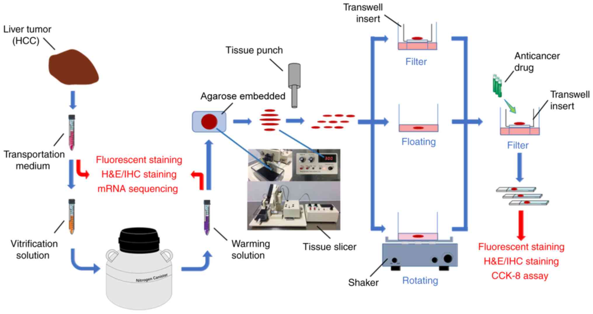
Biotechnology Co. Ltd.). Details are illustrated in Fig. 1. This investigation was approved
by the Ethics Committee of Renji Hospital and followed the
guidelines of The Declaration of Helsinki. All patients provided
written informed consent. The inclusion criteria were: i)
pathological diagnosis of HCC; ii) no patients had received any
prior treatment; iii) the maximum diameter of a single tumor was
more than 2 cm; iv) Child-Pugh score A or B. The exclusion criteria
were: i) Child-Pugh score C; ii) exceptional circumstances, such as
syphilis and acquired immune deficiency syndrome.
Cryopreservation and warming
procedures
All specimens were cut into 1-mm-thick slices in a
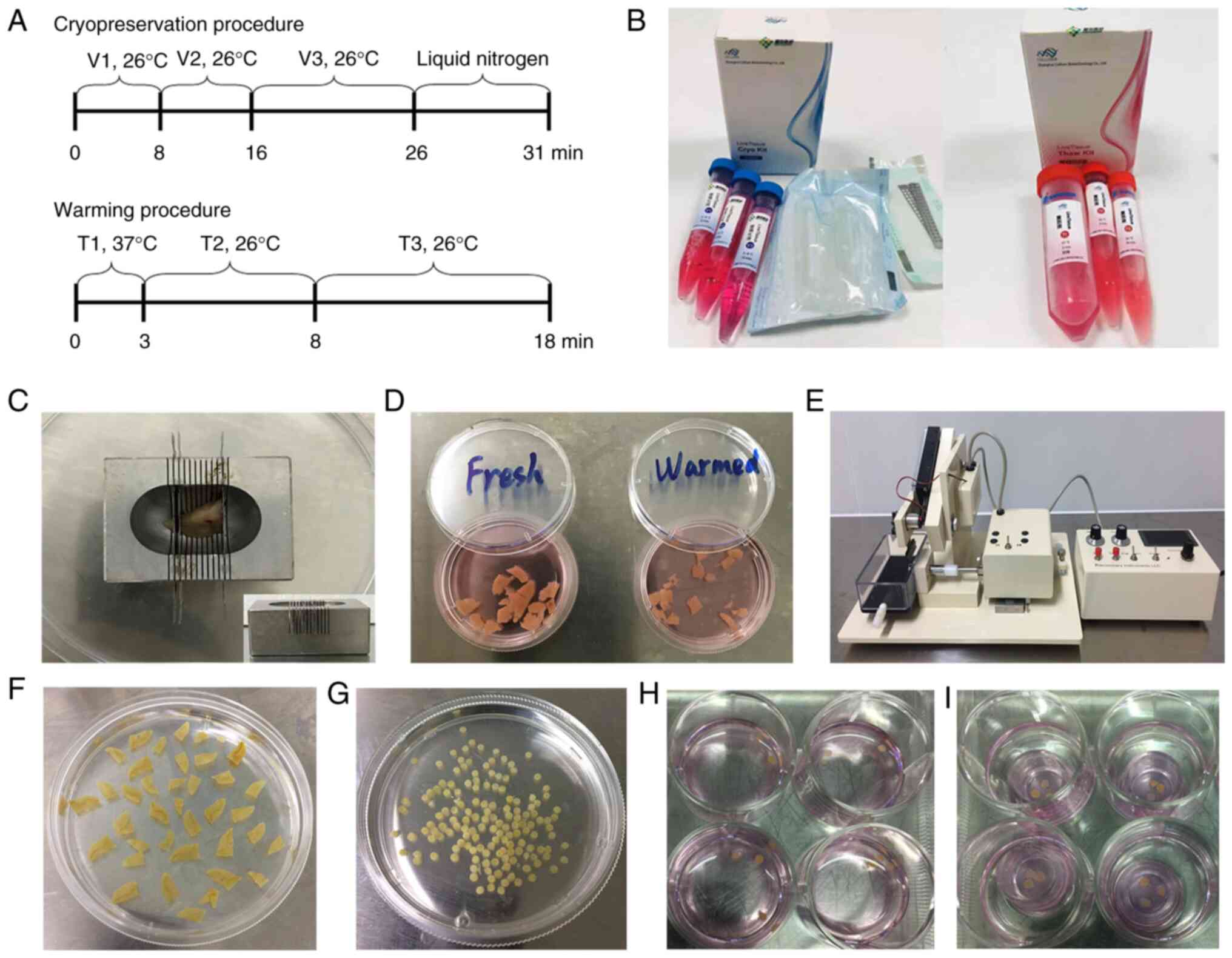
metal mold before cryopreservation (Fig. 2C). Cryopreservation solutions
(LT2601; Tissue Mate™) and warming solutions (LT2602; Tissue Mate™)
were provided by Celliver Biotechnology Co. Ltd. (Fig. 2B). For tissue cryopreservation,
vitrification solution 1 (V1), vitrification solution 2 (V2) and
vitrification solution 3 (V3) were pre-warmed in a 2~8°C water
bath. Fresh HCC tissues were cleaned twice with sterile PBS and
transferred into 10 ml V1, 10 ml V2 and 10 ml V3 for 8, 8, 10 min,
respectively. Tissues were then placed onto a thin metal strip and
submerged into liquid nitrogen for at least 5 min. Finally, the
strips with tissue were placed into frozen storage tubes and
preserved in the nitrogen canister. The tissue samples were stored
in the liquid nitrogen. For tissue warming, the frozen storage
tubes were removed from the nitrogen canister and the strips with
the cryopreserved biopsy tissues were quickly transferred into 30
ml warming solution 1 (T1), and incubated for 3 min in a 37°C water
bath. The tissues were then transferred into 10 ml warming solution
2 (T2) and 10 ml warming solution 3 (T3) for 5 and 10 min,
respectively, at room temperature. Warmed tissues were cleaned
twice with sterile PBS and kept on ice (Fig. 2D). The timeline of
cryopreservation and warming procedures are depicted in Fig. 2A.
Tissue slice preparation and
cultivation
Surgically resected specimens were cut into
300-µm-thick precision-cut slices using a microtome for
slice preparation (Bio-Gene Technology, Ltd.) (Fig. 2E). A thickness of 300 µm
was considered the most suitable thickness for HCC after several
early slicing pre-experiments (Fig.
2F). Parameter settings, such as the frequency and amplitude of
vibration slicing, were determined by the diverse cirrhosis degree
and tumor stage. Tissue slices (diameter, 2 mm) were then prepared
using a hand-held coring tool (Fig.
2G), and all the procedures were performed under sterile
conditions. One-third of the precision-cut slices were maintained
on Transwell inserts (pore size, 0.4 µm; Corning, Inc.)
(Fig. 2I). One-third of the
precision-cut slices were individually submerged in medium
(Fig. 2H) and incubation was
performed on a shaking platform (TYZD-III, QiQian Technology,
Ltd.). The remaining precision-cut slices were cultured statically
in medium as control (Fig. 2H).
Cultivation was performed in 12-well plates containing 450
µl DMEM medium (Gibco™; Thermo Fisher Scientific, Inc.) with
10% fetal bovine serum (Gibco™; Thermo Fisher Scientific, Inc.),
penicillin and streptomycin (100 U/ml; Gibco™; Thermo Fisher
Scientific, Inc.), and kept at 37°C in a humidified incubator with
5% CO2.
Cell Counting Kit-8 (CCK-8) assay
A CCK-8 assay (Dojindo Molecular Technologies, Inc.)
was used to evaluate the viability of tissue slices at each time
point (24, 48, 72, 96 h). DMEM (90 µl/well) and CCK-8
solution (10 µl/well) were added into 96-well plates. The
tissue slices were added one slice/well. The plates were maintained
at 37°C in a humidified incubator with 5% CO2 for 2 h.
The slices were removed from the 96-well plates and the plates were
transferred to microplate reader (Multiskan GO; Thermo Fisher
Scientific, Inc.). The absorbance at 450 nm was measured and three
wells were tested for each sample at each time point.
Calcein-AM cell viability assay and
Hoechst 33342 staining
The Live/Dead® Viability Assay kit
(Nanjing KeyGen Biotech Co.) and Hoechst 33342 (Beyotime Institute
of Biotechnology Co.) were stored at -20°C and allowed to warm to
room temperature prior to experimentation. The viability assay
stock reagents (calcein-AM, 4 mM) were diluted to 1 µM in
physiological solution and mixed with 2 µg/ml Hoechst 33342
stock reagents at room temperature for 30 min. Live cells are
characterized by a bright green fluorescent and cell nucleus are
blue. Representative images were captured with the Leica TCS SP8
confocal microscope (×20) (Leica Microsystems GmbH). The ratio of
living cells in the calcein-AM cell viability assay/Hoechst 33342
staining were calculated based on manual counting within 10 random
microscopic fields.
Hematoxylin and eosin
(H&E)/immunohistochemical (IHC) staining
Tumor slices were formalin-fixed, embedded in
paraffin and cut into 4-µm-thick sections. Paraffin sections
(4-µm) were stained with H&E at room temperature. IHC
staining was carried out by standard protocols. Briefly, sections
were de-waxed in xylene and rehydrated in graded ethanol, and
heat-mediated antigen retrieval of tissue sections was carried out
before being allowed to cool. Endogenous peroxidases were blocked
using 0.9-3% hydrogen peroxide for 10 min, and non-specific
antibody binding was blocked by incubation with serum-free blocking
solution or 10% normal serum block for 30 min. Tissue sections were
then incubated with the anti-Ki67 antibody (Ab15580; Abcam; 1:1,000
dilution), before being probed with the secondary antibodies Alexa
Fluor® 488 (Ab150077; Abcam; 1:1,000 dilution).
Antibodies were visualized using 3,3′-diaminobenzidine chromogen
and counterstained with Meyer's Hematoxylin for 2 min. Sections
were then dehydrated through graded alcohols, cleared in xylene and
mounted. Confocal laser scanning microscopy (magnification, ×20)
was performed using an Olympus Corp. BX51 instrument. The ratio of
proliferative cells in the Ki67 staining were calculated based on
manual counting within 10 random microscopic fields.
Experimental methods for mRNA
sequencing
RNA purity was assessed using the
kaiaoK5500® Spectrophotometer (Beijing Kaiao Technology
Development Co. Ltd.). RNA integrity and concentration were
assessed using the RNA Nano 6000 Assay kit and the Bioanalyzer 2100
system (Agilent Technologies, Inc.). A total amount of 2 µg
RNA/sample was used as input material for the RNA sample
preparations. Sequencing libraries were generated using
NEBNext® Ultra™ RNA Library Prep kit for
Illumina® (E7530L; New England BioLabs, Inc.), following
the manufacturer's recommendations, and index codes were added to
attribute sequences to each sample. Briefly, mRNA was purified from
the total RNA using poly-T oligo-attached magnetic beads.
Fragmentation was carried out using divalent cations under elevated
temperature in NEBNext® First Strand Synthesis Reaction
Buffer (5X) (New England BioLabs, Inc.). First-strand cDNA was
synthesized using random hexamer primer and RNase H. Second-strand
cDNA synthesis was subsequently performed using buffer, dNTPs, DNA
polymerase I and RNase H. The library fragments were purified with
QiaQuick PCR kits (Qiagen, Inc.) and elution with EB buffer, then
terminal repair, A-tailing and adapter adding were implemented. The
products were retrieved and PCR was performed, and then the library
was completed. The RNA concentration of the library was measured
using a Qubit® RNA Assay kit in Qubit® 3.0
(Thermo Fisher Scientific, Inc.) for preliminary quantification,
and then diluted to 1 ng/µl. Insert size was assessed using
the Agilent Bioanalyzer 2100 system (Agilent Technologies, Inc.),
and qualified insert size was accurately quantified using the
StepOnePlus™ Real-Time PCR System (Thermo Fisher Scientific, Inc.;
library valid concentration, >10 nM). The clustering of the
index-coded samples was performed on a cBot cluster generation
system using a HiSeq PE Cluster kit v4-cBot-HS (Illumina, Inc.)
according to the manufacturer's instructions. After cluster
generation, the libraries were sequenced on an Illumina, Inc.
platform and 150-bp paired-end reads were generated. The variations
in gene expression were detected by different colors in the heat
map.
Metabolic activity of pH/glucose/LDH
For the testing of the potential of hydrogen (pH),
we extract 15 µl culture medium from the slice culture
system using a detecting instrument (InLab Ultra Micro-ISM, Mettler
Toledo); Glucose was tested using a detecting instrument
(GlucCell™, Brookfield), using 3 µl of culture medium; For
LDH (lactate dehydrogenase), a detection kit (G1780, Promega Corp.)
in a 96-well plate was used. All the processes were conducted using
the operation manuals provided by the suppliers.
Drug sensitivity test in vitro
Drug testing commenced after 24 h of slice culture
and was performed for an additional 72 h. For drug testing of
slices in vitro, regorafenib (REG; MedChemExpress LLC) was
used and tested at a concentration of 5, 10, and 20 µM,
respectively. To investigate cell proliferation and tissue
morphology, the slices were incubated with CCK-8 solution and
stained with H&E/fluorescent dyes.
Statistical analysis
Statistical evaluations were performed using one-way
ANOVA with Scheffe's post hoc tests by IBM SPSS Statistics 22.0
(IBM Corp). P<0.05 was considered to indicate a statistically
significant difference. Three repeats were performed.
Results
Biological characteristics of HCC tissues
are maintained by vitrification-based cryopreservation and
precision-cut slice method
All of the fresh HCC specimens were obtained from
Renji Hospital Affiliated to Shanghai Jiao Tong University. The 30
patients included 23 men and 7 women with a mean age of 58 years.
The workflow was strictly performed by standard procedures, as
depicted in Fig. 1. The specific
explanation is provided in the Materials and methods section. Human
liver tissues were found to be very well sliceable and showed a
good reproducibility as well as tissue viability. The 1-mm-thick
HCC slices were cryopreserved and warmed according to the timeline
in Fig. 2A, using
cryopreservation solutions (Fig.
2B, left) and warming solutions (Fig. 2B, right). Thirty fresh specimens
were derived from 30 HCC patients. Half of each specimen was
processed by cryopreservation and warming procedures, and the
remaining tissues were used as the control group. The
300-µm-thick precision-cut slices were made and cultured
successfully (Fig. 2C-I).
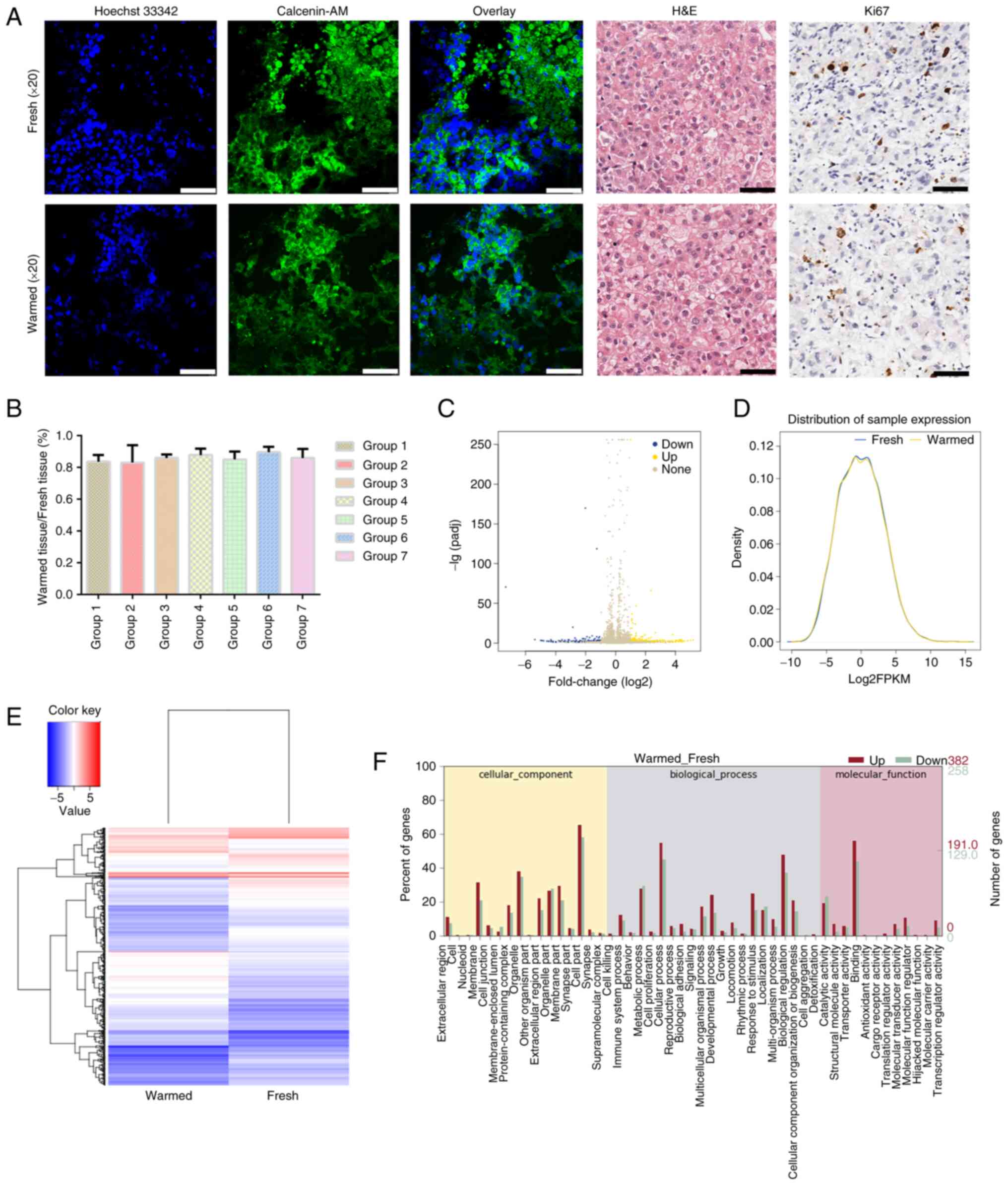
H&E staining of fresh tissue slices revealed no obvious
differences in the morphology when compared to the warmed tissue
slices. From the fluorescent and IHC staining, we found that the
living cell ratio was 93% in fresh tissues and 90% in warmed
tissues, which indicated that no obvious difference was detectable
between the fresh HCC and warmed HCC tissues (Fig. 3A and B). The heat map of the
cancer-associated genes indicated that the color of the left column
was mostly consistent with the right column (Fig. 3E). Only a small part of
differential gene expression was detected from the volcano plot
(Fig. 3C) and distribution of
sample expression (Fig. 3D).
According to the GO analysis, it was found that the differential
genes were closely related to cell metabolism (Fig. 3F). Original data were uploaded to
the Gene Expression Omnibus database (accession number GSE194095).
Therefore, the variations in gene expression between fresh and
warmed tissues were limited. These results confirmed that the
vitrification-based cryopreservation method was able to largely
maintain the biological activity and histological features of the
HCC tissues.
Medium composition and culture mode are
critical to tissue viability
In order to identify the best slice culture methods,
we optimized the slicing process with different culture methods and
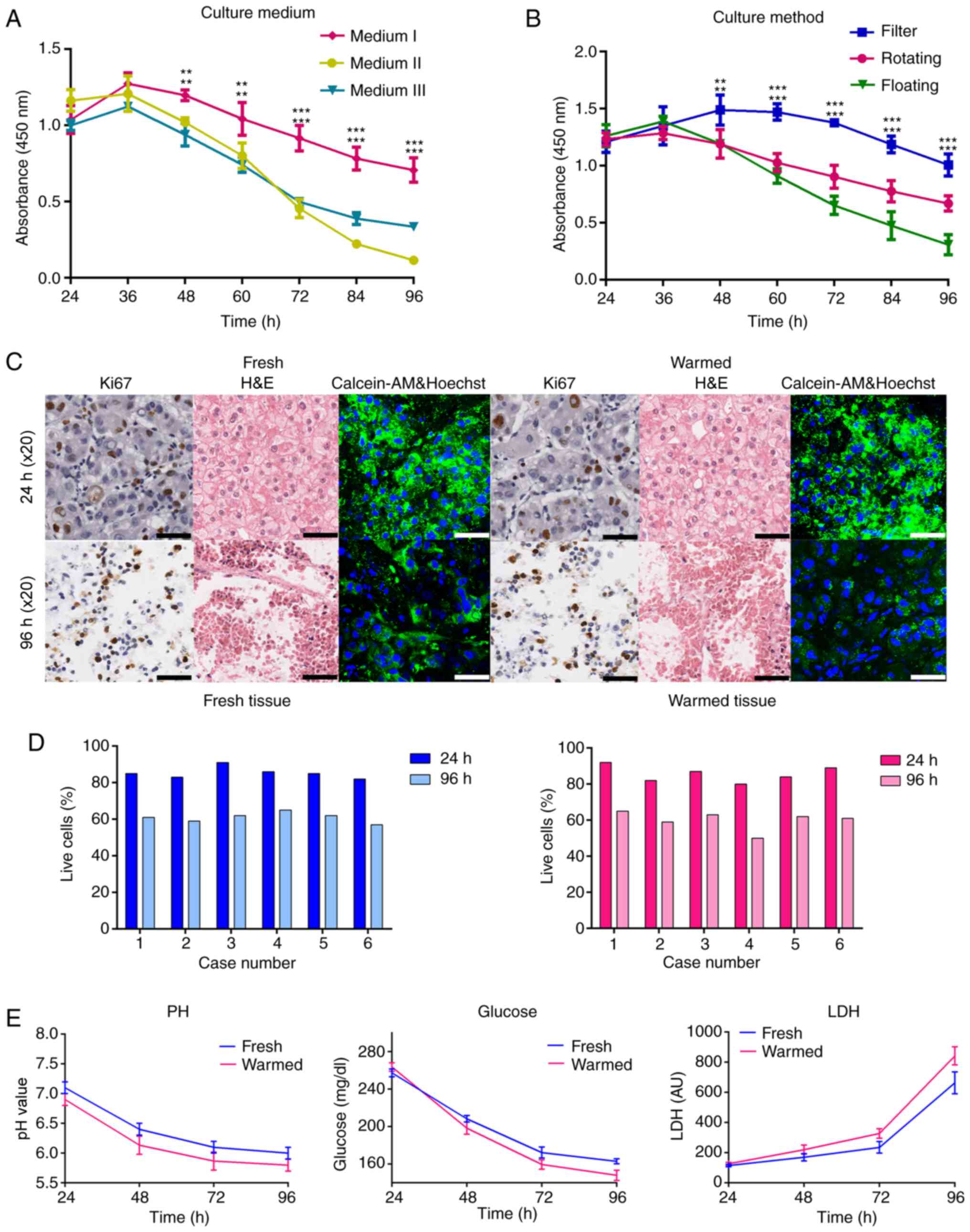
selected the optimal culture medium. Our results showed that Medium
I (DMEM with high glucose +10% FBS) could obviously maintain cell
viability, especially from 48 h (Fig. 4A and B); Medium II (1640+10% FBS)
and Medium III (DMEM/F12) were not suitable for the slice culture
(Fig. 4A). The filter cultures
were viable for up to 4 days and could receive a higher cell
viability than the floating and rotating cultures (Fig. 4B). Subsequently, we conducted
slice culture on Transwell insert with DMEM combined with 10% FBS
for 72 h. As determined from the fluorescent and IHC staining, the
living cell ratio was decreased slightly and a slight change in
tissue morphological features was detected. In addition, these
changes were observed both in fresh and warmed tissues (Fig. 4C and D). Moreover, we detected
that the levels of pH and glucose were decreased while LDH was
obviously increased (Fig.
4E).
Positive drug responses could be detected
in slice culture model
Drug testing commenced after 24 h of slice culture
and was performed for an additional 72 h. To study the activity of
anti-cancer drug REG in this tissue culture model, HCC slices were
treated with different concentrations of REG (5, 10 and 20
µM) for another 72 h as depicted in Fig. 5A. Our results revealed that 20
µM was the most obvious concentration with which to
significantly decrease the cell viability. Morphological staining
and viability assays both indicated that no obvious differences
were detectable after 24 h of slice culturing. However, compared
with the control group, both fresh and warmed tissue slices in the
drug treatment group cultured for 72 h showed a significant
decrease in cell viability. In addition, tissue slices in the drug
treatment group evidently lost the morphological structure of the
original tumor (Fig. 5B-E).
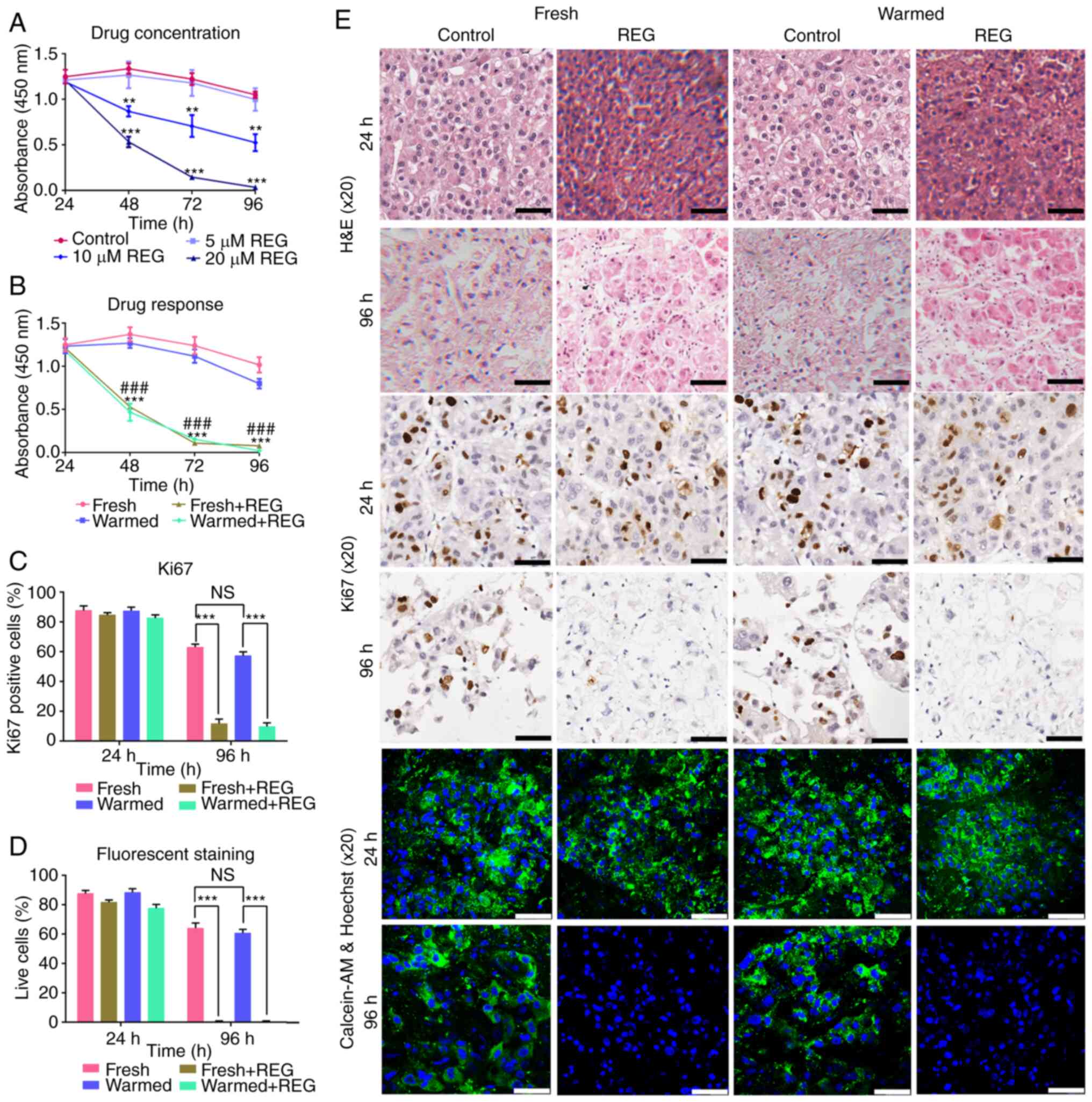 | Figure 5Positive drug responses in the slice
culture model. (A) HCC slices were treated with different
concentrations of REG (5, 10, and 20 µM) for 72 h. The
control group was statistically different from the 10 µM
group and 20 µM group, respectively. (B) CCK-8 cell
viability assay was conducted for 72 h. **P<0.01 and
***P<0.001, Fresh group vs. the Fresh + REG group;
###P<0.001 Warmed group vs. Warmed + REG group. (C)
The quantification of Ki67-positive cells, the proliferation rate
before and after cryopreservation, was not statistically different
as determined by Student's t-test. (D) The quantification of live
cells, the number of live cells before and after cryopreservation,
was not statistically different as determined by Student's
t-test.***P<0.001 and NS, not significant. The Fresh
group was statistically different from the Fresh + REG group and
the Warmed group was statistically different from the Warmed + REG
group in 5B-D. (E) Morphological staining and fluorescent staining
in fresh and warmed tissues during drug testing. Scale bars, 50
µm. HCC, hepatocellular carcinoma; REG, regorafenib. |
Discussion
Research has indicated that a tissue slice culture
system can be applied to perform preclinical and clinical studies
for medical research (12-15). In our research, we describe
precision-cut slice cultures as a novel model to perform ex
vivo experiments on hepatocellular carcinoma (HCC) tumors,
which preserves the three-dimensional structure of the tumor and
provides an alternative to in vivo experiments. Our study
was performed using standard procedures (Fig. 1). Studies have shown that there
may be a drastic difference between a drug effect on cancer cells
in a normal monolayer cell culture vs. a 3-D cell culture (13,16,17). This evidence indicates the
importance of normal tissue architecture and cell-cell
communications that clearly exist in vivo.
One way to maintain these features is the tissue
slice method, which was originally described for culture of breast
and colon tumors (18-20). It has several important
advantages. Firstly, the slice culture system provides the
possibility to investigate the relationship between tumor cells and
specific tumor microenvironments, which is suitable for the
evaluation of drug effects and many other biological studies
(21). Secondly, slice culture
systems may reduce the need for animal testing, since they provide
a biologically relevant platform for screening compounds. Normally,
the exact control of thickness will be beneficial for full
diffusion of nutrients and oxygen. The optimal thickness of slices
was found to be related to the different type of tissue (10,15,22). In order to determine the optimal
thickness of slicing and culturing, we optimized the slicing
process with a slicer and found that 300 µm was the most
suitable thickness for the HCC tumor slice after pre-experiments.
Previous studies have reported that viability and proliferation
could be retained for 3 to 7 days (10,23-25). Our results demonstrated that
slices (300-µm) cultured on filter inserts were viable for
up to 4 days. We did not characterize later time points, but there
were no significant signs of tissue deterioration after 4 days,
suggesting that extended incubation may be possible if required for
a specific functional assay.
In order to maintain the viability of the tissue and
improve the utilization of specimens, a standardized
vitrification-based cryopreservation method was developed. The
cryopreservation and warming procedures should be implemented
strictly in accordance with the time schedule. In fact, several
types of cells, such as embryo and stem cells have been
successfully vitrified (26,27). The results of our research showed
that no obvious difference was detected in the cell viability and
morphological characteristics of the original tumor before and
after cryopreservation. Gene expression analysis also showed that
no significant alterations in gene expression were introduced by
this cryopreservation method, except a slight alteration associated
with cell metabolism. As determined by pre-experiments, no
difference was induced by different lengths of preservation time in
liquid nitrogen after cryopreservation. These findings further
support the conclusion that vitrification is less damaging to cell
viability and function due to the minimal ice crystallization in
the process of cryopreservation (28).
To test and optimize the culture condition, the
different composition of medium and the different growth support
were compared. We adapted the culture medium for long-term
expansion of the slice, because composition of the culture medium
is highly important to maintain tumor slice viability. Similar as
observed with other slices (24), the filter culture was superior to
the rotating culture and floating culture. The reason may be
attributed to a better oxygen supply of the tissue in the filter
cultures. In the present research, tissue slices processed by a
microtome all showed evident responses to anticancer drugs. The
slice model therefore has tremendous potential in selecting
sensitive anticancer drugs via examining the cell morphology and
proliferation rate.
The present research demonstrated that HCC tissue
slices could be effectively cryopreserved, and the tumor biological
characteristics were well retained. The tissue slice model provides
a better predictability of cancer drug response and improves the
efficiency of precision or personalized treatment. Similar assays
can be developed to investigate other drugs. At present, human
tissue slice cultures have their limitations in regard to in
vitro cultivation time and low throughput. Accordingly, further
development is required to allow for high throughput analysis which
is not possible in the current experiments. In addition, the
cryopreserved method can also detect the toxicity of drugs to
normal cells, which can be an area of future research.
Availability of data and materials
The datasets used and/or analyzed during the current
study are available in the Gene Expression Omnibus database
(accession number GSE194095).
Authors' contributions
YZ made substantial contributions to the conception
and design of the study. ZYW made substantial contributions to the
analysis and interpretation of the data. HSJ was involved in
drafting the manuscript, and HDZ revised the draft critically for
important intellectual content, and these authors also contributed
to manuscript drafting and critical revisions on the intellectual
content. HXY made substantial contributions to the conception and
design of the study. JXF gave the final approval of the version to
be published. YZ, JXF and BZ validated the data generated in this
study. BZ agreed to be accountable for all aspects of the work in
ensuring that questions related to the accuracy or integrity of any
part of the work are appropriately investigated and resolved.
Ethics approval and consent to
participate
Patient-derived specimens were used in the research.
The manuscript does not contain experiments using animals. The
investigation was approved (2019-09-15) by Ethics Committee of
Renji Hospital, School of Medicine, Shanghai Jiao Tong University
(Shanghai, China) and all patients provided written informed
consent.
Patient consent for publication
Not applicable.
Competing interests
The authors declare that they have no competing
interests.
Acknowledgments
Not applicable.
Funding
This study was supported by the National Natural Science
Foundation of China (82070619).
Abbreviations:
|
HCC
|
hepatocellular carcinoma
|
|
H&E
|
hematoxylin and eosin
|
|
IHC
|
immunohistochemistry
|
|
LDH
|
lactate dehydrogenase
|
|
REG
|
regorafenib
|
|
CCK-8
|
Cell Counting Kit-8
|
References
|
1
|
Wang J, Mao Y, Liu Y, Chen Z, Chen M, Lao
X and Li S: Hepatocellular carcinoma in children and adolescents:
Clinical characteristics and treatment. J Gastrointest Surg.
21:1128–1135. 2017. View Article : Google Scholar : PubMed/NCBI
|
|
2
|
Huang A, Yang XR, Chung WY, Dennison AR
and Zhou J: Targeted therapy for hepatocellular carcinoma. Signal
Transduct Target Ther. 5:1462020. View Article : Google Scholar : PubMed/NCBI
|
|
3
|
Usui T, Sakurai M, Enjoji S, Kawasaki H,
Umata K, Ohama T, Fujiwara N, Yabe R, Tsuji S, Yamawaki H, et al:
establishment of a novel model for anticancer drug resistance in
three-dimensional primary culture of tumor microenvironment. Stem
Cells Int. 2016:70538722016. View Article : Google Scholar
|
|
4
|
Yu Y, Wang Y, Xiao X, Cheng W, Hu L, Yao
W, Qian Z and Wu W: MiR-204 inhibits hepatocellular cancer drug
resistance and metastasis through targeting NUAK1. Biochem Cell
Biol. 97:563–570. 2019. View Article : Google Scholar : PubMed/NCBI
|
|
5
|
Kumarasamy V, Vail P, Nambiar R,
Witkiewicz AK and Knudsen ES: Functional determinants of cell cycle
plasticity and sensitivity to CDK4/6 inhibition. Cancer Res.
81:1347–1360. 2021. View Article : Google Scholar
|
|
6
|
van de Wetering M, Francies HE, Francis
JM, Bounova G, Iorio F, Pronk A, van Houdt W, van Gorp J,
Taylor-Weiner A, Kester L, et al: Prospective derivation of a
living organoid biobank of colorectal cancer patients. Cell.
161:933–945. 2015. View Article : Google Scholar : PubMed/NCBI
|
|
7
|
Bruna A, Rueda OM, Greenwood W, Batra AS,
Callari M, Batra RN, Pogrebniak K, Sandoval J, Cassidy JW,
Tufegdzic-Vidakovic A, et al: A biobank of breast cancer explants
with preserved intra-tumor heterogeneity to screen anticancer
compounds. Cell. 167:260–274.e222. 2016. View Article : Google Scholar :
|
|
8
|
Naipal KA, Verkaik NS, Sanchez H, van
Deurzen CHM, den Bakker MA, Hoeijmakers JHJ, Kanaar R, Vreeswijk
MPG, Jager A and van Gent DC: Tumor slice culture system to assess
drug response of primary breast cancer. BMC Cancer. 16:782016.
View Article : Google Scholar : PubMed/NCBI
|
|
9
|
Hickman JA, Graeser R, de Hoogt R, Vidic
S, Brito C and Gutekunst M: Three-dimensional models of cancer for
pharmacology and cancer cell biology: Capturing tumor complexity in
vitro/ex vivo. Biotechnol J. 9:1115–1128. 2014. View Article : Google Scholar : PubMed/NCBI
|
|
10
|
Holliday DL, Moss MA, Pollock S, Lane S,
Shaaban AM, Millican-Slater R, Nash C, Hanby AM and Speirs V: The
practicalities of using tissue slices as preclinical organotypic
breast cancer models. J Clin Pathol. 66:253–255. 2013. View Article : Google Scholar
|
|
11
|
Zeng M, Yang QR, Fu GB, Zhang Y, Zhou X,
Huang WJ, Zhang HD, Li WJ, Wang ZY, Yan HX and Zhai B: Maintaining
viability and characteristics of cholangiocarcinoma tissue by
vitrification-based cryopreservation. Cryobiology. 78:41–46. 2017.
View Article : Google Scholar : PubMed/NCBI
|
|
12
|
Chadwick EJ, Yang DP, Filbin MG, Mazzola
E, Sun Y, Behar O, Pazyra-Murphy MF, Goumnerova L, Ligon KL, Stiles
CD and Segal RA: A brain tumor/organotypic slice co-culture system
for studying tumor microenvironment and targeted drug therapies. J
Vis Exp. 105:e533042015.
|
|
13
|
Kenny HA, Lal-Nag M, White EA, Shen M,
Chiang CY, Mitra AK, Zhang Y, Curtis M, Schryver EM, Bettis S, et
al: Quantitative high throughput screening using a primary human
three-dimensional organotypic culture predicts in vivo efficacy.
Nat Commun. 6:62202015. View Article : Google Scholar : PubMed/NCBI
|
|
14
|
Parajuli N and Doppler W: Precision-cut
slice cultures of tumors from MMTV-neu mice for the study of the ex
vivo response to cytokines and cytotoxic drugs. In Vitro Cell Dev
Biol Anim. 45:442–450. 2009. View Article : Google Scholar : PubMed/NCBI
|
|
15
|
Gerlach MM, Merz F, Wichmann G, Kubick C,
Wittekind C, Lordick F, Dietz A and Bechmann I: Slice cultures from
head and neck squamous cell carcinoma: A novel test system for drug
susceptibility and mechanisms of resistance. Br J Cancer.
110:479–488. 2014. View Article : Google Scholar :
|
|
16
|
Burdall SE, Hanby AM, Lansdown MR and
Speirs V: Breast cancer cell lines: Friend or foe? Breast Cancer
Res. 5:89–95. 2003. View
Article : Google Scholar : PubMed/NCBI
|
|
17
|
Boj SF, Hwang CI, Baker LA, Chio IIC,
Engle DD, Corbo V, Jager M, Ponz-Sarvise M, Tiriac H, Spector MS,
et al: Organoid models of human and mouse ductal pancreatic cancer.
Cell. 160:324–338. 2015. View Article : Google Scholar : PubMed/NCBI
|
|
18
|
van der Kuip H, Murdter TE, Sonnenberg M,
McClellan M, Gutzeit S, Gerteis A, Simon W, Fritz P and Aulitzky
WE: Short term culture of breast cancer tissues to study the
activity of the anticancer drug taxol in an intact tumor
environment. BMC Cancer. 6:862006. View Article : Google Scholar : PubMed/NCBI
|
|
19
|
Schwerdtfeger LA, Nealon NJ, Ryan EP and
Tobet SA: Human colon function ex vivo: Dependence on oxygen and
sensitivity to antibiotic. PLoS One. 14:e02171702019. View Article : Google Scholar :
|
|
20
|
Vaira V, Fedele G, Pyne S, Fasoli E, Zadra
G, Bailey D, Snyder E, Faversani A, Coggi G, Flavin R, et al:
Preclinical model of organotypic culture for pharmacodynamic
profiling of human tumors. Proc Natl Acad Sci USA. 107:8352–8356.
2010. View Article : Google Scholar : PubMed/NCBI
|
|
21
|
Vesci L, Carollo V, Roscilli G,
Aurisicchio L, Ferrara FF, Spagnoli L and Santis RD: Trastuzumab
and docetaxel in a preclinical organotypic breast cancer model
using tissue slices from mammary fat pad: Translational relevance.
Oncol Rep. 34:1146–1152. 2015. View Article : Google Scholar :
|
|
22
|
Grosso SH, Katayama ML, Roela RA, Nonogaki
S, Soares FA, Brentani H, Lima L, Folgueira MAAK, Waitzberg AFL,
Pasini FS, et al: Breast cancer tissue slices as a model for
evaluation of response to rapamycin. Cell Tissue Res. 352:671–684.
2013. View Article : Google Scholar : PubMed/NCBI
|
|
23
|
Unger FT, Bentz S, Kruger J, Rosenbrock C,
Schaller J, Pursche K, Spruessel A, Juhl H and David KA: Precision
cut cancer tissue slices in anti-cancer drug testing. J Mol
Pathophysiol. 4:1082015. View Article : Google Scholar
|
|
24
|
Davies EJ, Dong M, Gutekunst M, Närhi K,
van Zoggel HJAA, Blom S, Nagaraj A, Metsalu T, Oswald E,
Erkens-Schulze S, et al: Capturing complex tumour biology in vitro:
Histological and molecular characterisation of precision cut
slices. Sci Rep. 5:171872015. View Article : Google Scholar : PubMed/NCBI
|
|
25
|
Maund SL, Nolley R and Peehl DM:
Optimization and comprehensive characterization of a faithful
tissue culture model of the benign and malignant human prostate.
Lab Invest. 94:208–221. 2014. View Article : Google Scholar :
|
|
26
|
Karimi-Busheri F, Rasouli-Nia A and
Weinfeld M: Key issues related to cryopreservation and storage of
stem cells and cancer stem cells: Protecting biological integrity.
Adv Exp Med Biol. 951:1–12. 2016. View Article : Google Scholar : PubMed/NCBI
|
|
27
|
Ochota M, Wojtasik B and Niżański W:
Survival rate after vitrification of various stages of cat embryos
and blastocyst with and without artificially collapsed blastocoel
cavity. Reprod Domest Anim. 52(Suppl 2): S281–S287. 2017.
View Article : Google Scholar
|
|
28
|
Elder E, Chen Z, Ensley A, Nerem R,
Brockbank K and Song Y: Enhanced tissue strength in cryopreserved,
collagen-based blood vessel constructs. Transplant Proc.
37:4625–4629. 2005. View Article : Google Scholar
|


