Introduction
Esophageal carcinoma is the sixth most common cause
of cancer-related mortality, with an incidence in China accounting
for 50% of the cases worldwide (1).
Although chemotherapy, radiotherapy, surgery and biologically
targeted therapies continue to progress, the prognosis of
esophageal carcinoma patientS remains poor, with a 5-year survival
rate of <20% worldwide (2).
Accumulating evidence indicates that cancer cells with stem
cell-like properties have a potential for self-renewal and
differentiation, thereby driving tumorigenesis, resistance to
chemotherapy and/or radiotherapy (3).
Due to the existence of cancer stem cellS, conventional therapies
may not be able to effectively eliminate these cancer cells.
Therefore, novel biomarkers must be identified to improve the
prognosis of esophageal carcinoma. Recently, certain cell surface
markers have been identified as stem cell markers in cancer; among
these, CD133 is considered to be the most robust surface marker for
cancer stem cells to date.
CD133, also known as prominin-1, is a member of the
pentaspan transmembrane glycoprotein family (4). the CD133+ phenotype was first
used to identify and isolate malignant brain tumor stem cells.
CD133 is currently identified as a cancer stem cell marker in
various solid tumors, such as hepatocellular carcinoma (5), ovarian (6), colon (7)
and esophageal carcinoma (8).
Zimmerer et al (9) reported
that as few as 500 CD133+ melanoma cells were able to
form a tumor in NOD/SCID mice, whereas 100,000 CD133−
cells failed to do so; in addition, taxol induced apoptosis in
CD133− cells, but not in CD133+ cells. These
findings suggested that cancer stem cells have the ability to form
the bulk of a tumor cell population and confer resistance to
conventional therapy.
As regards esophageal carcinoma, the correlation
between CD133 expression and the clinicopathological parameters of
esophageal carcinoma is relatively unclear. In order to address
these issues, we performed a meta-analysis to determine the
association between CD133 expression and the clinicopathological
characteristics of esophageal carcinoma.
Materials and methods
Publication search
We performed a comprehensive search through web of
Science, PubMed and the China National Knowledge Infrastructure
(CNKI) databases for relevant articles using the following
keywords: (CD133 or prominin or AC133) and (outcome or survival or
prognosis) and (esophageal carcinoma or esophageal cancer) and
(neoplastic stem cells or cancer stem cell or tumor-initiating
cell), up to January 3, 2015. A manual search was also performed
through the bibliographies of relevant articles to identify studies
potentially eligible for inclusion. The title and abstract of each
study identified in the search was scanned to exclude any clearly
irrelevant articles.
Inclusion criteria
The following criteria were adopted for the included
studies: i) Diagnosis of esophageal carcinoma was proven by
histopathological methods; ii) The studies investigated CD133
expression in primary esophageal carcinoma tissues (obtained
surgically or bioptically); iii) the association between CD133
expression and clinicopathological parameters or prognosis was
analyzed; iv) The studies were published as full articles in
English or Chinese; and v) The articles were published as original
research. Reviews, comments, duplicated studies and articles
unrelated to our analysis were excluded.
Data extraction
All the data were extracted by two investigators
independently. If an agreement could not be reached, an expert was
invited to the discussion. Data tables were drawn to extract all
relevant data from texts, tables and figures of the included
papers, including first author's name, year of publication,
patient's country, tumor stage, number of patients, research
technique used, histopathological type and tumor location.
Statistical analysis
A meta-analysis was conducted oF the data collected
by using Review Manager 5.2 software. the P-values were two-sided,
with the significance level set at P<0.05. Odds ratios (ORs)
with 95% confidence intervalS (CIS) were used to evaluate the
association between the expression of the stem cell marker CD133
and the clinicopathological parameters of esophageal carcinoma,
including lymph node metastasis, clinical stage, histopathological
grade and depth of invasion. Heterogeneity across studies was
evaluated using the Q-test and P-values. ORs were calculated using
a random-effects model when the P-value was <0.05. Otherwise, a
fixed-effects model was used. Eggers funnel plotS were used to
assess publication bias using Stata 13.0 software (statacorp lp,
college station, TX, USA). P<0.05 was considered to be
representative of statistically significant publication bias.
Results
Study characteristics
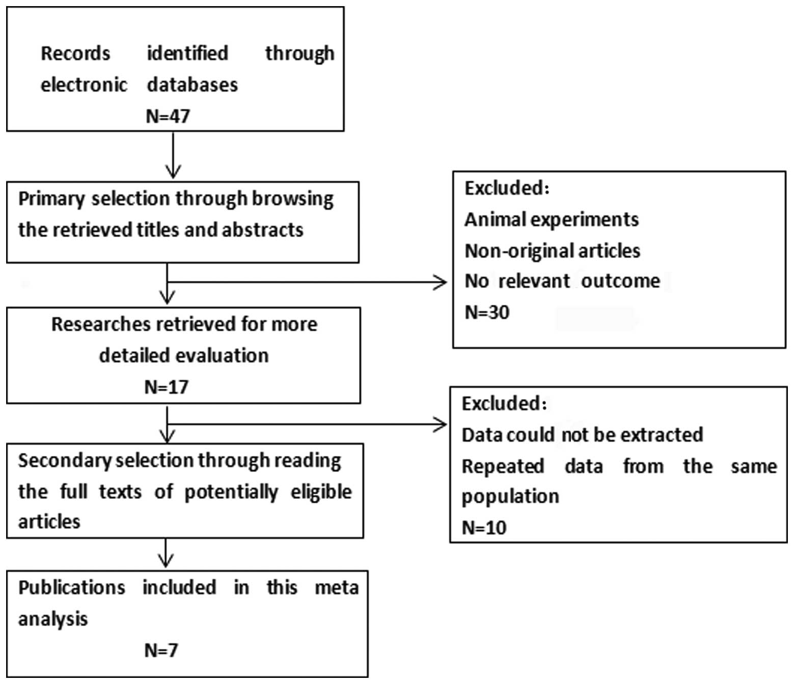
The characteristics of the eligible studies are
summarized in Table I. A total of 7
studies, published between 2009 and 2014, met the inclusion
criteria for this meta-analysis (Fig.
1). The total number of patients was 538, ranging from 28 to
136 patients per study. All the studies were based on the data of
retrospective analysEs. A total of 5 studies reported the
association between CD133 expression and lymph node metastasis; 5
studies reported the association between CD133 expression and
clinical stage; 7 studies reported the association between CD133
expression and histopathological grade; and 4 studies reported the
association between CD133 expression and depth of invasion.
Immunohistochemistry (IHC) was used to evaluate CD133 expression in
esophageal carcinoma specimens in all the studies.
 | Table I.Characteristics of studies included in
the meta-analysis. |
Table I.
Characteristics of studies included in
the meta-analysis.
| First author | Year | Language | Country | Tumor stage
(TNM) | Median age
(years) | Histopathological
type | Technique | No. of patients | Site | (Refs.) |
|---|
| Yang | 2010 | Chinese | China | I–IV | 52.8 | SCC | IHC | 90 | Esophagus or
bone | (10) |
| Cao | 2009 | Chinese | China | II–III | 54.3 | SCC | IHC | 68 | Esophagus or
bone | (11) |
| Fei | 2011 | Chinese | China | I–IV | 55.4 | SCC | IHC | 90 | Esophagus or
liver | (12) |
| Feng | 2014 | Chinese | China | I–IV | 68.8 | SCC | IHC | 28 | Esophagus or
bone | (13) |
| Wang | 2014 | Chinese | China | I–IV | 69 | SCC | IHC | 40 | Esophagus or
brain | (14) |
| Okamoto | 2013 | English | Japan | I–IV | 56.1 | SCC | IHC | 86 | Esophagus or
brain | (15) |
| Peng | 2012 | Chinese | China | III | 58 | SCC | IHC | 136 | Esophagus or
liver | (16) |
Main results of the meta-analysis
Correlation of CD133 with lymph node
metastasis
A total of 5 studies assessed the association
between CD133 expression and lymph node metastasis (Fig. 2). A positive CD133 expression was
significantly associated with lymph node metastasis (OR=3.09, 95%
CI: 1.93–4.95; P<0.00001), without any heterogeneity in the data
(χ2=5.58, I2=28%; P=0.23). These studies
indicated that CD133 overexpression is associated with the
prognosis of esophageal carcinoma.
Correlation of CD133 with clinical stages
A total of 5 studies assessed the association
between CD133 expression and clinical stage (Fig. 3). A positive CD133 expression was
significantly associated with clinical stage (OR=4.26, 95% CI:
1.55–11.73; P=0.005), with heterogeneity in the data
(χ2=13.84, I2=71%; P=0.008). In order to test
the heterogeneity in clinical stage, we performed sensitivity
analyses to assess the stability of the results. Our results
suggested that the sensitivity was low and the resultS were more
robust and credible. These studies indicated that CD133
overexpression is associated with the prognosis of esophageal
carcinoma.
Correlation of CD133 with histopathological
grade
A total of 7 studies assessed the association
between CD133 expression and histopathological grade (Fig. 4). There was A significant difference
between the well- and moderately differentiated esophageal
carcinoma group and the poorly differentiated esophageal carcinoma
group (OR=2.40, 95% CI: 1.16–4.94; P=0.02), with heterogeneity in
the data (χ2=14.75, I2=59%; P=0.02). In order
to test the heterogeneity in histopathological grade, we performed
sensitivity analyses to assess the stability of the results. Our
results suggested that the results were more robust and
credible.
Correlation of CD133 with depth of invasion
A total of 4 studies assessed the association
between CD133 expression and depth of invasion (Fig. 5). There were no significant
differences in the depth of invasion (OR=1.89, 95% CI: 0.42–8.43;
P=0.41) between different expression groups, indicating that higher
CD133 expression was not significantly associated with depth of
invasion in esophageal carcinoma.
Publication bias
Egger's testS were applied to estimate the
publication bias of the included studies (Fig. 6) and did not reveal any evidence of
obvious asymmetry in the overall meta-analysis of all the included
studies.
Discussion
Esophageal carcinoma is one of the most common and
most aggressive cancers worldwide and a leading cause of
cancer-related mortality in China (17). Surgical resection, which achieves
long-term survival of esophageal carcinoma patients, is considered
to be one of the standard treatments of esophageal carcinoma,
provided that the tumor is resectable (18). Although there has been significant
progress in the diagnosis and treatment of esophageal carcinoma,
its incidence and mortality rates remain high; therefore, new
therapies are urgently needed (19).
It has been reported that a rare subpopulation of cells with
special surface markers within esophageal carcinoma have the
potential to initiate and sustain tumor growth. these cells have
the exclusive properties of self-renewal and may give rise to all
the heterogeneous lineages of cancer cells that eventually
constitute the tumor bulk (20).
Cancer stem cells were first reported in acute myeloid leukemia, in
which a rare subset comprising 0.01–1% of the total population was
able to establish tumors when transplanted into mice with severe
combined immunodeficiency (SCID MICE), whereas the major cell
population could not (21). Since
then, cancer stem cells have been reported to promote solid tumor
development, including breast cancer (22), melanoma (23), hepatocellular carcinoma (5) and esophageal carcinoma (8). This new paradigm has remarkable
implications for cancer therapy, as it suggests that our currently
available therapies are more successful at eradicating non-cancer
stem cells rather than cancer stem cells (24,25).
Cancer stem cells exhibit major phenotypic and functional
heterogeneity, which may help distinguish them from cancer cells
and may lead to the identification of better targets for
therapeutic intervention (26).
Over the Last few years, several cell surface
markers have been identified as stem cell markers in cancer,
including CD133, CD90, CD271, CD44, CD24, ABCB5 and ALDH. CD133 is
a widely used marker for isolating cancer stem cells in a range of
solid tumors (27). CD133 is a member
of the cell membrane protein superfamily and has been used to
identify tumor-initiating cells as a specific marker in esophageal
carcinoma (28). In addition, it has
been reported that the presence of CD133+ cells was
associated with distinct clinicopathological characteristics in
esophageal carcinoma. It is notable that this association was
observed in our meta-analysis of CD133 expression with lymph node
metastasis, clinical stage and histopathological grade, suggesting
that CD133 may be a marker of poor prognosis in esophageal
carcinoma. There are also certain shortcomings in this study that
ought to be discussed. First, the number of included studies was
relatively small, with only 538 cases in total. Esophageal
carcinoma patients had received different treatments (perioperative
adjuvant therapy or curative surgical resection alone), and the
preoperative tnm stage varied. We were unable to assess these
potential confounders present in individual studies. Second,
potential publication bias was a major concern. We restricted our
systematic review to articles published in English or Chinese, as
other languages were not accessible to the readers. Third, in the
meta-analyses of prognostic factors, variability in definitions,
outcomes, measurements and experimental procedures may contribute
to between-study heterogeneity (29).
In conclusion, despite the abovementioned
limitations, this meta-analysis indicated that CD133 expression was
associated with the clinical parameters of esophageal carcinoma,
such as lymph node metastasis, clinical stage and histopathological
grade. Further studies on CD133 and its potential as a marker for
esophageal carcinoma prognosis in clinical practice are
required.
References
|
1
|
Shi HY, Zhu SC, Shen WB and Liu ML:
Pathological characteristics of esophageal cancer. Oncol Lett.
8:533–538. 2014.PubMed/NCBI
|
|
2
|
Tomochika S, Iizuka N, Watanabe Y, Tsutsui
M, Takeda S, Yoshino S, Ichihara K and Oka M: Increased serum
cell-free DNA levels in relation to inflammation are predictive of
distant metastasis of esophageal squamous cell carcinoma. Exp Ther
Med. 1:89–92. 2010.PubMed/NCBI
|
|
3
|
Tan Y, Chen B, Xu W, Zhao W and Wu J:
Clinicopathological significance of CD133 in lung cancer: A
meta-analysis. Mol Clin Oncol. 2:111–115. 2014.PubMed/NCBI
|
|
4
|
He A, Qi W, Huang Y, Feng T, Chen J, Sun
Y, Shen Z and Yao Y: CD133 expression predicts lung metastasis and
poor prognosis in osteosarcoma patients: A clinical and
experimental study. Exp Ther Med. 4:435–441. 2012.PubMed/NCBI
|
|
5
|
Bodzin AS, Wei Z, Hurtt R, Gu T and Doria
C: Gefitinib resistance in HCC Mahlavu cells: Upregulation of CD133
expression, activation of IGF-1R signaling pathway, and enhancement
of IGF-1R nuclear translocation. J Cell Physiol. 227:2947–2952.
2012. View Article : Google Scholar : PubMed/NCBI
|
|
6
|
Ferrandina G, Bonanno G, Pierelli L,
Perillo A, Procoli A, Mariotti A, Corallo M, Martinelli E, Rutella
S, Paglia A, et al: Expression of CD133-1 and CD133-2 in ovarian
cancer. Int J Gynecol Cancer. 18:506–514. 2008. View Article : Google Scholar : PubMed/NCBI
|
|
7
|
Kojima M, Ishii G, Atsumi N, Fujii S,
Saito N and Ochiai A: Immunohistochemical detection of CD133
expression in colorectal cancer: A clinicopathological study.
Cancer Sci. 99:1578–1583. 2008. View Article : Google Scholar : PubMed/NCBI
|
|
8
|
Hang D, Dong HC, Ning T, Dong B, Hou DL
and Xu WG: Prognostic value of the stem cell markers CD133 and
ABCG2 expression in esophageal squamous cell carcinoma. Dis
Esophagus. 25:638–644. 2012. View Article : Google Scholar : PubMed/NCBI
|
|
9
|
Zimmerer RM, Korn P, Demougin P, Kampmann
A, Kokemüller H, Eckardt AM, Gellrich NC and Tavassol F: Functional
features of cancer stem cells in melanoma cell lines. Cancer Cell
Int. 13:782013. View Article : Google Scholar : PubMed/NCBI
|
|
10
|
Yang AP: The expression of marker cancer
stem cells CD133 and Musashi-1 in human esophageal carcinoma and
its clinical significances. Southeast Univ. 15:6982010.(In
Chinese).
|
|
11
|
Cao YK: Relationship between CD133
expression and chemoradio therapy response in esophageal squamous
cell carcinoma. Zhongshan Univ. 16:752009.(In Chinese).
|
|
12
|
Fei ZH, Chen SX and Chen L: Expression and
significance of Bmi-1 and CD133 in esophageal squamous cell
carcinoma. Mod Pract Med. 23:3372011.(In Chinese).
|
|
13
|
Feng KX, Li SP, Liu XL, Zhou J, Yuan S,
Xie MH, Jing DS and Sun YZ: Expression of NF-κB, CD133 in
esophageal cancer and correlation with metastasis. Mod Oncol.
23:02062014.(In Chinese).
|
|
14
|
Wang YW, Zhang J and Feng G: Expression of
CD133 in esophageal squamous cell carcinoma and its clinical
significance. Shaanxi Med J. 43:14642014.(In Chinese).
|
|
15
|
Okamoto H, Fujishima F, Nakamura Y, et al:
Significance of CD133 expression in esophageal squamous cell
carcinoma. World J Surg Oncol. 11:512013.(In Chinese). View Article : Google Scholar : PubMed/NCBI
|
|
16
|
Peng J, Guo JJ, Ao X, Zhou TJ, Wang M, Li
YQ and Zhang HZ: Expression of CDl33 in the tissue of locally
advanced esophagus squamous cell cancer patients and its
significance. Chin J Exp Surg. 29:5412012.(In Chinese).
|
|
17
|
Ning Z, Zhu H, Li F, Liu Q, Liu G, Tan T,
Zhang B, Chen S, Li G, Huang D, et al: Tumor suppression by miR-31
in esophageal carcinoma is p21-dependent. Genes Cancer. 5:436–444.
2014.PubMed/NCBI
|
|
18
|
Rios-Galvez S, Meixueiro-Daza A and
Remes-Troche JM: Achalasia: a risk factor that must not be
forgotten for esophageal squamous cell carcinoma. BMJ Case Rep.
2015(pii): bcr20142044182015. View Article : Google Scholar : PubMed/NCBI
|
|
19
|
Shimizu Y, Takahashi M, Mizushima T, Ono
S, Mabe K, Ohnishi S, Kato M, Asaka M and Sakamoto N:
Chromoendoscopy with iodine staining, as well as narrow-band
imaging, is still useful and reliable for screening of early
esophageal squamous cell carcinoma. Am J Gastroenterol.
110:193–194. 2015. View Article : Google Scholar : PubMed/NCBI
|
|
20
|
Nguyen LV, Vanner R, Dirks P and Eaves CJ:
Cancer stem cells: An evolving concept. Nat Rev Cancer. 12:133–143.
2012.PubMed/NCBI
|
|
21
|
Bonnet D and Dick JE: Human acute myeloid
leukemia is organized as a hierarchy that originates from a
primitive hematopoietic cell. Nat Med. 3:730–737. 1997. View Article : Google Scholar : PubMed/NCBI
|
|
22
|
Al-Hajj M, Wicha MS, Benito-Hernandez A,
Morrison SJ and Clarke MF: Prospective identification of
tumorigenic breast cancer cells. Proc Natl Acad Sci USA.
100:3983–3988. 2003. View Article : Google Scholar : PubMed/NCBI
|
|
23
|
Klein WM, Wu BP, Zhao S, Wu H,
Klein-Szanto AJ and Tahan SR: Increased expression of stem cell
markers in malignant melanoma. Mod Pathol. 20:102–107. 2007.
View Article : Google Scholar : PubMed/NCBI
|
|
24
|
Lobo NA, Shimono Y, Qian D and Clarke MF:
The biology of cancer stem cells. Annu Rev Cell Dev Biol.
23:675–699. 2007. View Article : Google Scholar : PubMed/NCBI
|
|
25
|
Vermeulen L, Sprick MR, Kemper K, Stassi G
and Medema JP: Cancer stem cells - old concepts, new insights. Cell
Death Differ. 15:947–958. 2008. View Article : Google Scholar : PubMed/NCBI
|
|
26
|
Liu S, Liu C, Min X, Ji Y, Wang N, Liu D,
Cai J and Li K: Prognostic value of cancer stem cell marker
aldehyde dehydrogenase in ovarian cancer: A meta-analysis. PLoS
One. 8:e810502013. View Article : Google Scholar : PubMed/NCBI
|
|
27
|
Visvader JE and Lindeman GJ: Cancer stem
cells in solid tumours: Accumulating evidence and unresolved
questions. Nat Rev Cancer. 8:755–768. 2008. View Article : Google Scholar : PubMed/NCBI
|
|
28
|
Yao Q, Sun JG, Ma H, Zhang AM, Lin S, Zhu
CH, Zhang T and Chen ZT: Monitoring microRNAs using a molecular
beacon in CD133+/CD338+ human lung
adenocarcinoma-initiating A549 cells. Asian Pac J Cancer Prev.
15:161–166. 2014. View Article : Google Scholar : PubMed/NCBI
|
|
29
|
Simon R and Altman DG: Statistical aspects
of prognostic factor studies in oncology. Br J Cancer. 69:979–985.
1994. View Article : Google Scholar : PubMed/NCBI
|