Introduction
Oral squamous cell carcinoma (OSCC) is the most
common malignant tumor of the head and neck, which represents
approximately 90% of all oral neoplasms affecting the oral cavity
(1). OSCC is the 8th most common
cancer worldwide in terms of occurrence, which is more prevalent
(approximately 4%) in men than in women (2%) (2,3). An
increased incidence and prevalence of OSCC has been reported
particularly in developing countries in recent years. The annual
occurrence rate of oral cancer is about 300,000 worldwide (4–6), with
over 11,000 new cases each year in Japan (7). Despite recent advances in surgery and
chemoradiation therapies, only 50% of the OSCC patients survive 5
years after the diagnosis (8,9). OSCC
typically shows poor prognosis at the advanced stage of the
disease; and probably due to the heterogeneous nature of the
disease, it shows differential outcomes to the same treatment.
Because early diagnosis is crucial for the successful treatment of
OSCC, development of promising biomarkers is necessary for its
detection at an early stage (7).
Some cancers can develop resistance to a particular
chemotherapy that was effective initially. Although chemoresistance
can be caused by multiple mechanisms, the markers involved in the
chemoresistance-related mechanisms can help to predict the response
of OSCC to a certain chemotherapeutic agent. The efficacy of
neoadjuvant chemotherapy (NAC) for OSCC remains to be elucidated,
but it could be improved by the detection of the biomarkers related
to chemoresistance. Many researchers have identified new molecular
predictors and biomarkers that are useful for understanding the
response of tumors to certain anticancer agents. The detection of
thymidylate synthase (TS), dihydropyrimidine dehydrogenase (DPD),
thymidine phosphorylase (TP) and orotate phosphoribosyltransferase
(OPRT) as the predictive factors of the response to treatment with
5-fluorouracil (5-FU) is one such example (10,11).
Docetaxel (Doc) or Taxotere
(N-debenzoyl-N-tart-butoxycarbonyl-10-deacetyl taxol) is a
semi-synthetic taxane developed from a non-cytotoxic precursor of
10-deacetyl baccatin III obtained from the needles of the European
yew tree Taxus baccata L. Doc is an effective drug to combat
cancer and is used as a first-line treatment or as an adjuvant
therapy for various cancers including OSCC (12). Doc showed a 22.2% response rate as a
single-agent therapy in advanced/recurrent head and neck cancer
patients (13). Doc binds with
microtubules, thereby interrupting their normal function during
mitosis, which eventually causes cell death. Chemotherapeutic
agents with different mechanisms of antitumor activity (e.g., 5-FU,
cisplatin, etc.) than Doc are sometimes used with Doc as an
effective combined chemotherapy against various types of cancers.
We previously carried out a clinical trial of Doc and S-1
combination therapy against OSCC (14). Moreover, we treated patients with
locally advanced OSCC with Doc-containing regimens as an NAC in our
hospital, which showed promising results. Recently, we carried out
a microarray analysis of Doc-resistant OSCC cells established in
our laboratory and identified a few genes potentially related to
Doc resistance. One of those genes was Forkhead box protein M1
(FOXM1).
FOXM1, a member of the FOX family of transcription
factors, is characterized by a 100-amino acid winged-helix DNA
binding domain (15). FOXM1 is a
human proto-oncogene that plays a key role in cell cycle
progression, mitosis, differentiation and aging (16,17).
Moreover, it has already been reported that overexpression of FOXM1
is related to the development and progression of various cancers,
and it is often associated with a poor prognosis and poor outcome
in patients (18–20). Furthermore, FOXM1 amplification is
reported to be responsible for gefitinib-resistance in non-small
cell lung cancer (NSCLC) and for acquired resistance of herceptin
and paclitaxel in breast cancer (21,22).
Therefore, FOXM1 may be closely associated with the resistance of
cancers cells to various chemotherapeutic agents including Doc.
Several studies have reported the association between Doc
resistance and high expression of FOXM1 in different cancer types
(20,23,24).
However, the relationship between high expression of FOXM1 and Doc
resistance in OSCC is still unknown. We have to clarify further
whether FOXM1 expression can be clinically used as a predictive
factor for the response of OSCC patients to Doc-containing
chemotherapies.
In the present study, we tried to examine the
potential value of FOXM1 as a prognostic factor for OSCC patients
receiving a Doc-containing chemotherapy.
Materials and methods
Patients and specimens
In the present study, we retrospectively used tissue
samples taken from a total of 56 patients with OSCC who visited
Yamaguchi University Hospital between August 2004 and September
2012. Most of these patients were in stage II or III of OSCC and
were not diagnosed with distant metastasis at the first visit to
our hospital. All patients had a diagnosis of squamous cell
carcinoma and had not been treated for OSCC previously. The
clinicopathological characteristics of the patients are shown in
Table I. All patients received Doc
40–50 mg/m2 by superselective intra-arterial infusion on
day 1 and S-1 65 mg/m2 or tegafur/uracil (UFT) 300–400
mg/body on day 1–14 (14 days). Surgical operation was carried out
1–2 weeks after the administration of the combination chemotherapy
mentioned above. Tissue specimens were collected from all 56
patients by biopsy before they received any treatment. We performed
a surgical operation when the tumor was resectable. However, we
selected this chemotherapy with Doc for the patients who had a hope
of functional preservation (limited operation) or a refusal of
extended surgery after discussion with patients. So, the potential
for selection bias of patients is unavoidable. This study was
conducted according to the ethical standards of the Institutional
Review Board (IRB) of Yamaguchi University Hospital.
 | Table I.Patient characteristics (n=56). |
Table I.
Patient characteristics (n=56).
| Characteristics | No. of patients, n
(%) |
|---|
| Sex |
|
| Male | 34 (60.7) |
|
Female | 22 (39.3) |
| T
classification |
|
| 1 | 6 (10.7) |
| 2 | 29 (51.8) |
| 3 | 13 (23.2) |
| 4 | 8 (14.3) |
| N
classification |
|
| 0 | 46 (82.1) |
| 1 | 5 (8.9) |
| 2 | 4 (7.1) |
| 3 | 1 (1.8) |
| Stage |
|
| I | 5 (8.9) |
| II | 29 (51.8) |
|
III | 12 (21.4) |
| IV | 10 (17.9) |
| Tumor
differentiation |
|
|
Well | 38 (67.8) |
|
Moderately | 15 (26.8) |
|
Poorly | (3) 5.4 |
| Therapeutic
effect |
|
| CR | 11 (19.6) |
| PR | 37 (66.1) |
| SD | 8 (14.3) |
| Outcome |
|
|
Alive | 47 (83.9) |
|
Death | 9 (16.1) |
| FOXM1 expression in
tumor cell cytoplasm |
|
|
Low | 35 (62.5) |
|
High | 21 (37.5) |
| Age (years) | Median=67; |
|
| Min-max=30–83 |
Immunohistochemical staining and
evaluation
Tissue specimens were fixed in phosphate-buffered
10% formalin, embedded in paraffin, and 4 µm-thick tissue sections
were prepared. These tissue sections were deparaffinized in xylene
and rehydrated in graded ethanol (70–100%). After washing with
phosphate buffered saline (PBS), the sections were microwaved in an
antigen retrieval solution and allowed to cool down gradually.
After immersion of slides for 30 min in methanol containing 0.3%
H2O2 at room temperature, the sections were
washed again in PBS. A Dako REAL™ Peroxidase-Blocking
Solution (S2023, Dako; Agilent Technologies GmbH, Waldbronn,
Germany) was used for 15 min as a blocking reagent to reduce
nonspecific binding. Then the sections were incubated overnight at
4°C with anti-FOXM1 rabbit polyclonal antibody (1:250; ab137647,
Abcam, Cambridge, UK). After washing in PBS, a secondary antibody
solution (EnVision+ System HRP; Dako; Agilent Technologies GmbH)
was applied for 60 min at room temperature, and the sections were
incubated with diaminobenzidine using a REAL™
EnVision™ Detection System kit (K5007, Dako; Agilent
Technologies GmbH). After a tap-water wash, the sections were
lightly counterstained with hematoxylin (Muto Pure Chemicals,
Tokyo, Japan), immersed in graded alcohol (70–100%) and xylene and
finally mounted and cover slipped. In the case of negative
controls, the slides were incubated without any FOXM1 antibody.
We evaluated FOXM1 expression as the mean percentage
of positive tumor cells observed in at least five random fields of
each section at ×400 magnification. The intensity of the
FOXM1-immunoreaction was scored as follows: 1+, weak; 2+, moderate;
and 3+, intense. The final score or the FOXM1-immunohistochemical
staining score was calculated by multiplying the percentage of
positive tumor cells with the staining intensity (25). High expression was defined as a score
of ≥111.7 (the highest score for normal tissue including a
dysplastic lesion), and low expression was defined as a score of
<111.7. All the specimens were evaluated by three authors (KH,
TF and YU), who had no knowledge of the patient's clinical status.
The tissue samples were also stained with hematoxylin and eosin
(Wako Pure Chemical Industries, Ltd., Osaka, Japan) for
histological evaluation.
Statistical analysis
Fisher's exact test was used to estimate the
associations between FOXM1 and different clinicopathological
parameters of patients. The Kaplan-Meier method was used to
calculate overall survival (OS), and the log-rank test was used to
compare between different groups. Univariate and multivariate
analyses were performed using the Cox proportional hazards model.
P<0.05 was considered to indicate a statistically significant
difference. Statistical analyses were performed using the StatView
software (version 5.0J; SAS Institute, Inc., Cary, NC, USA).
Results
Patients and tumor
characteristics
Table I summarizes
the clinicopathological data of 56 OSCC patients who participated
in this study. All of the patients were treated with a
Doc-containing regimen. The median follow-up time was 8.6 years,
and the median age was 67 years (range 30–83 years). Clinical
stages I, II, III and IV were diagnosed in 5, 29, 12 and 10
patients, respectively. All tissue specimens were collected before
the primary treatment, and there were adequate histologic materials
available for immunohistochemical analysis of those patients.
FOXM1 expression in tumor cells and
clinicopathological features
Table II summarized
the association between the status of FOXM1 expression and the
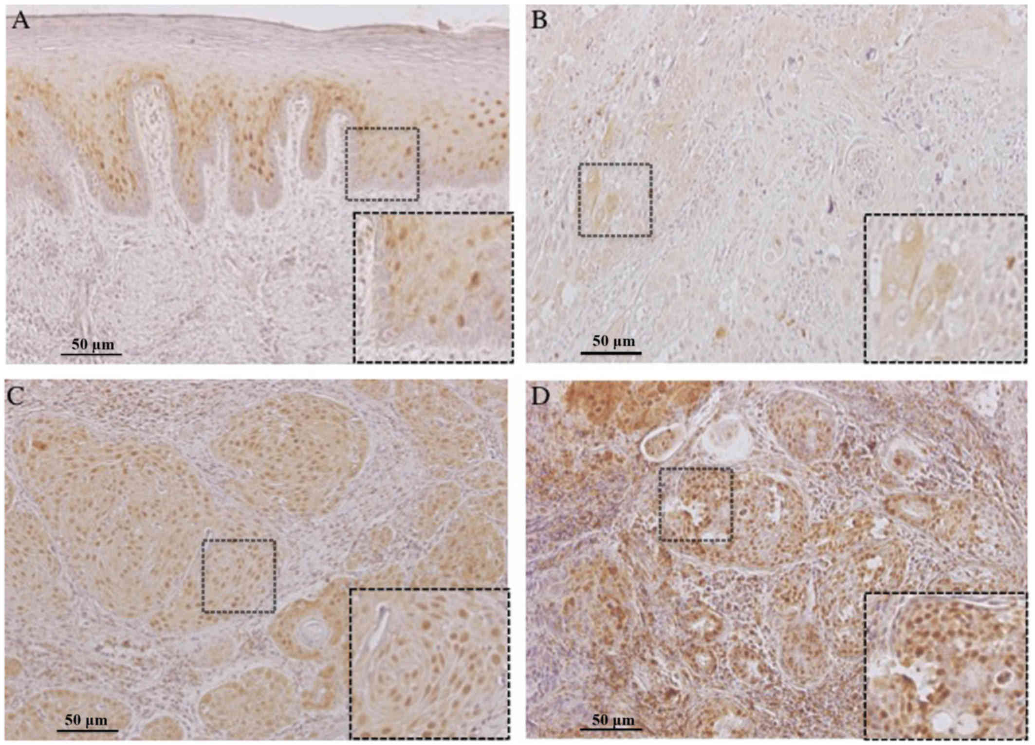
clinicopathological characteristics of the patients. FOXM1
expression was observed in the nucleus of normal oral tissues
adjacent to tumors and both in the cytoplasm and nucleus of OSCC
tumors (Fig. 1). In the case of
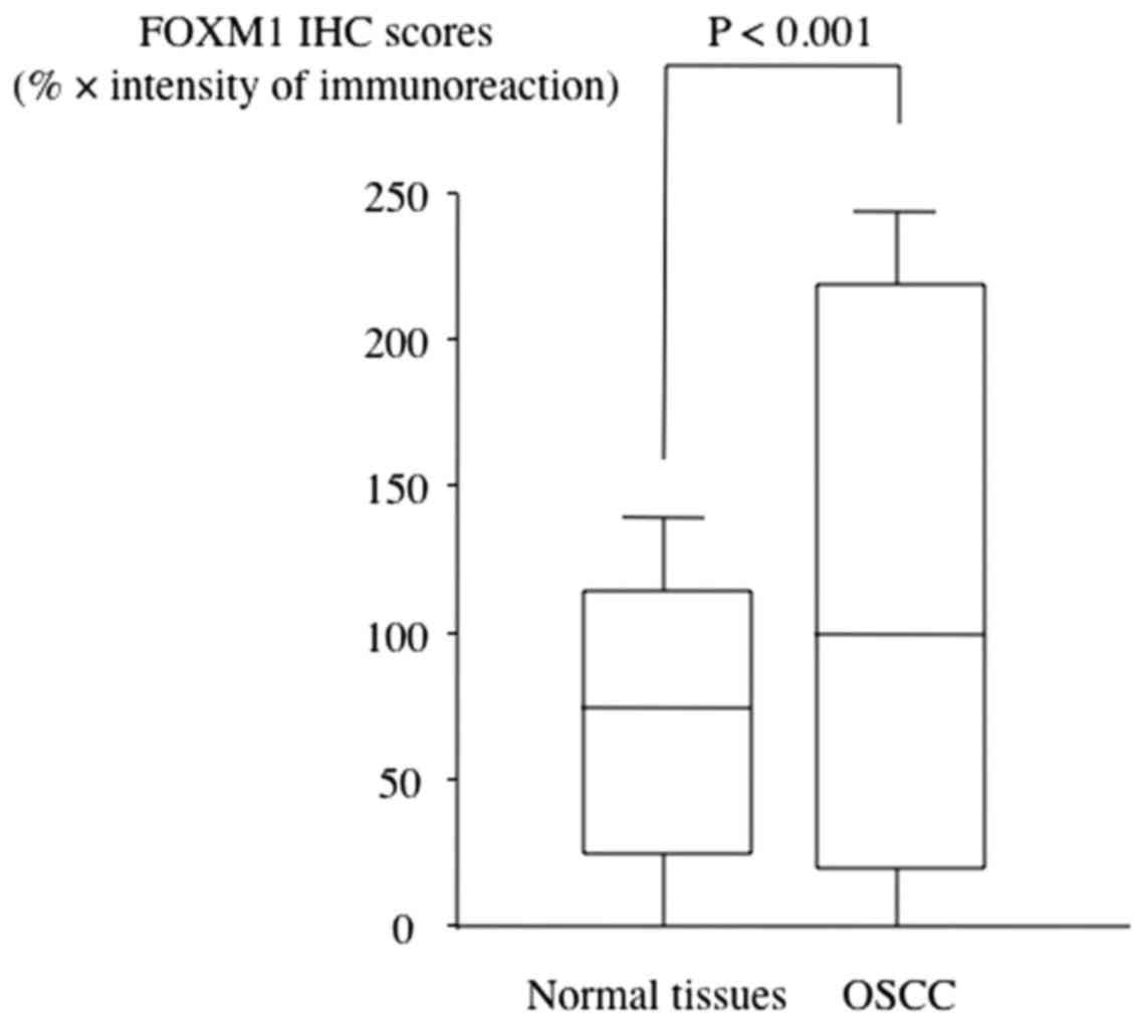
normal tissues adjacent to tumors, the immunohistochemistry scores
for FOXM1 ranged from 24.2 to 111.7 (mean, 74.2). The FOXM1
expression level in primary OSCCs ranged from 18.7 to 217.6 (mean,
98.7), which was significantly higher than those in normal oral
tissues (P<0.001, Fig. 2). Among
56 patients with OSCC, 35 patients (62.5%) showed low expression
(<111.7) of FOXM1 and 21 patients (37.5%) showed high expression
(≥111.7) (Table II). No correlation
was found between FOXM1 expression and sex, age, T classification
or tumor differentiation of OSCC patients. However, a significant
association was observed between FOXM1 expression and N
classification (P=0.0395), therapeutic efficacy (P=0.0040), stage
(P=0.0113) and patient outcome (P=0.0134; Table II).
| Table II.Associations between FOXM1 expression
and clinicopathological factors in oral squamous cell carcinoma
treated patients with a docetaxel-containing regimen. |
Table II.
Associations between FOXM1 expression
and clinicopathological factors in oral squamous cell carcinoma
treated patients with a docetaxel-containing regimen.
|
| FOXM1 expression in
tumor cells |
|
|---|
|
|
|
|
|---|
|
Characteristics | Low expression,
(n=35, 62.5%) | High expression
(n=21, 37.5%) | Total (n=56) | P-value |
|---|
| Sex |
|
|
| 0.7614 |
|
Male | 21 | 13 | 34 |
|
|
Female | 14 | 8 | 22 |
|
| Age (years) |
|
|
| 0.5307 |
|
≥65 | 19 | 14 | 33 |
|
|
<65 | 16 | 7 | 23 |
|
| T
classification |
|
|
| 0.0539 |
|
T1+T2 | 25 | 7 | 32 |
|
|
T3+T4 | 10 | 14 | 24 |
|
| N
classification |
|
|
| 0.0395 |
| 0 | 30 | 11 | 41 |
|
|
N1+N2+N3 | 5 | 10 | 15 |
|
| Stage |
|
|
| 0.0113 |
|
I+II | 23 | 4 | 27 |
|
|
III+IV | 12 | 17 | 29 |
|
| Tumor
differentiation |
|
|
| 0.0523 |
|
Well | 27 | 11 | 38 |
|
|
Moderately+Poorly | 8 | 10 | 18 |
|
| Therapeutic
efficacy |
|
|
| 0.004 |
|
CR+PR | 34 | 14 | 48 |
|
| SD | 1 | 7 | 8 |
|
| Outcome |
|
|
| 0.0134 |
|
Alive | 34 | 13 | 47 |
|
|
Death | 1 | 8 | 9 |
|
FOXM1 expression and survival
time
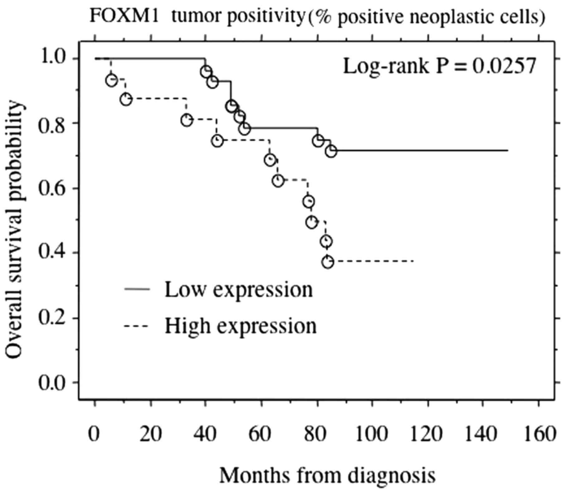
A total of 47 patients survived, and 9 patients died
during the study period with a median follow-up time of 8.6 years.
The relationship between FOXM1 expression and patients' OS was
analyzed by a Kaplan-Meier curve. There was a significant
association between high expression of FOXM1 in tumor cells and
shorter OS (P=0.0257; Fig. 3).
Moreover, a Cox proportional hazards model was applied to estimate
the effect of FOXM1 expression on OSCC patient survival. A
univariate Cox regression analysis identified T classification,
stage, tumor differentiation, therapeutic effect and the expression
of FOXM1 as significant prognostic factors. Using a multivariate
analysis, T classification, therapeutic effect and the expression
of FOXM1 were found to be independent prognostic factors for
overall survival (Table III).
Collectively, the results indicate that FOXM1 expression may act as
an independent predictor for poor patient prognosis.
| Table III.Univariate and multivariate analysis
of overall survival. |
Table III.
Univariate and multivariate analysis
of overall survival.
|
| Univariate
analysis |
| Multivariate
analysis |
|
|---|
|
|
|
|
|
|
|---|
| Variables | Hazard ratio | 95% CI | P-value | Hazard ratio | 95% CI | P-value |
|---|
| Sex |
| Male
vs. Female | 1.074 | 0.573–1.984 | 0.9746 |
|
|
|
| Age (years) |
| >65
vs. <65 | 1.172 | 0.648–1.867 | 0.6894 |
|
|
|
| T
classification |
| T3+T4
vs. T1+T2 | 2.687 | 1.056–6.834 | 0.0379 | 1.874 | 1.014–2.931 | 0.0463 |
| N
classification |
| N0 vs.
N1+N2+N3 | 0.99 | 0.353–2.788 | 0.9847 |
|
|
|
| Stage |
| Stage
III+IV vs. Stage I+II | 2.808 | 1.051–7.501 | 0.0394 | 2.576 | 0.745–6.945 | 0.0871 |
| Tumor
differentiation |
| Well
vs. Moderately+Poorly | 0.174 | 0.012–0.928 | 0.0427 | 0.132 | 0.010–0.847 | 0.1678 |
| Effect |
| CR+PR
vs. NC | 0.111 | 0.036–0.345 | 0.0001 | 0.105 | 0.021–0.282 | 0.0354 |
| FOXM1
expression |
| High
vs. Low | 2.765 | 1.087–7.033 | 0.0327 | 1.867 | 0.946–5.393 | 0.0472 |
Discussion
FOXM1 has vital roles in adult tissue homeostasis,
cell proliferation, cell differentiation, apoptosis and aging as
well as in the pathogenesis of human cancers (16,17,26).
Normal cells show a lower expression of FOXM1 than cancer cells.
Dysfunction of FOXM1 inhibits cell differentiation, which may
finally lead to the malignant transformation of undifferentiated
cells (27). Moreover, upregulated
expression of FOXM1 has been observed in a number of cancers
including hepatocellular carcinoma, breast cancer, non-small cell
lung carcinomas and glioblastomas as well as prostate, cervical and
gastric cancer (21,28–33).
Recent studies have strongly suggested that overexpression of FOXM1
could be correlated with cancer progression and might be a
potential prognostic biomarker for cancer patients (28–33). In
addition, the prognostic significance of FOXM1 expression is
clearly seen in various cancers including renal, liver, pancreatic
and lung cancer, by using The Cancer Genome Atlas or The Human
Protein Atlas. These days, Doc, a semisynthetic taxane drug with a
notable anticancer effect, has been extensively used to treat
various cancers. However, acquired resistance to Doc is one of the
major obstacles to the application of Doc containing regimens to
treat cancers. It was reported that FOXM1 is related to Doc
resistance in several cancer types; nevertheless, few studies have
investigated the association between FOXM1 and Doc resistance
(20,23,24).
In gastric cancer, FOXM1 is reported to mediate
Doc-resistance by upregulating the microtubule-destabilizing
protein stathmin (23). Okada et
al (20) also reported the
relationship between FOXM1 overexpression and Doc chemoresistance
in gastric cancer cells. Moreover, FOXM1 expression is also
associated with paclitaxel resistance in several cancers (26,34–36).
However, until now, no link between FOXM1 expression and Doc
resistance has been reported in OSCC.
In recent years, we have treated OSCC patients with
a Doc-containing regimen as an NAC (Doc plus S-1 or UFT) in our
hospital, but the number of patients in each trial was small. In
the present study, we aimed to investigate the usefulness of FOXM1
in predicting the response of these 56 OSCC patients to an NAC with
a Doc-containing regimen.
In this study, upregulated expression of FOXM1 was
detected in OSCC cells compared to normal tissues (Fig. 2). Moreover, overexpression of FOXM1
was significantly associated with therapeutic efficacy, N
classification and stage, patient outcome (Table II) and shorter OS (Fig. 3). We also observed that OSCC patients
with low expression of FOXM1 responded well (CR or PR) to NAC
treatments with a Doc-containing regimen than those with high FOXM1
expression (Table II).
Additionally, multivariate analysis showed that high expression of
FOXM1 was a predictive factor of reduced survival (P=0.0327)
(Table III). These findings
suggest that high expression of FOXM1 might be associated with Doc
resistance and poor prognosis in OSCC. Thus, examining the FOXM1
expression pattern in biopsy samples might help to determine the
most effective treatment strategies for OSCC patients.
FOXM1 promotes drug resistance in cancers by
targeting and mediating several molecules [e.g., XIAP, survivin,
nibrin or NBS1; kinesin-like protein (KIF) 20A; stathmin, etc.]
involved in DNA repair, metastasis, cell invasion, migration and
mitosis (23,36–38). It
is assumed that FOXM1 and Doc might have overlapping roles in the
progression of mitosis, and FOXM1 might target other molecules
involved in regulation of mitosis to ensure Doc resistance
(23). Therefore, identification of
those molecules is essential to understand the FOXM1-mediated
resistance of Doc. It was reported that agents that suppress FOXM1
expression can reverse the acquired docetaxel resistance in cancer
cells. For example, proteasome inhibitor thiostrepton and
cell-penetrating adenosine diphosphate ribosylation factor (ARF)
peptide are reported to inhibit the FOXM1 functions that lead to
the reversal of Doc resistance and reduced tumor cell proliferation
in vitro, respectively (23,39).
Therefore, the use of FOXM1 inhibitors might be promising as new
anticancer therapeutics in cancer patients with elevated FOXM1
expression or Doc resistance.
In this study we showed that FOXM1 could be a
potential prognostic marker for OSCC treated with a Doc-containing
regimen. Molecularly targeted therapies against FOXM1 may have
promising therapeutic benefits for the successful treatment of
cancer. Our results agree with most of the previous findings on the
association between FOXM1 overexpression and patients' response to
DOC based chemotherapies in different types of cancers. Further
studies are needed to clarify the clinical importance of FOXM1 and
to understand the detailed mechanism of Doc-resistance and FOXM1
expression in OSCC in both in vitro and in vivo
models.
Acknowledgements
The authors would like to thank to Dr Dan Cui
(Department of Pathology, Yamaguchi University Graduate School of
Medicine) for her helpful advice and suggestions in the
immunohistochemical analysis. They would also like to thank Mr. Reo
Kawano (Center for Clinical Research, Yamaguchi University
Hospital) for data and statistical analysis support and Professor
Yoichi Mizukami (Center for Gene Research, Yamaguchi University)
for his helpful advice and suggestions in the usage of The Cancer
Genome Atlas.
Funding
The present study was supported in part by a
Grant-in-Aid (grant no. 25861949) from the Japanese Ministry of
Education, Science and Culture.
Availability of data and materials
The datasets used and/or analyzed during the current
study are available from the corresponding author on reasonable
request.
Authors' contributions
KH designed the experimental study, analyzed the
data and wrote the manuscript. TF carried out the
immunohistochemical experiments, collected and evaluated the data
and assisted with writing and revising the manuscript. HM was
involved in data collection and analysis. KM collected and analyzed
the data, and revised and edited the manuscript.
Ethics approval and consent to
participate
The present study was approved by the ethical
standards of the Institutional Review Board (IRB) of Yamaguchi
University Hospital (Ref. H24-125). Due to the retrospective nature
of the present study, the requirement for informed consent was
waived by the IRB.
Patient consent for publication
Not applicable.
Competing interests
The authors declare that they have no competing
interests.
Glossary
Abbreviations
Abbreviations:
|
FOXM1
|
Forkhead box protein M1
|
|
SCC
|
squamous cell carcinoma
|
References
|
1
|
Lawoyin JO, Lawoyin DO and Aderinokun G:
Intra-oral squamous cell carcinoma in Ibadan: A review of 90 cases.
Afr J Med Med Sci. 26:187–188. 1997.PubMed/NCBI
|
|
2
|
Exarchos KP, Goletsis Y and Fotiadis DI: A
multiscale and multiparametric approach for modeling the
progression of oral cancer. BMC Med Inform Decis Mak. 12:1362012.
View Article : Google Scholar : PubMed/NCBI
|
|
3
|
Parkin DM, Bray F, Ferlay J and Pisani P:
Global cancer statistics, 2002. CA Cancer J Clin. 55:74–108. 2005.
View Article : Google Scholar : PubMed/NCBI
|
|
4
|
Rautava J, Luukkaa M, Heikinheimo K, Alin
J, Grenman R and Happonen RP: Squamous cell carcinomas arising from
different types of oral epithelia differ in their tumor and patient
characteristics and survival. Oral Oncol. 43:911–919. 2007.
View Article : Google Scholar : PubMed/NCBI
|
|
5
|
Funk GF, Karnell LH, Robinson RA, Zhen WK,
Trask DK and Hoffman HT: Presentation, treatment, and outcome of
oral cavity cancer: A National Cancer Data Base report. Head Neck.
24:165–180. 2002. View Article : Google Scholar : PubMed/NCBI
|
|
6
|
Mehrotra R, Singh MK, Pandya S and Singh
M: The use of an oral brush biopsy without computer-assisted
analysis in the evaluation of oral lesions: A study of 94 patients.
Oral Surg Oral Med Oral Pathol Oral Radiol Endod. 106:246–253.
2008. View Article : Google Scholar : PubMed/NCBI
|
|
7
|
Tanaka T, Tanaka M and Tanaka T: Oral
carcinogenesis and oral cancer chemoprevention: A review. Pathol
Res Int. 2011:4312462011. View Article : Google Scholar
|
|
8
|
Inagi K, Takahashi H, Okamoto M, Nakayama
M, Makoshi T and Nagai H: Treatment effects in patients with
squamous cell carcinoma of the oral cavity. Acta Otolaryngol Suppl.
25–29. 2002. View Article : Google Scholar : PubMed/NCBI
|
|
9
|
Shingaki S, Takada M, Sasai K, Bibi R,
Kobayashi T, Nomura T and Saito C: Impact of lymph node metastasis
on the pattern of failure and survival in oral carcinomas. Am J
Surg. 185:278–284. 2003. View Article : Google Scholar : PubMed/NCBI
|
|
10
|
Kamoshida S, Shiogama K, Shimomura R,
Inada K, Sakurai Y, Ochiai M, Matuoka H, Maeda K and Tsutsumi Y:
Immunohistochemical demonstration of fluoropyrimidine-metabolizing
enzymes in various types of cancer. Oncol Rep. 14:1223–1230.
2005.PubMed/NCBI
|
|
11
|
Miyoshi Y, Uemura H, Ishiguro H, Kitamura
H, Nomura N, Danenberg PV and Kubota Y: Expression of thymidylate
synthase, dihydropyrimidine dehydrogenase, thymidine phosphorylase,
and orotate phosphoribosyl transferase in prostate cancer. Prostate
Cancer Prostatic Dis. 8:260–265. 2005. View Article : Google Scholar : PubMed/NCBI
|
|
12
|
Bissery MC, Guénard D, Guéritte-Voegelein
F and Lavelle F: Experimental antitumor activity of taxotere (RP
56976, NSC 628503), a taxol analogue. Cancer Res. 51:4845–4852.
1991.PubMed/NCBI
|
|
13
|
Inuyama Y, Kataura A, Togawa K, Saijo S,
Satake B, Takeoda S, Konno A, Ebihara S, Sasaki Y, Kida A, et al:
Late phase II clinical study of RP56976 (docetaxel) in patients
with advanced/recurrent head and neck cancer. Gan To Kagaku Ryoho.
26:107–116. 1999.(In Japanese). PubMed/NCBI
|
|
14
|
Ueyama Y, Okafuji M, Harada K, Mano T,
Mihara M, Uchida K, Horinaga D and Wada N: Clinical phase I trial
of S-1 in the combination with DOC using super-selective
intra-arterial infusion with oral cancer. Gan To Kagaku Ryoho.
36:395–399. 2009.(In Japanese). PubMed/NCBI
|
|
15
|
Wierstra I and Alves J: FOXM1, a typical
proliferation-associated transcription factor. Biol Chem.
388:1257–1274. 2007. View Article : Google Scholar : PubMed/NCBI
|
|
16
|
Tuteja G and Kaestner KH: SnapShot:
Forkhead transcription factors I. Cell. 130:11602007. View Article : Google Scholar : PubMed/NCBI
|
|
17
|
Tuteja G and Kaestner KH: Forkhead
transcription factors II. Cell. 131:1922007. View Article : Google Scholar : PubMed/NCBI
|
|
18
|
Liu M, Dai B, Kang SH, Ban K, Huang FJ,
Lang FF, Aldape KD, Xie TX, Pelloski CE, Xie K, et al: FoxM1B is
overexpressed in human glioblastomas and critically regulates the
tumorigenicity of glioma cells. Cancer Res. 66:3593–3602. 2006.
View Article : Google Scholar : PubMed/NCBI
|
|
19
|
Yau C, Wang Y, Zhang Y, Foekens JA and
Benz CC: Young age, increased tumor proliferation and FOXM1
expression predict early metastatic relapse only for
endocrine-dependent breast cancers. Breast Cancer Res Treat.
126:803–810. 2011. View Article : Google Scholar : PubMed/NCBI
|
|
20
|
Okada K, Fujiwara Y, Takahashi T, Nakamura
Y, Takiguchi S, Nakajima K, Miyata H, Yamasaki M, Kurokawa Y, Mori
M and Doki Y: Overexpression of forkhead box M1 transcription
factor (FOXM1) is a potential prognostic marker and enhances
chemoresistance for docetaxel in gastric cancer. Ann Surg Oncol.
20:1035–1043. 2013. View Article : Google Scholar : PubMed/NCBI
|
|
21
|
Xu N, Zhang X, Wang X, Ge HY, Wang XY,
Garfield D, Yang P, Song YL and Bai CX: FoxM1 mediated resistance
to gefitinib in non-small-cell lung cancer cells. Acta Pharmacol
Sin. 33:675–681. 2012. View Article : Google Scholar : PubMed/NCBI
|
|
22
|
Carr JR, Park HJ, Wang Z, Kiefer MM and
Raychaudhuri P: FoxM1 mediates resistance to herceptin and
paclitaxel. Cancer Res. 70:5054–5063. 2010. View Article : Google Scholar : PubMed/NCBI
|
|
23
|
Li X, Yao R, Yue L, Qiu W, Qi W, Liu S,
Yao Y and Liang J: FOXM1 mediates resistance to docetaxel in
gastric cancer via up-regulating Stathmin. J Cell Mol Med.
18:811–823. 2014. View Article : Google Scholar : PubMed/NCBI
|
|
24
|
Li X, Qiu W, Liu B, Yao R, Liu S, Yao Y
and Liang J: Forkhead box transcription factor 1 expression in
gastric cancer: FOXM1 is a poor prognostic factor and mediates
resistance to docetaxel. J Transl Med. 11:2042013. View Article : Google Scholar : PubMed/NCBI
|
|
25
|
Lok GT, Chan DW, Liu VW, Hui WW, Leung TH,
Yao KM and Ngan HY: Aberrant activation of ERK/FOXM1 signaling
cascade triggers the cell migration/invasion in ovarian cancer
cells. PLoS One. 6:e237902011. View Article : Google Scholar : PubMed/NCBI
|
|
26
|
Laoukili J, Stahl M and Medema RH: FoxM1:
At the crossroads of ageing and cancer. Biochim Biophys Acta.
1775:92–102. 2007.PubMed/NCBI
|
|
27
|
Wang Z, Banerjee S, Kong D, Li Y and
Sarkar FH: Down-regulation of Forkhead Box M1 transcription factor
leads to the inhibition of invasion and angiogenesis of pancreatic
cancer cells. Cancer Res. 67:8293–8300. 2007. View Article : Google Scholar : PubMed/NCBI
|
|
28
|
Kalinichenko VV, Major ML, Wang X,
Petrovic V, Kuechle J, Yoder HM, Dennewitz MB, Shin B, Datta A,
Raychaudhuri P and Costa RH: Foxm1b transcription factor is
essential for development of hepatocellular carcinomas and is
negatively regulated by the p19ARF tumor suppressor. Genes Dev.
18:830–850. 2004. View Article : Google Scholar : PubMed/NCBI
|
|
29
|
Bektas N, Haaf At, Veeck J, Wild PJ,
Lüscher-Firzlaff J, Hartmann A, Knüchel R and Dahl E: Tight
correlation between expression of the Forkhead transcription factor
FOXM1 and HER2 in human breast cancer. BMC Cancer. 8:422008.
View Article : Google Scholar : PubMed/NCBI
|
|
30
|
Gialmanidis IP, Bravou V, Amanetopoulou
SG, Varakis J, Kourea H and Papadaki H: Overexpression of hedgehog
pathway molecules and FOXM1 in non-small cell lung carcinomas. Lung
Cancer. 66:64–74. 2009. View Article : Google Scholar : PubMed/NCBI
|
|
31
|
Kalin TV, Wang IC, Ackerson TJ, Major ML,
Detrisac CJ, Kalinichenko VV, Lyubimov A and Costa RH: Increased
levels of the FoxM1 transcription factor accelerate development and
progression of prostate carcinomas in both TRAMP and LADY
transgenic mice. Cancer Res. 66:1712–1720. 2006. View Article : Google Scholar : PubMed/NCBI
|
|
32
|
Chan DW, Yu SY, Chiu PM, Yao KM, Liu VW,
Cheung AN and Ngan HY: Over-expression of FOXM1 transcription
factor is associated with cervical cancer progression and
pathogenesis. J Pathol. 215:245–252. 2008. View Article : Google Scholar : PubMed/NCBI
|
|
33
|
Li Q, Zhang N, Jia Z, Le X, Dai B, Wei D,
Huang S, Tan D and Xie K: Critical role and regulation of
transcription factor FoxM1 in human gastric cancer angiogenesis and
progression. Cancer Res. 69:3501–3509. 2009. View Article : Google Scholar : PubMed/NCBI
|
|
34
|
Wang X, Wu E, Wu J, Wang TL, Hsieh HP and
Liu X: An antimitotic and antivascular agent BPR0L075 overcomes
multidrug resistance and induces mitotic catastrophe in
paclitaxel-resistant ovarian cancer cells. PLoS One. 8:e656862013.
View Article : Google Scholar : PubMed/NCBI
|
|
35
|
Zhao F, Siu MK, Jiang L, Tam KF, Ngan HY,
Le XF, Wong OG, Wong ES, Gomes AR, Bella L, et al: Overexpression
of forkhead box protein M1 (FOXM1) in ovarian cancer correlates
with poor patient survival and contributes to paclitaxel
resistance. PLoS One. 9:e1134782014. View Article : Google Scholar : PubMed/NCBI
|
|
36
|
Khongkow P, Gomes AR, Gong C, Man EP,
Tsang JW, Zhao F, Monteiro LJ, Coombes RC, Medema RH, Khoo US and
Lam EW: Paclitaxel targets FOXM1 to regulate KIF20A in mitotic
catastrophe and breast cancer paclitaxel resistance. Oncogene.
35:990–1002. 2016. View Article : Google Scholar : PubMed/NCBI
|
|
37
|
de Moraes Nestal G, Delbue D, Silva KL,
Robaina MC, Khongkow P, Gomes AR, Zona S, Crocamo S, Mencalha AL,
Magalhães LM, et al: FOXM1 targets XIAP and Survivin to modulate
breast cancer survival and chemoresistance. Cell Signal.
27:2496–2505. 2015. View Article : Google Scholar : PubMed/NCBI
|
|
38
|
Khongkow P, Karunarathna U, Khongkow M,
Gong C, Gomes AR, Yagüe E, Monteiro LJ, Kongsema M, Zona S, Man EP,
et al: FOXM1 targets NBS1 to regulate DNA damage-induced senescence
and epirubicin resistance. Oncogene. 33:4144–4155. 2014. View Article : Google Scholar : PubMed/NCBI
|
|
39
|
Radhakrishnan SK, Bhat UG, Hughes DE, Wang
IC, Costa RH and Gartel AL: Identification of a chemical inhibitor
of the oncogenic transcription factor forkhead box M1. Cancer Res.
66:9731–9735. 2006. View Article : Google Scholar : PubMed/NCBI
|