Introduction
With current advancements in computer science,
artificial intelligence (AI) has made remarkable progress recently.
The hypothetical moment in time when AI becomes so advanced that
humanity undergoes a dramatic and irreversible change (1) is likely to arrive in this century. AI
has already exceeded human experts in the field of games with
perfect information, such as Go (2),
showing us novel tactics. Therefore, since AI can recognize some
information that conventional procedures cannot detect, it may
provide a more precise diagnosis in practical medicine. AI may also
be able to support clinicians in practical medicine, reducing the
time and effort necessary. The aim of the present study was to
investigate the feasibility of applying deep learning, a type of
AI, for gynecological clinical practice.
Uterine cervical cancer continues to be a major
public health problem. Cervical cancer is the third most common
female cancer and the leading cause of cancer-related mortality
among women in Eastern, Western and Middle Africa, Central America,
South-Central Asia and Melanesia. New methodologies of cervical
cancer prevention should be made available and accessible to women
of all countries (3).
Colposcopy is a well-established tool for examining
the cervix under magnification (4–6). When
lesions are treated with acetic acid diluted to 3–5%, colposcopy
can detect and recognize cervical intraepithelial neoplasia (CIN).
Classification systems, such as the Bethesda System in 2002 are
used to categorize lesions as high-grade squamous intraepithelial
lesions (HSIL) or low-grade SIL (LSIL) (7,8). HSIL
and LSIL were previously referred to as CIN2/CIN3 and CIN1,
respectively. In clinical practice, it is important for clinicians
to distinguish HSIL from LSIL in biopsy specimens, since further
examination or treatment, such as conization, may be required for
HSIL. Expert gynecologic oncologists spend much time and effort to
provide precise colposcopy findings.
For these reasons, we explored whether AI can
evaluate colposcopy findings as well as a gynecologic oncologist.
In the present study, we applied deep learning with a convolutional
neural network to the realm of AI to develop an original classifier
for predicting HSIL or LSIL from colposcopy images. Deep learning
is becoming very popular among machine learning methods, such as
logistic regression (9), naive Bayes
(10), nearest neighbor (11), random forest (12) and neural network (13). The classifier program was developed
using supervised deep learning with a convolutional neural network
(14) that tried to mimic the visual
cortex of the mammal brain (15–23), in
order to categorize colposcopy images as either HSIL or LSIL. The
present study demonstrated the effective use of the AI colposcopy
image classifier in predicting HSIL or LSIL by comparing the
colposcopic diagnosis to that of gynecologic oncologists.
Patients and methods
Patients
This retrospective study used fully deidentified
patient data and was approved by the Institutional Review Board of
Shikoku Cancer Center (approval no. 2017-81). This study was
explained to the patients, who underwent cervical biopsy by
gynecologic oncologists at Shikoku Cancer Center from January 1,
2012 to December 31, 2017. Patients were also directed to a website
with additional information, including an opt-out option for the
study. The Institutional Review Board of Shikoku Cancer Center
approved the opt-out option for patients to choose to withdraw from
this study. Gynecologic oncologists at the Shikoku Cancer Center
determined the biopsy in routine conventional practice when
necessary. A total of 330 patients were enrolled in this study.
Images
Colposcopy images of lesions processed with acetic
acid prior to biopsy were captured, cropped to a square and saved
in JPEG format. The images were used retrospectively as the input
data for deep learning. Gynecologists biopsied the most advanced
lesion, the pathological results of which were revealed later.
Preparation for AI
All deidentified images stored offline were
transferred to our AI-based system. Each image was cropped to a
square and then saved. Twenty percent of the images were randomly
selected as the test dataset, and the rest were used as the
training dataset. Next, 20% of the training dataset was used as the
validation dataset, and the rest was used to train the
AI-classifier. Thus, the training, validation and test datasets did
not overlap. That way, the AI classifier was trained by a training
dataset and simultaneously validated and then tested for the test
dataset. The number of training datasets was augmented, as is often
done in computer science, in a process known as data augmentation.
The training dataset was augmented in this study because the image
processing of the arbitrary degrees of rotation can lead to images
being included in the same category of different vector data.
AI-classifier
We developed classifier programs using supervised
deep learning with a convolutional neural network (14,19). We
tested a lot of convolutional neural networks by varying L2
regularization (24,25) and the architectures consisted of a
combination of convolution layers with kernels (26–28),
pooling layers (29–32), flattened layers (33), linear layers (34,35),
rectified linear unit layers (36,37) and
a softmax layer (38,39) that demonstrated the probability of
LSIL or HSIL from an image (Table
I). We also tested the ResNet-50 network (40), which performed very well in the
ImageNet Large Scale Visual Recognition Challenge (41) and Microsoft common objects in context
MS-COCO (42) competition. We
modified the ResNet-50, the first layer of which was replaced with
the convolutional layer with a kernel size of 4, stride size of 2,
padding size of 2, and input image size of 111×111 pixels, which is
the minimum size for the ResNet-50. The last layer of the ResNet-50
was also replaced with a linear layer, followed by a softmax layer
with an output with a vector size of 2.
 | Table I.Architectures of the top classifier
that exhibited the highest accuracy. |
Table I.
Architectures of the top classifier
that exhibited the highest accuracy.
| Layers |
Supplementations |
|---|
| Convolution
layer | Output channels;
64, Kernel size; 3×3 |
| ReLU | N/A |
| Pooling layer | Kernel size;
2×2 |
| Convolution
layer | Output channels;
64, Kernel size; 3×3 |
| ReLU | N/A |
| Pooling layer | Kernel size;
2×2 |
| Flatten layer | N/A |
| Linear layer | Size;
29 |
| ReLU | N/A |
| Linear layer | 2 |
| Softmax layer | N/A |
Cross-validation (43–45), a
powerful method for model selection, was applied to identify the
optimal method of machine learning. The suitable number of images
for the training data was investigated by evaluating the accuracy
and variances using the 5-fold cross-validation method. This
calculation procedure reveals the optimal number of training data
and can be used to avoid overfitting (46–51),
which is a modeling error that occurs when a classifier is too
closely fit to a limited set of data points. After the optimal
number of training data was obtained, the classifier that showed
the best accuracy was selected, as is standard practice in computer
science. Conventional colposcopy diagnosis and AI colposcopy
diagnosis for test dataset were compared.
Development environment
The following development environment was used in
the present study: A Mac running OS X 10.14.3 (Apple, Inc.) and
Mathematica 11.3.0.0 (Wolfram Research).
Statistical analysis
The laboratory and AI-classifier data were compared.
The two proportion between gynecologic oncologists and the
classifier using deep learning was compared by two-proportion
z-test. The agreements among the conventional colposcopy, the AI
classifier and pathological results were evaluated by Cohen's Kappa
(52) coefficients. The formula to
calculate Cohen's kappa for two raters is as follows:
(Aobserved - Aexpected by
chance)/(1- Aexpected by chance)
where:
Aobserved = the relative observed
agreement among raters,
Aexpected by chance = the hypothetical
probability of chance agreement. Mathematica 11.3.0.0 (Wolfram
Research) was used for all statistical analyses.
Results
Profiles of pathological diagnosis and
colposcopy
The pathological diagnoses and corresponding number
of patients were as follows: LSIL, 97; HSIL, 213; squamous cell
carcinoma, 12; microinvasive squamous cell carcinoma, 1;
adenocarcinoma, 5; adenocarcinoma in situ, 2. A total of 310 images
of both pathological LSIL and HSIL were used, due of the limited
number of available images. Among the 213 pathological HSIL cases,
177, 32, 3 and 1 received a conventional colposcopy diagnosis by
gynecologists of HSIL, LSIL, invasive cancer and cervicitis,
respectively. Among the 97 pathological LSIL cases, 22, 70 and 5
received a conventional colposcopy diagnosis by gynecologists of
HSIL, LSIL and cervicitis, respectively (Table II) The accuracy, sensitivity,
specificity, positive predictive value, negative predictive value
and Youden's J index (53) for HSIL,
as determined by gynecologists were 0.797 (247/310), 0.831
(177/213), 0.773 (75/97), 0.889 (117/199), 0.686 (70/102) and
0.604, respectively (Table
III).
| Table II.Charactersitics of the 330 patients
that underwent colposcopy and biopsy by gynecologic
oncologists. |
Table II.
Charactersitics of the 330 patients
that underwent colposcopy and biopsy by gynecologic
oncologists.
| Patient
characteristics | Pathological HSIL
(n=213) | Pathological LSIL
(n=97) |
|---|
| Age (years) |
|
|
| Mean ±
SD | 31.66±5.01 | 33.75±8.94 |
|
Median | 32 | 33 |
|
Range | 19-46 | 19-62 |
| HPV |
|
|
| Type 16
positive | 75 | 2 |
| Type 18
positive | 5 | 2 |
| Type 16
and 18 positive | 1 | 0 |
|
Positive, but not type 16 or
18 | 123 | 33 |
|
Negative | 6 | 6 |
| Not
available | 3 | 54 |
| Colposcopic
diagnosis |
|
|
|
HSIL | 177 | 22 |
|
LSIL | 32 | 70 |
|
Cervicitis | 1 | 5 |
|
Invasive cancer | 3 | 0 |
| Table III.Comparison between gynecologic
oncologists and the top classifier using deep learning. |
Table III.
Comparison between gynecologic
oncologists and the top classifier using deep learning.
| Variable | Gynecologic
oncologists | AI |
|---|
| Accuracy | 0.797
(247/310) | 0.823 (51/62) |
| Sensitivity | 0.831
(177/213) | 0.800 (36/45) |
| Specificity | 0.773 (75/97) | 0.882 (15/17) |
| Positive predictive
value | 0.889
(177/199) | 0.947 (36/38) |
| Negative predictive
value | 0.686 (70/102) | 0.625 (15/24) |
| Youden's J
index | 0.604 | 0.682 |
AI-classifier results
The best accuracy for HSIL was 0.823 (51/62), when
the number of the augmented training data set was 1,488, the value
of L2 regularization 0.175, the number of layers of the
architecture 11 (Table II) and the
image size 70×70 pixels. The sensitivity, specificity, positive
predictive value, negative predictive value and Youden's J index
were 0.800 (36/45), 0.882 (15/17), 0.947 (36/38), 0.625 (15/24) and
0.682, respectively (Table III).
The accuracy, sensitivity, specificity, positive predictive value,
negative predictive value and Youden's J index of the best modified
ResNet-50 were 0.790 (49/62), 0.847 (39/46), 0.625 (10/16), 0.867
(39/45), 0.588 (10/17) and 0.472, respectively. The original
conventional neural network was better than the modified ResNet-50,
although not significantly. There were no differences between the
gynecologic oncologists and the best AI in accuracy, sensitivity,
specificity, positive or negative predictive value, as determined
by a proportional test.
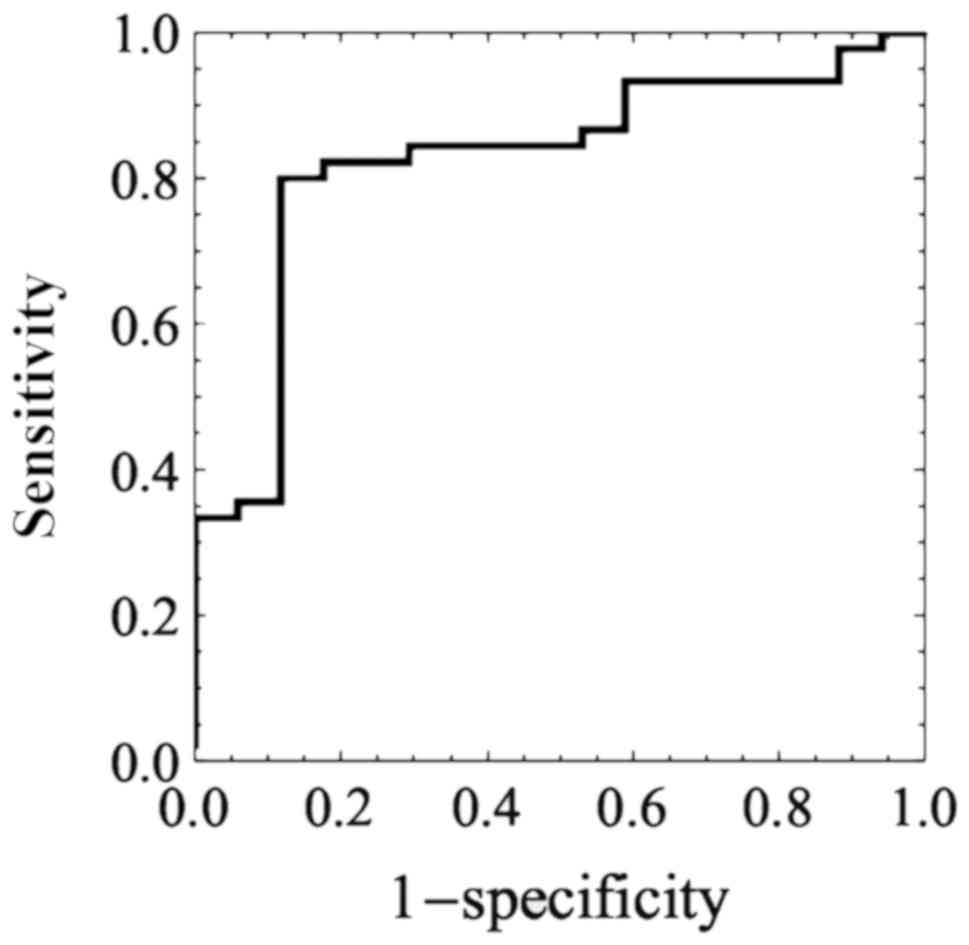
Using confidence score, the area under the
receiver-operating characteristic curve (ROC) of the best
classifier for predicting HSIL was 0.824±0.052 (mean ± SE), and the
95% confidence interval 0.721–0.928. The ROC curve is shown in
Fig. 1. The optimal cut-off point
was 0.692.
Comparison of AI-classifier with
conventional colposcopy
The association among conventional colposcopy, the
AI classifier and pathological results for the test dataset that
was 20% of patients of both pathological HSIL and LSIL diagnosed by
punch biopsy are shown in Tables
IV–VI. Cohen's Kappa (52) coefficients of the conventional
colposcopy and pathological results, the AI classifier and
pathological results, and the conventional colposcopy and the AI
classifier were 0.691, 0.561 and 0.437 (all P<0.0001),
respectively. All were more than moderate agreements (54). Conventional colposcopy showed better
agreement than the AI, although the difference was not significant.
Classification took less than 0.2 sec per image.
| Table IV.Conventional colposcopy diagnosis and
pathological results of the test data set. |
Table IV.
Conventional colposcopy diagnosis and
pathological results of the test data set.
|
| Conventional
colposcopy diagnosis |
|---|
|
|
|
|---|
| Lesion type | HSIL | LSIL |
|---|
| Pathological
HSIL | 39 | 6 |
| Pathological
LSIL | 2 | 15 |
| Table VI.Conventional colposcopy diagnosis and
AI colposcopy diagnosis for test data set. |
Table VI.
Conventional colposcopy diagnosis and
AI colposcopy diagnosis for test data set.
|
| AI colposcopy
diagnosis |
|---|
|
|
|
|---|
|
| HSIL | LSIL |
|---|
| Conventional
colposcopy HSIL | 32 | 9 |
| Conventional
colposcopy LSIL | 7 | 14 |
Discussion
We developed a classifier using deep learning with
convolutional neural networks using images of cervical SILs to
predict the pathological diagnosis. In the present study, the
accuracy values achieved by the classifier and by gynecologic
oncologists were 0.823 and 0.797, respectively (Table III). The sensitivity values of the
classifier and gynecologic oncologists were 0.800 and 0.831,
respectively. The specificity values of the classifier and
gynecologic oncologists were 0.882 and 0.773, respectively. The
accuracy and specificity of the classifier were superior to those
of gynecologic oncologists, although the difference was not
significant. Only moderate agreement was obtained between
conventional colposcopy diagnosis and AI colposcopy diagnosis with
the Kappa value of 0.437. McHugh reported that Cohen suggested 0.41
might be acceptable and the Kappa result be interpreted as follows:
Values ≤0 as indicating no agreement and 0.01–0.20 as none to
slight, 0.21–0.40 as fair, 0.41–0.60 as moderate, 0.61–0.80 as
substantial and 0.81–1.00 (54).
Thus, the Kappa value of 0.437 was acceptable. But, AI colposcopy
that might have a potential would not be an alternative to
conventional colposcopy without further studies.
Several reports have used AI (55) for deep learning with convolutional
neural networks in medicine (56).
The accuracy values of this method with deep learning have been
published and include 0.997 for the histopathological diagnosis of
breast cancer (57), 0.90–0.83 for
the early diagnosis of Alzheimer's disease (58), 0.83 for urological dysfunctions
(59), 0.72 (60) and 0.50 (61) for colposcopy, 0.68–0.70 for
localization of rectal cancer (62),
0.83 for the diagnostic imaging of orthopedic trauma (63), 0.98 for the morphological quality of
blastocysts and evaluation by an embryologist (64), 0.65 for predicting live birth without
aneuploidy from a blastocyst image (65) and 0.64–0.88 for predicting live birth
from a blastocyst image of patients by age (66).
Several studies have reported a limitation of
conventional colposcopy. An investigation of the accuracy of
colposcopically-directed biopsy reported a total biopsy failure
rate, comprising both non-biopsy and incorrect selection of biopsy
site, of 0.20 in CIN1, 0.11 in CIN2 and 0.09 in CIN3 (67). The colposcopic impression of
high-grade CIN had a sensitivity of 0.54 and a specificity of 0.88,
as determined by 9 expert colposcopists in 100 cervigrams (68). The sensitivity of an online
colpophotographic assessment of high-grade disease (CIN2 and CIN3)
by 20 colposcopists was 0.39 (69).
Thus, conventional colposcopy does not provide good sensitivity,
even by colposcopists. By contrast, the accuracy and sensitivity
reported in this study for predicting HSIL from colposcopy images
using deep learning was 0.823 and 0.800, respectively, which
appears favorable. Since the classifier was not trained in
colposcopy findings such as acetowhite epithelium, mosaic,
punctuation, it may recognize some features of cervical SILs by
itself in high-dimensional space. It is possible that the
AI-classifier may recognize features that colposcopists do not,
such as complexity of the shape of the lesion, relative or absolute
brightness of acetowhite, distribution of punctuation density, and
quantitative marginal evaluation of borders. The pathological
results were obtained and defined by punch biopsy in this study, as
it was not ethically recommended for patients with LSIL (CIN1)
diagnosed by colposcopy to undergo conization or hysterectomy. If
the pathological results were defined by conization or
hysterectomy, the advanced lesion would have been be revealed, and
thus both conventional colposcopy and the AI classifier may have
demonstrated different results. In the present study, we only tried
to compare the effectiveness of AI with that of conventional
colposcopy for SIL. When the AI will be used to predict more
advanced diseases, such as squamous cell carcinoma, adenocarcinoma
and AIS, the pathological diagnosis should be provided by not punch
biopsy but conization or hysterectomy.
In clinical practice, it is important for clinicians
to distinguish HSIL from LSIL in biopsy specimens. Further
examination or treatment, such as conization, may be required for
HSIL. When a reliable classifier indicates HSIL from colposcopy
images in clinical practice, the clinician should consider biopsy.
The accuracy values of the classifier and gynecologists for
detecting HSIL were 0.823 and 0.797, respectively. The classifier
might help untrained clinicians to avoid or reduce the risks of
overlooking HSIL. When the AI-classifier can perform higher in
terms of accuracy, sensitivity and specificity for classifying
HSIL/LSIL, clinicians will be able to perform more precise
practice, referencing AI. Furthermore, a gynecologist could reduce
the time and effort it takes to become a colposcopy expert and, as
a result, improve in other skills, training and activities.
The architecture of the neural network has
progressed. The LeNet study published in 1998 (70) consisted of 5 layers. AlexNet,
published in 2012 (38), consisted
of 14 and Google Net, published in 2014 (35), was constructed from a combination of
micro networks. ResNet-50, published in 2015 (41), consisted of modules with a shortcut
process. The Squeeze-and-Excitation Networks were published in 2017
(71). AI used for image recognition
is still being developed. Progress in AI will allow us to achieve
better results. Image information is one of the parameters that
need to be investigated. Only 15×15 pixels are used to detect
cervical cancer (72). In a
colposcopy study (61), it was
reported that the accuracy for images of 150×150 pixels was better
than those for 32×32 or 300×300 pixels. Hence, image size remains
an issue. We used 70×70 and 111×111 pixels for our images, in order
to use the original neural networks and the modified ResNet-50,
respectively. The original conventional neural network was better
than the modified ResNet-50, although not significantly. We believe
that a pixel size of 70×70 falls within the acceptable range.
Regularization values are also important parameters for
constructing a good classifier that avoids overfitting. If the
regularization value is too low, overfitting occurs. If the value
is too large, the classifier will not be trained well. Choosing the
appropriate number of training datasets is also very important. If
the number of training datasets is too high, the accuracy will be
lower and more variances will be observed. The validation dataset,
as well as L2 regularization, also prevent overfitting. The
appropriate number of training datasets must be achieved to obtain
a good classifier. More varied patterns of images may be needed for
datasets. Ordinarily, 500–1,000 images are prepared for each class
during image classification with deep learning (61,73).
Such a large number of datasets will improve the accuracy and
specificity of the classifier with deep learning.
In the present study, a classifier was developed
based on deep learning, which used images of uterine cervical SILs
to predict pathological HSIL/LSIL. Its accuracy was 0.823. Although
further study may be required to validate the classifier, we
demonstrated that AI may have a clinical use in colposcopic
examinations and may provide benefits to both patients and medical
personnel.
Acknowledgements
Not applicable.
Funding
No funding was received.
Availability of data and materials
The datasets generated and/or analyzed during the
present study are not publicly available, since data sharing is not
approved by the Institutional Review Board of Shikoku Cancer Center
(approval no. 2017-81).
Authors' contributions
YM designed the current study, performed AI
programming, produced classifiers by AI, performed statistical
analysis and wrote the manuscript. KT performed clinical
intervention, data entry and collection, designed the current
study, and critically revised the manuscript. TM designed the
current study and critically revised the manuscript.
Ethics approval and consent to
participate
The protocol for the present retrospective study
used fully deidentified patient data and was approved by the
Institutional Review Board of Shikoku Cancer Center (approval no.
2017-81). The study protocol was explained to the patients who
underwent cervical biopsy at the Shikoku Cancer Center from January
1, 2012 to December 31, 2017. Patients were also directed to a
website with additional information, including an opt-out option,
allowing them to not participate. Written informed consent for was
not required, according to the guidance of the Ministry of
Education, Culture, Sports, Science and Technology of Japan.
Patient consent for publication
The current study was explained to the patients who
underwent cervical biopsy at the Shikoku Cancer Center from January
1, 2012 to December 31, 2017. Patients were also directed to a
website with additional information, including an opt-out option
that let them know they had the right to refuse publication.
Competing interests
YM and TM declare that they have no competing
interests. KT reports personal fees from Taiho Pharmaceuticals,
Chugai Pharma, AstraZeneca, Nippon Kayaku, Eisai, Ono
Pharmaceutical, Terumo Corporation and Daiichi Sankyo, outside of
the submitted work.
References
|
1
|
Müller VC and Bostrom N: Future progress
in artificial intelligence: A survey of expert opinion. In:
Fundamental Issues of Artificial IntelligenceSpringer; Berlin: pp.
555–572. 2016
|
|
2
|
Silver D, Schrittwieser J, Simonyan K,
Antonoglou I, Huang A, Guez A, Hubert T, Baker L, Lai M, Bolton A,
et al: Mastering the game of Go without human knowledge. Nature.
550:354–359. 2017. View Article : Google Scholar : PubMed/NCBI
|
|
3
|
Arbyn M, Castellsagué X, de Sanjosé S,
Bruni L, Saraiya M, Bray F and Ferlay J: Worldwide burden of
cervical cancer in 2008. Ann Oncol. 22:2675–2686. 2011. View Article : Google Scholar : PubMed/NCBI
|
|
4
|
Garcia-Arteaga JD, Kybic J and Li W:
Automatic colposcopy video tissue classification using higher order
entropy-based image registration. Comput Biol Med. 41:960–970.
2011. View Article : Google Scholar : PubMed/NCBI
|
|
5
|
Kyrgiou M, Tsoumpou I, Vrekoussis T,
Martin-Hirsch P, Arbyn M, Prendiville W, Mitrou S, Koliopoulos G,
Dalkalitsis N, Stamatopoulos P and Paraskevaidis E: The up-to-date
evidence on colposcopy practice and treatment of cervical
intraepithelial neoplasia: The Cochrane colposcopy and cervical
cytopathology collaborative group (C5 group) approach. Cancer Treat
Rev. 32:516–523. 2006. View Article : Google Scholar : PubMed/NCBI
|
|
6
|
O'Neill E, Reeves MF and Creinin MD:
Baseline colposcopic findings in women entering studies on female
vaginal products. Contraception. 78:162–166. 2008. View Article : Google Scholar : PubMed/NCBI
|
|
7
|
Waxman AG, Chelmow D, Darragh TM, Lawson H
and Moscicki AB: Revised terminology for cervical histopathology
and its implications for management of high-grade squamous
intraepithelial lesions of the cervix. Obstet Gynecol.
120:1465–1471. 2012. View Article : Google Scholar : PubMed/NCBI
|
|
8
|
Darragh TM, Colgan TJ, Cox JT, Heller DS,
Henry MR, Luff RD, McCalmont T, Nayar R, Palefsky JM, Stoler MH, et
al: The lower anogenital squamous terminology standardization
project for HPV-associated lesions: Background and consensus
recommendations from the college of American pathologists and the
American society for colposcopy and cervical pathology. J Low Genit
Tract Dis. 16:205–242. 2012. View Article : Google Scholar : PubMed/NCBI
|
|
9
|
Dreiseitl S and Ohno-Machado L: Logistic
regression and artificial neural network classification models: A
methodology review. J Biomed Inform. 35:352–359. 2002. View Article : Google Scholar : PubMed/NCBI
|
|
10
|
Ben-Bassat M, Klove KL and Weil MH:
Sensitivity analysis in Bayesian classification models:
Multiplicative deviations. IEEE Trans Pattern Anal Mach Intell.
2:261–266. 1980. View Article : Google Scholar : PubMed/NCBI
|
|
11
|
Friedman JH, Baskett F and Shustek LJ: An
algorithm for finding nearest neighbors. IEEE Trans Comput.
24:1000–1006. 1975. View Article : Google Scholar
|
|
12
|
Breiman L: Random forests. Mach Lean.
45:5–32. 2001. View Article : Google Scholar
|
|
13
|
Rumelhart D, Hinton G and Williams R:
Learning representations by back-propagating errors. Nature.
323:533–536. 1986. View
Article : Google Scholar
|
|
14
|
Bengio Y, Courville A and Vincent P:
Representation learning: A review and new perspectives. IEEE Trans
Pattern Anal Mach Intell. 35(1): 798–828. 2013.
|
|
15
|
Fukushima K: Neocognitron: A
self-organizing neural network model for a mechanism of pattern
recognition unaffected by shift in position. Biol Cybern.
36:193–202. 1980. View Article : Google Scholar : PubMed/NCBI
|
|
16
|
Hubel DH and Wiesel TN: Receptive fields
and functional architecture of monkey striate cortex. J Physiol.
195:215–243. 1968. View Article : Google Scholar : PubMed/NCBI
|
|
17
|
Hubel DH and Wiesel TN: Receptive fields
of single neurones in the cat's striate cortex. J Physiol.
148:574–591. 1959. View Article : Google Scholar : PubMed/NCBI
|
|
18
|
Schmidhuber J: Deep learning in neural
networks: An overview. Neural Netw. 61:85–117. 2015. View Article : Google Scholar : PubMed/NCBI
|
|
19
|
LeCun Y, Bottou L, Orr GB and Müller KR:
Efficient BackPropNeural Networks: Tricks of the Trade. Springer;
Berlin: 1998
|
|
20
|
LeCun Y, Bottou L, Bengio Y and Haffner P:
Gradient-based learning applied to document recognition. Proc IEEE.
86:2278–2324. 1998. View
Article : Google Scholar
|
|
21
|
LeCun Y, Boser B, Denker JS, Henderson D,
Howard RE, Hubbard W and Jackel LD: Backpropagation applied to
handwritten zip code recognition. Neural Computation. 1:541–551.
1989. View Article : Google Scholar
|
|
22
|
Serre T, Wolf L, Bileschi S, Riesenhuber M
and Poggio T: Robust object recognition with cortex-like
mechanisms. IEEE Trans Pattern Anal Mach Intell. 29:411–426. 2007.
View Article : Google Scholar : PubMed/NCBI
|
|
23
|
Wiatowski T and Bölcskei H: A mathematical
theory of deep convolutional neural networks for feature
extraction. IEEE Trans Inf Theory. 64:1845–1866. 2018. View Article : Google Scholar
|
|
24
|
Srivastava N, Hinton G, Krizhevsky A,
Sutskever I and Salakhutdinov R: Dropout: A simple way to prevent
neural networks from overfitting. J Mach Lean Res. 15:1929–1958.
2014.
|
|
25
|
Nowlan SJ and Hinton GE: Simplifying
neural networks by soft weight-sharing. Neural Comput. 4:473–493.
1992. View Article : Google Scholar
|
|
26
|
Bengio Y: Learning deep architectures for
AI. Found Trends Mach Lean. 2:1–127. 2009. View Article : Google Scholar
|
|
27
|
Mutch J and Lowe DG: Object class
recognition and localization using sparse features with limited
receptive fields. Int J Comput Vision. 80:45–57. 2008. View Article : Google Scholar
|
|
28
|
Neal RM: Connectionist learning of belief
networks. Artificial Intell. 56:71–113. 1992. View Article : Google Scholar
|
|
29
|
Ciresan D, Meier U, Masci J, Gambardella
LΜ and Schmidhuber J: Flexible, high performance convolutional
neural networks for image classification. IJCAI Proc Int Joint Conf
Artificial Intell. 22:1237–1242. 2011.
|
|
30
|
Scherer D, Müller A and Behnke S:
Evaluation of pooling operations in convolutional architectures for
object recognition. In: Artificial Neural Networks (ICANN)
2010Lecture Notes in Computer Science. Diamantaras K, Duch W and
Iliadis LS: Springer; Berlin: pp. 92–101. 2010, View Article : Google Scholar
|
|
31
|
Huang FJ and LeCun Y: Large-scale learning
with SVM and convolutional for generic object categorization.
Computer vision and pattern recognition. IEEE Comput Soc Conf.
1:284–291. 2006.
|
|
32
|
Jarrett K, Kavukcuoglu K, Ranzato MA and
LeCun Y: What is the best multi-stage architecture for object
recognition?2009 IEEE 12th International Conference on Computer
Vision. ICCV 2009; Kyoto, Japan: pp. 2146–2153. 2009, View Article : Google Scholar
|
|
33
|
Zheng Y, Liu Q, Chen E, Ge Y and Zhao JL:
Time series classification using multi-channels deep convolutional
neural networksLi F, Li G, Hwang S, Yao B and Zhang Z: Web-Age
Information Management, WAIM 2014. Springer; Cham: pp. 298–310.
2014
|
|
34
|
Mnih V, Kavukcuoglu K, Silver D, Rusu AA,
Veness J, Bellemare MG, Graves A, Riedmiller M, Fidjeland AK,
Ostrovski G, et al: Human-level control through deep reinforcement
learning. Nature. 518:529–533. 2015. View Article : Google Scholar : PubMed/NCBI
|
|
35
|
Szegedy C, Liu W, Jia Y, Sermanet P, Reed
S, Anguelov D, et al: Going deeper with convolutions. IEEE
Conference on Computer Vision and Pattern Recognition (CVPR). 1–9.
2015.
|
|
36
|
Glorot X, Bordes A and Bengio Y: Deep
sparse rectifier neural networks. Proc Fourteenth Int Conf
Artificial Intell Stat. 315–323. 2011.
|
|
37
|
Nair V and Hinton G: Rectified linear
units improve restricted Boltzmann machines. Proc Int Conf Mach
Lean. 807–814. 2010.
|
|
38
|
Krizhevsky A, Sutskever I and Hinton GE:
Imagenet classification with deep convolutional neural networks.
Adv Neural Inf Proc Syst. 1097–1105. 2012.
|
|
39
|
Bridle JS: Probabilistic interpretation of
feedforward classification network outputs, with relationships to
statistical pattern recognitionNeurocomputing. Soulié FF and
Hérault J: Springer; Berlin: 1990, View Article : Google Scholar
|
|
40
|
He K, Zhang X, Ren S and Sun J: Deep
residual learning for image recognition. arXiv: 1512.03385.
2015.
|
|
41
|
Russakovsky O, Deng J, Su H, Krause J,
Satheesh S, Ma S, Huang Z, Karpathy A, Khosla A, Bernstein M and
Berg AC: Imagenet large scale visual recognition challenge. Int J
Comput Vision. 115:211–252. 2015. View Article : Google Scholar
|
|
42
|
Lin, Tsung-Yi, Michael Maire, Serge
Belongie, James Hays, Pietro Perona, Deva Ramanan, Piotr Dollár and
C. Lawrence Zitnick: Microsoft coco: Common objects in contextIn
European conference on computer vision. pp. 740–755. Springer;
Cham: 2014
|
|
43
|
Kohavi R: A study of cross-validation and
bootstrap for accuracy estimation and model selection. Proc Int
Joint Conf Artificial Intell. 2:1137–1143. 1995.
|
|
44
|
Schaffer C: Selecting a classification
method by cross-validation. Mach Lean. 13:135–143. 1993. View Article : Google Scholar
|
|
45
|
Refaeilzadeh P, Tang L and Liu H:
Cross-validationEncyclopedia of Database Systems. Liu L and Özsu
MT: Springer; New York: 2009
|
|
46
|
Yu L, Chen H, Dou Q, Qin J and Heng PA:
Automated melanoma recognition in dermoscopy images via very deep
residual networks. IEEE Trans Med Imaging. 36:994–1004. 2017.
View Article : Google Scholar : PubMed/NCBI
|
|
47
|
Caruana R, Lawrence S and Giles CL:
Overfitting in neural nets: Backpropagation, conjugate gradient,
and early stopping. Adv Neural Inf Proc Syst. 402–408. 2001.
|
|
48
|
Baum EB and Haussler D: What size net
gives valid generalization? Neural Comput. 1:151–160. 1989.
View Article : Google Scholar
|
|
49
|
Geman S, Bienenstock E and Doursat R:
Neural networks and the bias/variance dilemma. Neural Comput.
4:1–58. 1992. View Article : Google Scholar
|
|
50
|
Krogh A and Hertz JA: A simple weight
decay can improve generalization. Adv Neural Inf Proc Syst.
4:950–957. 1992.
|
|
51
|
Moody JE: The effective number of
parameters: An analysis of generalization and regularization in
nonlinear learning systems. Adv Neural Inf Proc Syst. 4:847–854.
1992.
|
|
52
|
Cohen J: A coefficient of agreement for
nominal scales. Educ Psychol Meas. 20:37–46. 1960. View Article : Google Scholar
|
|
53
|
Youden WJ: Index for rating diagnostic
tests. Cancer. 3:32–35. 1950. View Article : Google Scholar : PubMed/NCBI
|
|
54
|
McHugh ML: Interrater reliability: the
kappa statistic. Biochem Med (Zagreb). 22:276–282. 2012. View Article : Google Scholar : PubMed/NCBI
|
|
55
|
Miyagi Y, Fujiwara K, Oda T, Miyake T and
Coleman RL: Development of new method for the prediction of
clinical trial results using compressive sensing of artificial
intelligence. J Biostat Biometric. 3:2022018.
|
|
56
|
Abbod MF, Catto JW, Linkens DA and Hamdy
FC: Application of artificial intelligence to the management of
urological cancer. J Urol. 178:1150–1156. 2007. View Article : Google Scholar : PubMed/NCBI
|
|
57
|
Litjens G, Sánchez CI, Timofeeva N,
Hermsen M, Nagtegaal I, Kovacs I, Hulsbergen-van de Kaa C, Bult P,
van Ginneken B and van der Laak J: Deep learning as a tool for
increased accuracy and efficiency of histopathological diagnosis.
Sci Rep. 6:262862016. View Article : Google Scholar : PubMed/NCBI
|
|
58
|
Ortiz A, Munilla J, Górriz JM and Ramírez
J: Ensembles of deep learning architectures for the early diagnosis
of the Alzheimer's disease. Int J Neural Syst. 26:16500252016.
View Article : Google Scholar : PubMed/NCBI
|
|
59
|
Gil D, Johnsson M, Chamizo JMG, Paya AS
and Fernandez DR: Application of artificial neural networks in the
diagnosis of urological disfunctions. Expert Syst Appl.
36:5754–5760. 2009. View Article : Google Scholar
|
|
60
|
Simões PW, Izumi NB, Casagrande RS, Venson
R, Veronezi CD, Moretti GP, da Rocha EL, Cechinel C, Ceretta LB,
Comunello E, et al: Classification of images acquired with
colposcopy using artificial neural networks. Cancer Inform.
13:119–124. 2014. View Article : Google Scholar : PubMed/NCBI
|
|
61
|
Sato M, Horie K, Hara A, Miyamoto Y,
Kurihara K, Tomio K and Yokota H: Application of deep learning to
the classification of images from colposcopy. Oncol Lett.
15:3518–3523. 2018.PubMed/NCBI
|
|
62
|
Trebeschi S, van Griethuysen JJM,
Lambregts DMJ, Lahaye MJ, Parmar C, Bakers FCH, Peters NHGM,
Beets-Tan RGH and Aerts HJWL: Deep learning for fully-automated
localization and segmentation of rectal cancer on multiparametric
MR. Sci Rep. 7:53012017. View Article : Google Scholar : PubMed/NCBI
|
|
63
|
Olczak J, Fahlberg N, Maki A, Razavian AS,
Jilert A, Stark A, Sköldenberg O and Gordon M: Artificial
intelligence for analyzing orthopedic trauma radiographs. Acta
Orthop. 88:581–586. 2017. View Article : Google Scholar : PubMed/NCBI
|
|
64
|
Khosravi P, Kazemi E, Zhan Q, Toschi M,
Makmsten J, Hickman C, Meseguer M, Rosenwaks Z, Elemento O,
Zaninovic N and Hajirasouliha I: Robust automated assessment of
human blastocyst quality using deep learning. bioRxiv 394882.
2018.
|
|
65
|
Miyagi Y, Habara T, Hirata R and Hayashi
N: Feasibility of artificial intelligence for predicting live birth
without aneuploidy from a blastocyst image. Reprod Med Biol.
18:204–211. 2019. View Article : Google Scholar : PubMed/NCBI
|
|
66
|
Miyagi Y, Habara T, Hirata R and Hayashi
N: Feasibility of deep learning for predicting live birth from a
blastocyst image in patients classified by age. Reprod Med Biol.
18:190–203. 2019. View Article : Google Scholar : PubMed/NCBI
|
|
67
|
Sideri M, Garutti P, Costa S, Cristiani P,
Schincaglia P, Sassoli de Bianchi P, Naldoni C and Bucchi L:
Accuracy of colposcopically directed biopsy: Results from an online
quality assurance programme for colposcopy in a population-based
cervical screening setting in Italy. BioMed Res Int.
2015:6140352015. View Article : Google Scholar : PubMed/NCBI
|
|
68
|
Sideri M, Spolti N, Spinaci L, Sanvito F,
Ribaldone R, Surico N and Bucchi L: Interobserver variability of
colposcopic interpretations and consistency with final histologic
results. J Low Genit Tract Dis. 8:212–216. 2004. View Article : Google Scholar : PubMed/NCBI
|
|
69
|
Massad LS, Jeronimo J, Katki HA and
Schiffman M; National Institutes of Health/American Society for
Colposcopy and Cervical Pathology Research Group, : The accuracy of
colposcopic grading for detection of high grade cervical
intraepithelial neoplasia. J Low Genit Tract Dis. 13:137–144. 2009.
View Article : Google Scholar : PubMed/NCBI
|
|
70
|
LeCun Y, Haffner P, Bottou L and Bengio Y:
Object recognition with gradient-based learning. In Shape, contour
and grouping in computer visionSpringer; Berlin, Heidelberg:
1999
|
|
71
|
Hu J, Shen L and Sun G:
Squeeze-and-excitation networks. Proceedings of the IEEE conference
on computer vision and pattern recognition. 7132–7141. 2018.
|
|
72
|
Kudva V, Prasad K and Guruvare S:
Automation of detection of cervical cancer using convolutional
neural networks. Crit Rev Biomed Eng. 46:135–145. 2018. View Article : Google Scholar : PubMed/NCBI
|
|
73
|
Esteva A, Kuprel B, Novoa RA, Ko J,
Swetter SM, Blau HM and Thrun S: Dermatologist-level classification
of skin cancer with deep neural networks. Nature. 542:115–118.
2017. View Article : Google Scholar : PubMed/NCBI
|