Introduction
The increasing use and application of broad-spectrum
antibiotics has led to increased reports of bacterial drug
resistance and clinical infection. At present, drug-resistant
bacteria, including methicillin-resistant Staphylococcus
aureus, multidrug-resistant Streptococcus pneumoniae,
vancomycin-resistant Enterococcus, multidrug-resistant
Baumanii, Klebsiella pneumoniae, Escherichia
coli and Pseudomonas aeruginosa are increasingly being
detected in clinical and laboratory settings (1). Appearance of pan-resistant bacteria,
including Cray Borrelia, which produces New Delhi Metallo
β-lactamase-1, an enzyme responsible for generating resistance
(2), is causing an even greater
challenge to humans. Therefore, study of clinical bacterial drug
resistance and mechanisms and the development of solutions to
bacterial resistance is important. Previously, Gram-negative
bacilli AmpC β-lactamase was understood to be generated by
chromosome mediation only. However, Papanicolaou et
al(3) identified
plasmid-mediated AmpC β-lactamase (MIR-1) in a strain of Cray
Borrelia bacterium isolated from a patient in Rhode Island
Hospital (USA) in 1989. Following this, the plasmid-mediated AmpC
enzyme was rapidly identified in a number of other countries. At
present, more than 20 plasmid-mediated AmpC enzymes have been
identified. The plasmid-mediated AmpC enzyme is resistant to
antibiotics, enabling the plasmid to continue to express the
protein at high levels and carry multiple drug-resistant genes.
Plasmid-mediated drug resistance is transmitted between members of
the same or different bacterial species. Transmission is rapid and
performed at a range of distances. Therefore, understanding of the
mechanism and elucidation of solutions to plasmid-mediated AmpC
enzyme resistance is crucial.
P. aeruginosa is one of the main bacterial
strains associated with hospital infections, including burns,
cystic pulmonary fibrosis and ventilator-associated infections. In
addition, P. aeruginosa is resistant to multiple antibiotics
and is associated with high rates of mortality (4–6). A
number of drug resistance mechanisms have been identified in P.
aeruginosa, including production of β-lactamase
(extended-spectrum β-lactamases or AmpC) (7), alteration of penicillin-binding
protein expression (8), formation
of biomembranes, expression of cytomembrane permeability barriers
(9,10) or active multidrug efflux pumps,
including mex-ABoprM (11) and
alterations in channel protein expression, including oprD (11,12).
At present, P. aeruginosa plasmid-mediated AmpC β-lactamase
has not been studied. The present study aimed to investigate
plasmid-mediated AmpC in clinically-isolated P. aeruginosa
through identification of the genetype of a drug-resistant strain,
to aid clinical antibiotic applications.
Materials and methods
Specimens
In the present study, 108 strains of P.
aeruginosa were clinically isolated from the sputum, urine,
feces, ascites, hydrothorax, arthroedema, cerebrospinal fluid and
other specimens of hospitalized patients in the ICU, respiratory,
cardiology, infection, burns and pediatrics departments in the
First Affiliated Hospital of Xin Xiang Medical University and Xiang
Ya Hospital Center South University. Bacterial isolation, culture
and identification was conducted according to the National Guide to
Clinical Laboratory Procedures (2nd edition) and bacterial
identification was conducted using an automatic bacterial
identification analyzer. E. coli ATCC25922 and P.
aeruginosa ATCC27853 were used as quality-control strains and
Aerobacter cloacae 029M was the used as a positive control
strain as it is known to express the AmpC enzyme. The study was
evaluated and approved by the ethics committees of Xin Xiang
Medical University (XinXiang, China) and Xiang-Ya Medical College
of Central South University (Changsha, China). Informed patient
consent was obtained from the patient or the patient’s family.
Primary screening test of specimens
Isolated P. aeruginosa was inoculated onto a
blood plate and cultured at 37°C overnight. The following day,
cultures were suspended in 0.9% stroke-physiological saline
solution (Maxwell turbidity, 0.5) and coated uniformly onto M-H
plates with sterile cotton swabs. Cefoxitin (FOX), ceftazidime
(CAZ), cefotaxime/clavulanic acid (CTX/CA) and imipenem (IMP)
drug-sensitive slips were symmetrically placed onto the M-H plates
with bacterial suspension using sterile forceps and cultured
overnight at 37°C. The next day, bacteriostatic rings of
drug-sensitive slips were observed and the diameters of
bacteriostatic rings were measured with a vernier caliper.
According to criteria prepared by the National Clinical Laboratory
Standardization Committee of America (13), bacteriostatic ring diameters of
≤18, ≤21, ≤22 and ≥13 mm for FOX, CTX/CA, CAZ and IMP,
respectively, were indicative of strains expressing the AmpC
enzyme.
Preparation of AmpC enzyme
P. aeruginosa stains identified by screening
to express the AmpC enzyme were inoculated into 10 ml LB broth and
cultured overnight in a thermostat shaker at a speed of 80–100 rpm
at 37°C. Subsequently, the culture mixture was centrifuged in a
refrigerated centrifuge at 4°C to remove the supernatant. Bacterial
pellets were frozen for 30 min at −80°C and then thawed in a water
bath. The freeze-thaw cycle was performed 5 times. Following this,
bacteria were suspended in 1.5 ml PBS buffer (0.01 M) in an EP
tube, mixed uniformly by agitation and centrifuged at low
temperature to obtain the supernatant. The supernatant contained
the AmpC enzyme and was filtered using a 0.22-μm microporous
membrane. Next, the filtrate was inoculated onto the blood plate
and cultured overnight at 37°C. Bacterial growth was observed and
negative filtrates were stored at −20°C.
Three-dimensional test
E. coli strain ATCC25922 was suspended in
solution (Maxwell turbidity, 0.5) and uniformly coated with sterile
cotton swabs onto M-H plates. A FOX drug-sensitive slip (30 μg) was
applied to the center of the plate and a sterile surgical blade was
used to cut a small groove with a width of ~1–2 cm, 3 mm away from
the slip edge. In each small groove, 10–20 μl AmpC enzyme extract
of each test bacterium was added, avoiding extract overflow. The
extract was cultured overnight at 37°C for 18–24 h prior to
observation. The Bacillus levans (0.29 M) strain was used as
a positive control as it is known to produce the AmpC enzyme. If
the cefoxitin three-dimensional test shows a positive result, it
suggests that the test bacterium generated the AmpC enzyme.
Plasmid extraction and
electrophoresis
P. aeruginosa plasmids with positive
three-dimensional test results were extracted by the SDS alkaline
lysis method and then run on a 1% agarose gel. Electrophoresis was
performed using 0.5X TBE electrophoresis buffer and images were
captured under UV lamp.
PCR amplification
Plasmid-mediated AmpC β-lactamase-positive strains
were divided into 6 populations according to chromosome source: i)
ACC (Hafinia alvei); ii) FOX (unknown source); iii) MOX
(unknown source); iv) DHA (Morganella morganii); v) CIT
(Citrobacter freundii); or vi) EBC population (A.
cloacae). Primer design was performed using AmpC enzyme gene
sequences identified in previous reports and primer 5.0 software
(Table I).
 | Table IAmpC enzyme primers. |
Table I
AmpC enzyme primers.
| AmpC enzyme | Primer | Sequence (5′-3′) | Length (bp) | Genbank no. |
|---|
| MOX-1 MOX2 CMY-1 | MOX | GCT TGA GCG GTA AAC
GAG TG | 171 | D 13304 |
| CMY-8 CMY-11 | | GAA TGT GCT GCC TGG
GTG | | |
| LAT-1-LAT-4 | CIT | AGG GAT TAG GCT GGG
AGA TG | 251 | X 78117 |
| CMY-7 BIL-1 | | GAC ACG GAC AGG GTT
AGG ATA G | | |
| DHA-1 DHA-2 | DHA | CCG TCT CCG TAA AGG
GTA AGC | 396 | Y 1641 |
| | CGG TCA GAG CAC CAA
ACA GG | | |
| ACC | ACC | GTT ATC CGT GAT TAC
CTG TC | 248 | AJ 133121 |
| | TAC TCA GCG AAC CCA
CTT | | |
| MIR-1 ACT-1 | EBC | GAA ATC CGC CAC ATC
CTG | 462 | M 37839 |
| | CCG TAA TAG CAG TTC
AAG GGA | | |
| FOX-1-FOX-5b | FOX | CT ACA GTG CGG GTG
GTT | 297 | X 77455 |
| | CGG GCT TAT CTT CCT
TCG | | |
PCR system
Total PCR volume was 50 μl and contained the
following: 5 μl 10X PCR buffer solution, 1 μl dNTP (10 mmol/l), 2
μl positive and reverse primers (100 pmol/μl), 4 μl
MgCl2 (25 mmol/l), 0.4 μl Taq enzyme (2 units), 4
μl template and 31.6 μl ddH2O. For each run, 6 reactions
were performed. With the exception of primers, all other reaction
components and volumes were constant. PCR conditions were as
follows: initial denaturation for 3 min at 94°C; 30 cycles of
denaturation for 30 sec at 94°C, annealing for 30 sec at 58°C,
extension for 30 sec at 72°C; and a final extension for 2 min at
72°C. Agarose gel electrophoresis of the PCR amplification products
was performed and images were captured using a gel imaging
instrument.
Sequencing and analysis
Sequencing of PCR amplification products was
performed by the Shanghai Biological Engineering Technology and
Services Co., Ltd. (Shanghai, China). PubMed BLAST sequence
alignments were analyzed with DNAssist software.
Results
Three-dimensional test
Primary screening tests of K-B drug-sensitive slips
of 108 strains of clinically-isolated P. aeruginosa
demonstrated that 30 strains were AmpC enzyme-positive.
Three-dimensional test of FOX confirmed that 28 strains were AmpC
enzyme-positive.
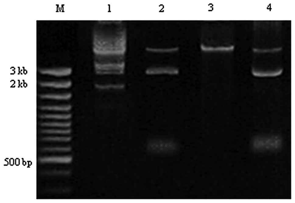
Electrophoresis
The 28 positive strains of P. aeruginosa were
analyzed by electrophoresis following plasmid extraction.
Electrophoretic analysis revealed bands in 22 strains. Single and
double bands were identified corresponding to 2 kb and/or 5 kb.
Single and double bands were observed in 7 and 15 strains,
respectively (Fig. 1).
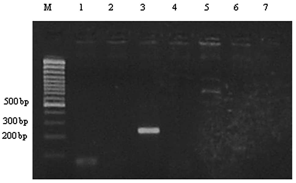
PCR amplification
Purified plasmid DNA was used for PCR amplification
templates. For each DNA template, 6 reactions were performed. An
amplification product was obtained using one pair of primers of a
total of 6 primer pairs. This primer was of CIT type and the target
length was 251 bp (Fig. 2). Within
22 strains of plasmid-positive P. aeruginosa, a single
purified PCR amplification product was positive for this band.
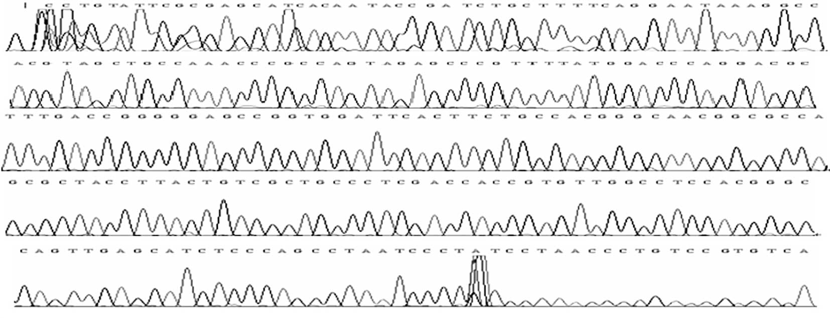
Sequencing analysis
The positive strain revealing a target band of 251
bp was sequenced. The sequencing results are shown in Fig. 3.
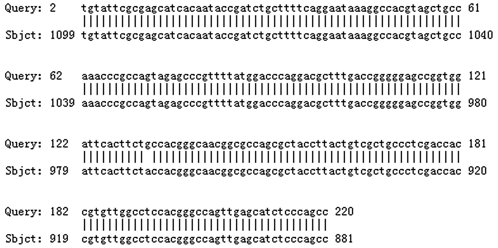
Homology comparison
The sequenced product (2–220 bp) was identified to
exhibit 99.5% (218/219) homology to the A. cloacae K9973
β-lactamase AmpC gene (881–1099 bp) The total length of the
β-lactamase AmpC gene sequence in A. cloacae is 1165 bp
(Fig. 4).
Discussion
P. aeruginosa is one of the main bacterial
species responsible for severe hospital infections (14) and is resistant to a number of
antibacterial drugs. P. aeruginosa has several
drug-resistant mechanisms: 1) the generatation of β-lactamase
enzymes; 2) efflux pumps (e.g. mex2oprM); 3) target-site or outer
membrane modifications. Multiple drug resistance is usually the
result of the chromosomal and plasmid-encoded AmpC enzymes
(15–17). Drug resistance cannot currently be
detected by routine antibiotic sensitivity tests and the detection
of the process is complex (18).
At present, no universal test exists for the detection of AmpC
β-lactamase; however, the three-dimensional test was previously
reported to be the most suitable detection method available
(19).
In the present study, 108 strains of P.
aeruginosa were screened using K-B drug-sensitive slips. An
AmpC enzyme-positive result was observed in 30 strains and the
three-dimensional test of FOX was performed, identifying 28 strains
as AmpC enzyme-positive. The detection coincidence rate of the two
methods for AmpC enzyme-positive stains was 93.3%.
The three-dimensional test identified 28 AmpC
enzyme-positive strains of P. aeruginosa. Following this,
electrophoresis of plasmid extracts identified representative
plasmid bands in 22 strains. Six strains were negative and had lost
or did not carry plasmids initially. PCR amplification analysis
identified positive plasmid-mediated AmpC enzyme expression in a
single strain, demonstrating that the plasmid-mediated AmpC enzyme
was present in P. aeruginosa; however, the proportion of
plasmid-positive strains carrying the AmpC enzyme gene was low. It
is possible that the P. aeruginosa strains that the
three-dimensional test showed as positive could produce the
majority of the chromosomal-mediated AmpC enzyme.
Previous studies on plasmid-mediated AmpC
β-lactamase are largely restricted to the bacterila strains A.
cloacae and Cray Borrelia(20–22).
A limited number of studies have analyzed drug resistance in P.
aeruginosa plasmid-mediated AmpC β-lactamase. In 2003, Shahid
et al(23) isolated P.
aeruginosa strains exhibiting plasmid-mediated AmpC enzyme from
burns patients in an intensive care hospital in Northern India. In
the domestic environment, a single strain of E. coli was
previously observed to exhibit ACT-1 plasmid AmpC enzyme. However,
no domestic reports on P. aeruginosa plasmid-mediated AmpC
enzyme expression currently exist. The present study identified a
single strain of P. aeruginosa plasmid-mediated AmpC enzyme
expression using plasmid extraction, PCR amplification, product
sequencing and BLAST homology comparison. A 99.5% homology with the
A. cloacae K9973-β-lactamase AmpC gene sequence was
identified. The overall length of A. cloacae
K9973-β-lactamase AmpC gene sequence was 1,165 bp, consistent with
the gene sequence of A. cloacae K9973-generating CMY-7 AmpC
enzyme previously identified by Lee et al(24). The present study is the first
reported identification of plasmid-mediated AmpC β-lactamase
expression in P. aeruginosa.
References
|
1
|
Lister PD, Wolter DJ and Hanson ND:
Antibacterial-resistant Pseudomonas aeruginosa: clinical
impact and complex regulation of chromosomally encoded resistance
mechanisms. Clin Microbiol Rev. 22:582–610. 2009.PubMed/NCBI
|
|
2
|
Yong D, Toleman MA, Giske CG, et al:
Characterization of a new metallo-lactamase gene, bla NDM-1 and a
novel erythromycin esterase gene carried on a unique genetic
structure in Klebsiella pneumoniae sequence type 14 from
India. Antimicrob Agents Chemother. 53:5046–5054. 2009. View Article : Google Scholar : PubMed/NCBI
|
|
3
|
Papanicolaou GA, Medeiros AA and Jacoby
GA: Novel plasmid-mediated beta-lactamase (MIR-1) conferring
resistance to oxyimino- and alpha-methoxy beta-lactams in clinical
isolates of Klebsiella pneumoniae. Antimicrob Agents
Chemother. 34:2200–2209. 1990. View Article : Google Scholar : PubMed/NCBI
|
|
4
|
Driscoll JA, Brody SL and Kollef MH: The
epidemiology, pathogenesis and treatment of Pseudomonas
aeruginosa infections. Drugs. 67:351–368. 2007. View Article : Google Scholar : PubMed/NCBI
|
|
5
|
Obritsch MD, Fish DN, MacLaren R and Jung
R: National surveillance of antimicrobial resistance in
Pseudomonas aeruginosa isolates obtained from intensive care
unit patients from 1993 to 2002. Antimicrob Agents Chemother.
48:4606–4610. 2004.PubMed/NCBI
|
|
6
|
Zhanel GG, DeCobry M, Adam H, et al:
Prevalence of antimicrobial-resistant pathogens in Canadian
hospitals: results of the Canadian Ward Surveillance Study (CANWARD
2008). Antimicrob Agents Chemother. 54:4684–4693. 2010. View Article : Google Scholar : PubMed/NCBI
|
|
7
|
Weldhagen GF, Poirel L and Nordmann P:
Ambler class A extended-spectrum beta-lactamases in Pseudomonas
aeruginosa: novel developments and clinical impact. Antimicrob
Agents Chemother. 47:2385–2392. 2003. View Article : Google Scholar : PubMed/NCBI
|
|
8
|
Moyá B, Zamorano L, Juan C, Ge Y and
Oliver A: Affinity of the new cephalosporin CXA-101 to
penicillin-binding proteins of Pseudomonas aeruginosa.
Antimicrob Agents Chemother. 54:3933–3937. 2010.PubMed/NCBI
|
|
9
|
Burmolle M, Thomsen TR, Fazli M, et al:
Biofilmsin chronic infections - a matter of opportunity -
monospecies biofilms in multispecies infections. FEMS Immunol Med
Microbiol. 59:324–336. 2010.PubMed/NCBI
|
|
10
|
del Pozo JL and Patel R: The challenge of
treating biofilm-associated bacterial infections. Clin Pharmacol
Ther. 82:204–209. 2007.PubMed/NCBI
|
|
11
|
Pfeifer Y, Cullik A and Witte W:
Resistance to cephalosporins and carbapenems in gram-negative
bacterial pathogens. Int J Med Microbiol. 300:371–379. 2010.
View Article : Google Scholar : PubMed/NCBI
|
|
12
|
Nikaido H: Outer membrane barrier as a
mechanism of antimicrobial resistance. Antimicrob Agents Chemother.
33:1831–1836. 1989. View Article : Google Scholar : PubMed/NCBI
|
|
13
|
Wikler MA, Cockerill FR, Craig WA, et al:
Methods for Dilution Antimicrobial Susceptibility Tests for
Bacteria that Grow Aerobically. 7th edition. Clinical and
Laboratory Standards Institute; Wayne, PA: 2006
|
|
14
|
Vincent JL: Nosocomial infections in adult
intensive care units. Lancet. 361:2068–2077. 2003. View Article : Google Scholar : PubMed/NCBI
|
|
15
|
Carmeli Y, Troillet N, Eliopoulos GM and
Samore MS: Emergence of antibiotic-resistant Pseudomonas
aeruginosa: comparison of risk associated with different
antipseudomonal agents. Antimicrob Agents Chemother. 43:1379–1382.
1999.PubMed/NCBI
|
|
16
|
Cavallo JD, Fabre R, Leblanc F,
Nicolas-Chanoine MH and Thabaut A: Antibiotic susceptibility and
mechanisms of beta-lactam resistance in 1310 strains of
Pseudomonas aeruginosa: a French multicentre study (1996). J
Antimicrob Chemother. 46:133–136. 2000. View Article : Google Scholar : PubMed/NCBI
|
|
17
|
Livermore DM: Multiple mechanisms of
antimicrobial resistance in Pseudomonas aeruginosa: our
worst nightmare? Clin Infect Dis. 34:634–640. 2002. View Article : Google Scholar : PubMed/NCBI
|
|
18
|
Thomson KS: Controversies about
extended-spectrum and AmpC beta-lactamases. Emerg Infect Dis.
7:333–336. 2001. View Article : Google Scholar : PubMed/NCBI
|
|
19
|
Tan TY, Ng LS, He J, Koh TH and Hsu LY:
Evaluation of screening methods to detect plasmid mediated AmpC in
Escherichia coli, Klebsiella pneumoniae and
Proteus mirabilis. Antimicrob Agents Chemother. 53:146–149.
2009. View Article : Google Scholar : PubMed/NCBI
|
|
20
|
Abdalhamid B, Wickman PA and Hanson ND:
Correlation of ampC induction with PBP binding in Enterobacter
cloacae. In: 45th Intersci Conf Antimicrob Agents Chemother;
Washington DC. pp. C1–2211. 2005
|
|
21
|
Kuga A, Okamoto R and Inoue M: AmpR gene
mutations that greatly increase class C beta-lactamase activity in
Enterobacter cloacae. Antimicrob Agents Chemother.
44:561–567. 2000. View Article : Google Scholar : PubMed/NCBI
|
|
22
|
Pfeifer Y, Cullik A and Witte W:
Resistance to cephalosporins and carbapenems in Gram-negative
bacterial pathogens. Int J Med Microbiol. 300:371–379. 2010.
View Article : Google Scholar : PubMed/NCBI
|
|
23
|
Shahid M, Malik A and Sheeba:
Multidrug-resistant Pseudomonas aeruginosa strains
harbouring R-plasmids and AmpC beta-lactamases isolated from
hospitalised burn patients in a tertiary care hospital of North
India. FEMS Microbiol Lett. 228:181–186. 2003.
|
|
24
|
Lee SH, Kim JY, Shin SH, et al:
Dissemination of SHV-12 and characterization of new AmpC-type
beta-lactamases genes among clinical isolates of
Enterobacter Species in Korea. J Clin Microbiol.
41:2477–2482. 2003. View Article : Google Scholar : PubMed/NCBI
|