Introduction
Adenoid cystic carcinoma (AdCC) is a rare major and
minor salivary gland malignancy characterized by cellular and
histopathological heterogeneity and a high incidence of distant
metastasis in its early stage and local recurrence (1). With a high aggressive potential, AdCC
is able to invade blood vessels and nerves even at an early stage
(2). The current preventive and
therapeutic methods for AdCC include chemotherapy, targeted agents
and surgery. Unfortunately, these therapies only offer partial
benefits and rarely improve the outcome (3). Therefore, novel therapeutic
strategies are urgently required.
Epidermal growth factor receptor (EGFR) is a
membrane-bound receptor. EGFR belongs to the ErbB family of
receptors, which comprises EGFR (ErbB1), HER2/neu (ErbB2), ErbB3
and ErbB4 (4). EGFR is frequently
overexpressed and mutated in various types of tumor, including lung
cancer, oral squamous cell carcinoma and breast cancer (5,6).
Anti-EGFR therapeutic approaches, specifically receptor blocking
monoclonal antibodies and small molecule tyrosine kinase
inhibitors, prolong tumor stabilization (7). EGFR-signaling pathways are also
implicated in cell survival, proliferation, apoptotic resistance
and invasion (8). Although EGFR
has been extensively investigated in the context of AdCC, the
association between EGFR and angiogenesis remains to be
elucidated.
Angiogenesis is considered to be an important event
during the progression of AdCC, due to the fact that blood vessels
provide the nutrients to support tumor growth and also provide an
entry site into the circulation for cancerous cells that have
detached from the tumor mass, leading to distant metastasis
(9). In contrast with normal
vessels, vessels in tumors exhibit different characteristics. They
are tortuous and dilated with excessive branching, shunts and have
an uneven diameter (10). The
walls of these tumor vessels are characterized by the absence of or
discontinuous basement membrane and widened interendothelial
junctions (11). With these
defects, tumor vessels exhibit high vascular permeability and
contribute to tumor metastasis (9). Generally, the adhesion molecule CD31
(platelet endothelial cell adhesion molecule) is used as a marker
to indicate the presence of blood vessels. However, it has been
reported that tumor vessels also highly express melanoma cell
adhesion molecule (CD146), a member of the immunoglobulin gene
superfamily (12). CD146 is
expressed in >90% of cutaneous melanomas (13) and previous studies have
demonstrated that the expression level of CD146 is associated with
invasion and predicted metastatic potential of melanoma cells as
well as AdCC (14,15). Genetic and pharmacological studies
have also revealed that CD146 is implicated in important distinct
aspects of biological processes in blood vessels and may correspond
to endothelial permeability, therefore it may be used as a
biomarker for pathological angiogenesis (16,17).
Hypoxia-inducible factor-1α (HIF-1α), the main transcription factor
involved in angiogenesis, was also analyzed in the present study
(18,19).
In the present study, the expression of EGFR was
determined in prospectively collected tumor tissues from a cohort
of AdCC and pleomorphic adenoma (PMA) patients treated with
surgery; the expression of EGFR was also detected in normal parotid
glands. The association between CD146, HIF-1α, CD31 and EGFR was
examined.
Materials and methods
Ethics statement
The present study was approved by the Medical Ethics
Committee of the Hospital of Stomatology, Wuhan University (Wuhan,
China) and was performed according to the the Declaration of
Helsinki guidelines on experimentation involving human subjects.
Written informed consent was obtained from participants.
Patient samples and tissue
microarray
A panel of AdCC and PMA at the Department of Oral
and Maxillofacial Surgery, School and Hospital of Stomatology Wuhan
University was identified by two independent pathologists according
to the 2006 World Health Organization classification system
(20). Tumor tissue microarrays
were constructed in collaboration with Shanghai Biochip Co., Ltd.
(Shanghai, China) and included 74 AdCC [cribriform pattern, 28;
tubular pattern, 26; solid pattern, 20 as described previously
(21)], 12 PMA and 18 normal
salivary gland (NSG) tissues.
Immunohistochemistry and scoring
system
Immunohistochemistry was performed as previously
described (22). Briefly, all
slides were rehydrated and antigen retrieval was performed using
sodium citrate (pH=6.0) in a pressure cooker with the exception of
EGFR (EDTA buffer, pH=8.4). All slides were blocked with endogenous
peroxidase with 3% hydrogen peroxide and blocked non-specific
protein with 2.5% bovine serum albumin in phosphate-buffered
saline. The following primary antibodies were used: Rabbit IgG EGFR
(4267; 1:200; Cell Signaling Technology, Danvers, MA, USA), rabbit
monoclonal HIF-1α, (ab190197; 1:200; Epitomics, Burlingame, CA,
USA), rabbit polyclonal CD146 (17564-1-AP; 1:400; ProteinTech
Group, Inc., Chicago, IL, USA), rabbit polyclonal CD31 (ab28364;
1:200; Epitomics) and slides were incubated at 4°C overnight with
the diluted primary antibody. Slides were incubated with
biotin-labeled secondary antibody [UltraSensitive™ S-P kit
(mouse/rabbit); Fuzhou Maixin Biotechnology Co., Ltd., Fuzhou,
China] and streptavidin peroxidase, visualized by
3,3′-diaminobenzidine and counterstained with hematoxylin. All
slides were scanned using the Aperio ScanScope CS whole slice
scanner (Aperio Technologies, Vista, CA, USA) with background
substrate. The settings of the Aperio MVD algorithm were modified
to allow identification of all CD31 stained blood vessels based on
brown thresholds. The positive result was quantified using Aperio
Quantification software (version 9.1; Aperio Technologies) for
membrane, nuclear or pixel quantification and the membrane v9
algorithm was used to quantify the membranous expression of EGFR.
Histoscores were calculated using the formula described previously
(23).
Hierarchical clustering and data
visualization
Histoscores were converted into scaled values
centered on zero in Microsoft Excel as described previously
(22). Cluster 3.0 (http://bonsai.ims.u-tokyo.ac.jp/~mdehoon/software/cluster)
with average linkage based on Pearson's correlation coefficient was
used to achieve the hierarchical analysis and visualized via the
Java TreeView 1.0.5 (http://jtreeview.sourceforge.net/).
Statistical analysis
Data analysis was performed using GraphPad Prism
5.00 for Windows (GraphPad Software, Inc., La Jolla, CA, USA).
One-way analysis of variance followed by Tukey's post-hoc test or
Bonferroni multiple comparison was used to analyze the differences
in immunohistochemical staining. The correlated expression of these
markers was calculated using two-tailed Pearson's correlation
following confirmation of the sample with Gaussian distribution.
All values are expressed as the mean ± standard error of the mean.
P<0.05 was considered to indicate a statistically significant
difference.
Results
Association between the expression of
EGFR and CD31 in NSG, PMA and AdCC tissues
The expression of EGFR was initially evaluated by
immunohistochemical staining. Representative immunostained EGFRs in
NSG, PMA and AdCC are shown in Fig.
1A. The expression of EGFR was increased in AdCC tissues
compared with NSG and PMA tissues. EGFR-positive cases presented a
membranous pattern in NSG tissues. By contrast, EGFR-positive
samples in PMA and AdCC tissues demonstrated a mixed cell
cytoplasmic and membranous pattern. Additionally, EGFR was strongly
expressed in the edges of the cancer nests in the three subtypes of
AdCC (Fig. 1A). As shown in
Fig. 1B, the staining intensity of
CD31 was increased in NSG, PMA and AdCC tissues. By calculating
weak positive and strong positive cases as positive, 63 out of 74
AdCC, 3 out of 12 PMA and 3 out of 18 NSG tissues were found to be
EGFR positive (Fig. 1C). Among the
subtype groups of AdCC, the difference in expression of EGFR was
not significant. Notably, the staining of EGFR in cribriform and
tubular forms was evidently stronger than that in solid forms
(Fig. 1D). To determine the
association between EGFR and angiogenesis, CD31 was subjected to
immunohistochemical staining. To verify whether or not EGFR is
involved in AdCC angiogenesis, the two-tailed Pearson's correlation
was conducted and immunohistochemical staining scores were
analyzed. The levels of EGFR were positively correlated with the
expression of CD31 (P<0.01, r=0.2618, n=104), suggesting a
possible role of the EGFR-signaling pathway in AdCC
angiogenesis.
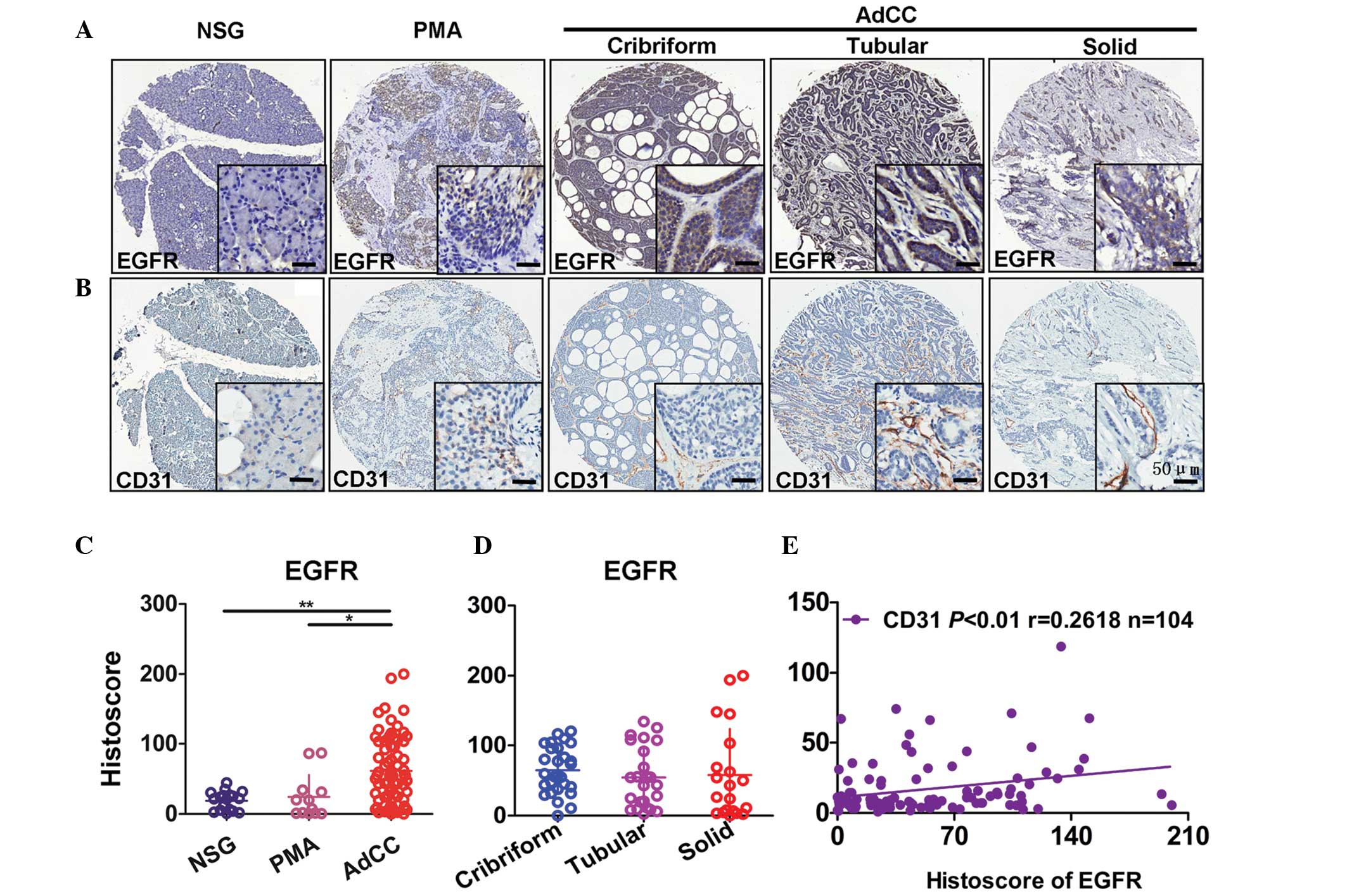 | Figure 1Association between the expression of
EGFR and CD31 in NSG, PMA and AdCC tissues. Representative
immunohistochemical staining of (A) EGFR membranous expression and
(B) CD31 membranous expression in human NSG, PMA and cribriform,
tubular or solid type AdCC tissues. Scale bar=50 µm.
Quantification of EGFR expression levels in (C) human NSG, PMA and
AdCC tissues and (D) subtypes of AdCC using an AperioScanscope
scanner and software. Data were analyzed by Graph Pad Prism 5
software. Data are presented as the mean ± standard error of the
mean. *P<0.05, AdCC vs. PMA tissues;
**P<0.01, AdCC vs. NSG tissues. (E) Correlation
between EGFR and CD31 expression levels in human NSG, PMA and AdCC
tissues (P<0.01, r=0.2618, n=104) using two-tailed Pearson's
test. EGFR, epidermal growth factor receptor; NSG, normal salivary
gland; PMA, polymorphism adenoma; AdCC, adenoid cystic
carcinoma. |
Expression of HIF-1α and CD146 in NSG,
PMA and AdCC tissues
Considering the significant role of HIF-1α in
angiogenesis in various types of cancer and the pathological
angiogenesis biomarker, CD146, in tumor-derived angiogenesis,
HIF-1α and CD146 immunostaining was performed to examine the
expression of CD146 and HIF-1α in AdCC, PMA and NSG tissues. The
results demonstrated that CD146 was expressed at the membrane in
NSG, PMA and AdCC tissues. CD146 was strongly stained at the
membrane in AdCC tissues, which were distributed in the
interstitial tissues and were also highly expressed in the inner
epithelial ductal cells of tubular pattern and irregular cancer
nests of cribriform form (Fig.
2A). In the NSG tissue, HIF-1α staining was present in the
cytoplasm and the nucleus of the cell, whereas, in PMA and AdCC
tissues, HIF-1α staining was largely restricted to the nuclear area
(Fig. 2B). Notably, the levels of
CD146 in the solid form of AdCC were significantly higher than
those in the cribriform subtype. However, the difference between
the mean levels of CD146 in the tubular subtype and the two other
subtypes of AdCC was not significant (Fig. 2C). By quantifying
immunohistochemical staining, the results demonstrated that the
expression of HIF-1α and CD146 was significantly increased in AdCC
(Fig. 2C) compared with NSG and
PMA tissues. The results also suggested that 42 out of 74 were
HIF-1α positive and 46 out of 74 were CD146 positive. In NSG
tissues, 6 out of 18 were HIF-1α positive and 4 out of 18 were
CD146 positive. In PMA tissues, 3 out of 12 were HIF-1α positive
and 5 out of 12 were CD146 positive (Fig. 2C). Hypoxia is a widespread
phenomenon in the three subtypes of AdCC as HIF-1α staining
demonstrated no significance in all of the three subtypes (Fig. 2C).
 | Figure 2Expression of HIF-1α and CD146 in NSG,
PMA and AdCC tissues. Representative immunohistochemical staining
of (A) CD146 membranous expression and (B) HIF-lα cytoplasmic and
nuclear expression in human NSG, PMA and cribriform, tubular or
solid type AdCC tissues. Scale bar=50 µm. (C) Quantification
of HIF-1α and CD146 expression levels in human NSG, PMA and AdCC
tissues and subtypes of AdCC using an AperioScanscope scanner and
software. Data were analyzed using Graph Pad Prism 5 software. Data
are expressed as the mean ± standard error of the mean.
*P<0.05, AdCC vs. PMA tissues in CD146, AdCC vs. NSG
tissues in HIF1α, solid vs. cribriform subtype tissues in CD146;
***P<0.001, AdCC vs. NSG tissues in CD146, AdCC vs.
PMA tissues in HIF1α. EGFR, epidermal growth factor receptor; NSG,
normal salivary gland; PMA, polymorphism adenoma; AdCC, adenoid
cystic carcinoma; HIF-1α, hypoxia-inducible factor-1α. |
Close correlations among HIF-1α, CD146
and EGFR in AdCC
Since EGFR and CD31 were demonstrated to be
significantly correlated with each other, the correlation between
the expression of CD31, HIF-1α, CD146 and EGFR was measured in AdCC
tissues, in which the two-tailed Pearson's correlation was
performed. Pearson's correlation of cases with interpretable scores
of CD31 and HIF-1α (P<0.001, r=0.5236, n=104) demonstrated a
positive correlation (Fig. 3A).
Similar results were also observed with CD31 and CD146 (Fig. 3B; P<0.001, r=0.3346, n=104).
These results suggested that HIF-1α and CD146 may be involved in
the angiogenesis of AdCC. To verify the association between HIF-1α
and CD146 with EGFR in AdCC, the present study then performed a
correlation analysis between HIF-1α and CD146 with EGFR. The levels
of HIF-1α and CD146 were positively correlated with the expression
of EGFR (P<0.05, r=0.2154, n=104, P<0.001, r=0.4701, n=104,
respectively; Fig. 4A). The
expression of HIF-1α was positively correlated with that of CD146
(Fig. 4B; P<0.001, r=0.3491,
n=104). These associations between angiogenic factors in human AdCC
were displayed in a visual image (Fig.
4C), which was obtained by hierarchical clustering.
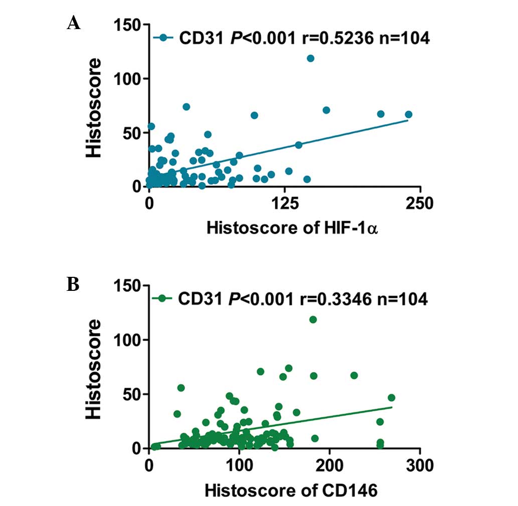 | Figure 3HIF-1α and CD146 may be involved in
angiogenesis in AdCC. Correlation and regression of HIF-1α and
CD146 in human NSG, PMA and AdCC tissues. (A) Correlation between
HIF-1α with CD31 expression levels in human NSG, PMA and AdCC
tissues (P<0.001, r=0.5236, n=104). (B) Correlation between
CD146 with CD31 expression levels in human NSG, PMA and AdCC
tissues (P<0.001, r=0.3346, n=104) using two-tailed Pearson's
test. HIF-1α, hypoxia-inducible factor-1α; NSG, normal salivary
gland; PMA, polymorphism adenoma; AdCC, adenoid cystic
carcinoma. |
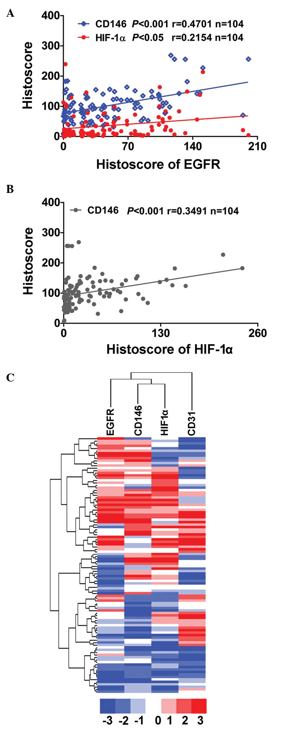 | Figure 4Overexpression of EGFR is correlated
with HIF-1α and CD146 in human AdCC tissue. (A) Expression of EGFR
was positively correlated with HIF1-α and CD146 (P<0.05,
r=0.2154, n=104; P<0.001, r=0.4701, n=104, respectively) and the
(B) expression of HIF-1α was significantly correlated with CD146
(P<0.001, r=0.3491, n=104) in human NSG, PMA and AdCC tissues by
analyzing the tissue microarray immunohistochemical staining. (C)
Hierarchical clustering of immunohistochemical results of human
AdCC with EGFR, HIF-1α and CD146 (statistics including AdCC tissue
only n=74). EGFR, epidermal growth factor receptor; NSG, normal
salivary gland; PMA, polymorphism adenoma; AdCC, adenoid cystic
carcinoma; HIF-1α, hypoxia-inducible factor-1α. |
Discussion
Angiogenesis is a fundamental event in various
physiological and pathological processes, including embryo
implantation the menstrual cycle, rheumatic disease and cancer
(9). Tumor angiogenesis is a key
mechanism for tumor growth and metastasis. Hence, targeting
angiogenesis has been regarded as a promising therapy for cancer
and other angiogenesis-associated diseases (9). Although agents that inhibit tumor
angiogenesis have been applied, the regulatory mechanisms of
angiogenesis remain to be elucidated (24). In the present study, the important
role of EGFR in the angiogenesis of human AdCC was uncovered by
immunohistochemical analysis. The results suggested that EGFR was
highly expressed in human salivary gland AdCC tissues compared with
in PMA and NSG tissues. In addition, EGFR levels were positively
correlated with the expression of HIF-1α, CD146 and CD31 in human
AdCC. These results revealed that EGFR is possibly involved in
angiogenesis by affecting the expression of HIF-1α and CD146 in
human AdCC.
In the present study, CD31 and CD146 were used as
angiogenic markers in order to analyze the blood vessel
distribution and status in this carcinoma. A higher expression of
CD31 and CD146 was identified in the AdCC tissues compared with in
the PMA and NSG tissues. CD31 is a common endothelial cell marker
that is widely used to monitor blood vessels, while CD146 is a
structural component of interendothelial junctions and is
particularly highly expressed in pathological vessels (25). It was reported that CD146 could
bind to vascular endothelial growth factor receptor (VEGFR)-2 and
mediate the phosphorylation of VEGFR-2, as well as the downstream
signaling pathways Akt/p38 and MAPK/NF-κB, which promote
endothelial cell migration and microvascular formation in tumors,
therefore promoting the development of tumors (26). Additionally, CD146 positive
endothelial cells were considered to be fully undifferentiated,
revealing distinguished characteristics compared with
differentiated endothelial cells (17). Therefore, CD146 was selected as a
tumor-associated endothelial biomarker in the present study. CD146
positive cells were found in the interstitial tissues of AdCC,
suggesting the formation of pathological blood vessels in human
AdCC. Notably, strong staining of CD146 was identified in the inner
epithelial ductal cells of tubular pattern and irregular cancer
nests of cribriform form. Since CD146 was found to be highly
expressed in cutaneous melanoma, a severe type of malignant cancer
due to its high rate of distant metastasis, CD146 blockade may
inhibit tumor growth and metastasis of human melanoma (27). Therefore, the high expression of
CD146 in AdCC tumor cells may explain the invasive tendency of AdCC
and early hematogenous metastasis. Currently, CD146 targeting
therapies have been proposed. It was reported that treatment with
microRNA-329 in a mouse model decreased excessive CD146 expression
in blood vessels, which could significantly repress the
neovascularization of tumors and therefore attenuate tumor growth
(28). Furthermore, anti-CD146
monoclonal antibody was demonstrated to inhibit angiogenesis by
suppressing NF-κB activation (29). By examining the association between
EGFR and CD146 in AdCC, the present study found that EGFR was
significantly correlated with CD146, suggesting CD146 may be
regulated by EGFR. However, this requires further investigation in
the future.
By contrast, a significant correlation was
identified between HIF-1α and CD146, as well as HIF-1α and EGFR.
Hypoxia was considered to be the most important microenvironment in
tumor development (30). By
regulating the transcription of angiogenic factors, including VEGF,
basic fibroblast growth factor and matrix metalloproteinase-9,
HIF-1α could promote tumor-derived angiogenesis (31). In the present study, the positive
nuclear immunoreactivity staining of HIF-1α was distributed in all
three forms of AdCC. In addition, the simultaneous high EGFR levels
and HIF-1α expression were observed in numerous cases of AdCC and
the correlation analysis suggested a significant association
between EGFR with HIF-1α. These data indicated that EGFR may have
potentially contributed to HIF-1α nuclear translocation as
previously reported (32).
Cetuximab, an anti-EGFR antibody used for cancer therapy, was
reported to be able to sensitize human head and neck squamous cell
carcinoma cells to radiation in part through inhibiting
radiation-induced upregulation of HIF-1α (33). In addition, HIF-1α was verified to
be required in HER2/neu (ERBB2)-mediated mammary tumor growth and
anoikis resistance (34). This
suggested that the EGFR signaling pathway acts as an upstream
regulator for HIF-1α and that targeting EGFR may be beneficial for
cancer treatment by affecting HIF-1α expression and translocation.
The ERBB-receptor network exemplifies the pathogenic ability of
aberrations in biological information transfer and is considered as
one of the most extensively investigated areas of signal
transduction (35). EGFR
reportedly exhibits positive AdCCs (67–85%) (36,37),
in accordance with the results of the present study. Another study
in our laboratory suggested that targeting EGFR could repress the
invasion and distant metastasis of human AdCC via downregulating
epithelial-mesenchymal transition and related anoikis resistance
(38). In addition, as discussed
previously, targeting EGFR could be beneficial for cancer therapy
by repressing cell growth, as well as inducing the apoptosis of
cancer cells. The results of the present study suggest that
targeting EGFR may suppress HIF-1α-mediated tumor angiogenesis. In
addition, considering the remediability of EGFR, indicating that
EGFR could be targeted by monoclonal antibodies or small
therapeutic molecules, including cetuximab and gefitinib and
therefore treat cancer, it may be a potential option for the
treatment of AdCC.
In conclusion, the present study verified that the
expression of EGFR was highly correlated with CD31, CD146 and
HIF-1α in human AdCC and also suggested a possible association
between EGFR and tumor-derived angiogenesis in this tumor. Thus,
targeting EGFR may provide a possible novel therapeutic strategy
for the treatment of human AdCC.
Acknowledgments
This study was supported by the National Natural
Science Foundation of China (grant nos. 81072203 and 81272963) to
Mr. Z. J. Sun, (grant no. 81371106) to Mrs. L. Zhang, (grant no.
81272946) to Mr. W. F. Zhang and (grant nos. 81170977 and 81371159)
to Mr. Y. F. Zhao.
References
|
1
|
Ellington CL, Goodman M, Kono SA, Grist W,
Wadsworth T, et al: Adenoid cystic carcinoma of the head and neck:
Incidence and survival trends based on 1973–2007 surveillance,
epidemiology and end results data. Cancer. 118:4444–4451. 2012.
View Article : Google Scholar : PubMed/NCBI
|
|
2
|
Bhayani MK, Yener M, El-Naggar A, Garden
A, Hanna EY, et al: Prognosis and risk factors for early-stage
adenoid cystic carcinoma of the major salivary glands. Cancer.
118:2872–2878. 2012. View Article : Google Scholar
|
|
3
|
Laurie SA, Ho AL, Fury MG, Sherman E and
Pfister DG: Systemic therapy in the management of metastatic or
locally recurrent adenoid cystic carcinoma of the salivary glands:
A systematic review. Lancet Oncol. 12:815–824. 2011. View Article : Google Scholar
|
|
4
|
Fantin VR and Abraham RT: Self-eating
limits EGFR-dependent tumor growth. Cell. 154:1184–1186. 2013.
View Article : Google Scholar : PubMed/NCBI
|
|
5
|
Kitano H, Chung JY, Ylaya K, Conway C,
Takikita M, et al: Profiling of phospho-AKT, phospho-mTOR,
phospho-MAPK and EGFR in non-small cell lung cancer. J Histochem
Cytochem. 62:335–346. 2014. View Article : Google Scholar : PubMed/NCBI
|
|
6
|
Wu M, Yuan Y, Pan YY and Zhang Y: Combined
gefitinib and pemetrexed overcome the acquired resistance to
epidermal growth factor receptor tyrosine kinase inhibitors in
non-small cell lung cancer. Mol Med Rep. 10:931–938.
2014.PubMed/NCBI
|
|
7
|
Wu M, Yuan Y, Pan YY and Zhang Y:
Antitumor activity of combination treatment with gefitinib and
docetaxel in EGFR-TKI-sensitive, primary resistant and acquired
resistant human non-small cell lung cancer cells. Mol Med Rep.
9:2417–2422. 2014.PubMed/NCBI
|
|
8
|
Fan QW, Cheng CK, Gustafson WC, Charron E,
Zipper P, et al: EGFR phosphorylates tumor-derived EGFRvIII driving
STAT3/5 and progression in glioblastoma. Cancer Cell. 24:438–449.
2013. View Article : Google Scholar : PubMed/NCBI
|
|
9
|
Carmeliet P and Jain RK: Angiogenesis in
cancer and other diseases. Nature. 407:249–257. 2000. View Article : Google Scholar : PubMed/NCBI
|
|
10
|
Hashizume H, Baluk P, Morikawa S, McLean
JW, Thurston G, et al: Openings between defective endothelial cells
explain tumor vessel leakiness. Am J Pathol. 156:1363–1380. 2000.
View Article : Google Scholar : PubMed/NCBI
|
|
11
|
Goel S, Duda DG, Xu L, Munn LL, Boucher Y,
et al: Normalization of the vasculature for treatment of cancer and
other diseases. Physiol Rev. 91:1071–1121. 2011. View Article : Google Scholar : PubMed/NCBI
|
|
12
|
Garcia S1, Dalès JP and Charafe-Jauffret
E: Poor prognosis in breast carcinomas correlates with increased
expression of targetable CD146 and c-Met and with proteomic
basal-like phenotype. Hum Pathol. 38:830–841. 2007. View Article : Google Scholar : PubMed/NCBI
|
|
13
|
Stopp S, Bornhauser M, Ugarte F, Wobus M,
Kuhn M, et al: Expression of the melanoma cell adhesion molecule in
human mesenchymal stromal cells regulates proliferation,
differentiation and maintenance of hematopoietic stem and
progenitor cells. Haematologica. 98:505–513. 2013. View Article : Google Scholar :
|
|
14
|
Chen W, Zhang HL, Jiang YG, Li JH, Liu BL,
et al: Inhibition of CD146 gene expression via RNA interference
reduces in vitro perineural invasion on ACC-M cell. J Oral Pathol
Med. 38:198–205. 2009. View Article : Google Scholar : PubMed/NCBI
|
|
15
|
Xie S, Luca M, Huang S, Gutman M, Reich R,
et al: Expression of MCAM/MUC18 by human melanoma cells leads to
increased tumor growth and metastasis. Cancer Res. 57:2295–2303.
1997.PubMed/NCBI
|
|
16
|
Malyszko J, Malyszko JS, Brzosko S,
Wolczynski S and Mysliwiec M: Adiponectin is related to CD146, a
novel marker of endothelial cell activation/injury in chronic renal
failure and peritoneally dialyzed patients. J Clin Endocrinol
Metab. 89:4620–4627. 2004. View Article : Google Scholar : PubMed/NCBI
|
|
17
|
Jiang T, Zhuang J, Duan H, Luo Y, Zeng Q,
et al: CD146 is a coreceptor for VEGFR-2 in tumor angiogenesis.
Blood. 120:2330–2339. 2012. View Article : Google Scholar : PubMed/NCBI
|
|
18
|
Li J, Xu Y, Long XD, Wang W, Jiao HK, et
al: Cbx4 governs HIF-1α to potentiate angiogenesis of
hepatocellular carcinoma by its SUMO E3 ligase activity. Cancer
Cell. 25:118–131. 2014. View Article : Google Scholar : PubMed/NCBI
|
|
19
|
Hughes JM, Groot AJ, van der Groep P,
Sersansie R, Vooijs M, et al: Active HIF-1 in the normal human
retina. J Histochem Cytochem. 58:247–254. 2010. View Article : Google Scholar :
|
|
20
|
Thompson L: World Health Organization
classification of tumours: pathology and genetics of head and neck
tumours. Ear Nose Throat J. 85:742006.PubMed/NCBI
|
|
21
|
Sun ZJ, Chen G, Hu X, Zhang W, Liu Y, et
al: Activation of PI3K/Akt/IKK-alpha/NF-kappaB signaling pathway is
required for the apoptosis-evasion in human salivary adenoid cystic
carcinoma: Its inhibition by quercetin. Apoptosis. 15:850–863.
2010. View Article : Google Scholar : PubMed/NCBI
|
|
22
|
Sun ZJ, Chen G, Zhang W, Hu X, Huang CF,
et al: Mammalian target of rapamycin pathway promotes tumor-induced
angiogenesis in adenoid cystic carcinoma: Its suppression by
isoliquiritigenin through dual activation of c-Jun NH2-terminal
kinase and inhibition of extracellular signal-regulated kinase. J
Pharmacol Exp Ther. 334:500–512. 2010. View Article : Google Scholar : PubMed/NCBI
|
|
23
|
Sun ZJ, Zhang L, Hall B, Bian Y, Gutkind
JS, et al: Chemopreventive and chemotherapeutic actions of mTOR
inhibitor in genetically defined head and neck squamous cell
carcinoma mouse model. Clin Cancer Res. 18:5304–5313. 2012.
View Article : Google Scholar : PubMed/NCBI
|
|
24
|
Welti J, Loges S, Dimmeler S and Carmeliet
P: Recent molecular discoveries in angiogenesis and antiangiogenic
therapies in cancer. J Clin Invest. 123:3190–3200. 2013. View Article : Google Scholar : PubMed/NCBI
|
|
25
|
Kratzer A, Chu HW, Salys J, Moumen Z,
Leberl M, et al: Endothelial cell adhesion molecule CD146:
Implications for its role in the pathogenesis of COPD. J Pathol.
230:388–398. 2013. View Article : Google Scholar : PubMed/NCBI
|
|
26
|
Kebir A, Harhouri K, Guillet B, Liu JW,
Foucault-Bertaud A, et al: CD146 short isoform increases the
proangiogenic potential of endothelial progenitor cells in vitro
and in vivo. Circ Res. 107:66–75. 2010. View Article : Google Scholar : PubMed/NCBI
|
|
27
|
Ye Z, Zhang C, Tu T, Sun M, Liu D, et al:
Wnt5a uses CD146 as a receptor to regulate cell motility and
convergent extension. Nat Commun. 4:28032013. View Article : Google Scholar : PubMed/NCBI
|
|
28
|
Wang P, Luo Y, Duan H, Xing S, Zhang J, et
al: MicroRNA 329 suppresses angiogenesis by targeting CD146. Mol
Cell Biol. 33:3689–3699. 2013. View Article : Google Scholar : PubMed/NCBI
|
|
29
|
Bu P, Gao L, Zhuang J, Feng J, Yang D, et
al: Anti-CD146 monoclonal antibody AA98 inhibits angiogenesis via
suppression of nuclear factor-kappaB activation. Mol Cancer Ther.
5:2872–2878. 2006. View Article : Google Scholar : PubMed/NCBI
|
|
30
|
Suzuki M, Shinohara F and Rikiishi H:
Zebularine-induced reduction in VEGF secretion by HIF-1α
degradation in oral squamous cell carcinoma. Mol Med Rep.
1:465–471. 2008.PubMed/NCBI
|
|
31
|
Zhu C, Liu X, Wang S, Yan X, Tang Z, et
al: Hepatitis C virus core protein induces hypoxia-inducible factor
1α-mediated vascular endothelial growth factor expression in Huh751
cells. Mol Med Rep. 9:2010–2014. 2014.PubMed/NCBI
|
|
32
|
Secades P, de Santa-María IS, Merlo A,
Suarez C and Chiara MD: In vitro study of normoxic epidermal growth
factor receptor-induced hypoxia-inducible factor-1-alpha, vascular
endothelial growth factor, and BNIP3 expression in head and neck
squamous cell carcinoma cell lines: Implications for anti-epidermal
growth factor receptor therapy. Head Neck. May 2–2014.Epub ahead of
print.
|
|
33
|
Lu H, Liang K, Lu Y and Fan Z: The
anti-EGFR antibody cetuximab sensitizes human head and neck
squamous cell carcinoma cells to radiation in part through
inhibiting radiation-induced upregulation of HIF-1α. Cancer Lett.
322:78–85. 2012. View Article : Google Scholar : PubMed/NCBI
|
|
34
|
Whelan KA, Schwab LP, Karakashev SV,
Franchetti L, Johannes GJ, et al: The oncogene HER2/neu (ERBB2)
requires the hypoxia-inducible factor HIF-1 for mammary tumor
growth and anoikis resistance. J Biol Chem. 288:15865–15877. 2013.
View Article : Google Scholar : PubMed/NCBI
|
|
35
|
Yarden Y and Pines G: The ERBB network: At
last, cancer therapy meets systems biology. Nat Rev Cancer.
12:553–563. 2012. View
Article : Google Scholar : PubMed/NCBI
|
|
36
|
Sequeiros-Santiago G, García-Carracedo D,
Fresno MF, Suarez C, Rodrigo JP, et al: Oncogene amplification
pattern in adenoid cystic carcinoma of the salivary glands. Oncol
Rep. 21:1215–1222. 2009.PubMed/NCBI
|
|
37
|
Vered M, Braunstein E and Buchner A:
Immunohistochemical study of epidermal growth factor receptor in
adenoid cystic carcinoma of salivary gland origin. Head Neck.
24:632–636. 2002. View Article : Google Scholar : PubMed/NCBI
|
|
38
|
Jia J, Zhang W, Liu JY, Chen G, Liu H, et
al: Epithelial mesenchymal transition is required for acquisition
of anoikis resistance and metastatic potential in adenoid cystic
carcinoma. PLoS One. 7:e515492012. View Article : Google Scholar : PubMed/NCBI
|


