Introduction
Breast cancer (BC) is one of the most common
malignancies in women worldwide and the incidence has increased
continuously in recent years (1,2).
Every year, there are ~1.7 million new cases of breast cancer
diagnosed globally, and >30% of patients succumb to BC (3). In spite of great advances in
diagnosis and therapy for the disease, the overall 5-year survival
rate for patients with BC remains low (4,5).
Thus, it is important to explore effective diagnostic biomarkers
and therapeutic targets to improve the prognosis of patients with
BC.
Cyclin-dependent kinases regulatory subunit 2 (CKS2)
was identified in 1990, and the gene is located at chromosome 9q22
(6). CKS2 is a member of the cell
cycle dependent protein kinase subunits family, which is involved
in cell cycle regulation (7,8). A
previous study demonstrated that CKS2 may serve critical roles in
early embryonic development and somatic cell division (9). Increasing numbers of studies
demonstrate that CKS2 may exert functions in tumor progression. For
example, Chen et al (10)
reported that CKS2 expression was increased and acted as a
biomarker for predicting superficial bladder cancer progression to
muscle-invasive cancer. Lin et al (11) demonstrated that depletion of CKS2
expression compromised cell proliferation and enhanced
chemotherapy-induced apoptosis in HepG2 cells. Wang et al
(12) reported that CKS2 was
upregulated and correlated with poor overall survival in patients
with BC.
The present study explored CKS2 expression in
patients with BC based on a number of public databases, to
illustrate its prognostic and potential therapeutic value.
Moreover, the functions of CKS2 in BC progression were determined.
The present results demonstrated that CKS2 may serve as a
prognostic biomarker and potential therapeutic target in BC
treatment.
Materials and methods
Oncomine database analysis
The Oncomine gene expression array database
(www.oncomine.org) was used to assess the CKS2
mRNA expression levels in four BC datasets, including Sørlie Breast
(13), Curtis Breast (14), Sørlie Breast 2 (15) and The Cancer Genome Atlas (TCGA;
http://cancergenome.nih.gov/) Breast. In
the present study, CKS2 mRNA expression in BC samples and normal
individuals was compared using a Student's t-test.
Human Protein Atlas
CKS2 protein expression levels in BC tissues and
normal tissues were reviewed in the Human Tissue Atlas (http://www.proteinatlas.org/) (16).
bcGenExMiner v4.0
Breast Cancer Gene-Expression Miner v4.0
(bcGenExMiner v4.0), a statistical mining tool of published
annotated genomic data including 36 annotated genomic datasets and
three classical mining functions: Expression, prognosis, and
correlation (17,18). The correlation between CKS2
expression and the risk of any event of relapse (AE) or metastatic
relapse (MR) in patients with BC was determined by univariate Cox
analysis.
Kaplan-Meier analysis
Kaplan-Meier Plotter (www.kmplot.com) is an online database including gene
expression data and clinical data (19). In the present study, Kaplan-Meier
Plotter was used to evaluate the overall survival (OS),
relapse-free survival (RFS) and distant metastasis-free survival
(DMFS) of patients with BC. Individuals were separated into two
groups based on median gene expression; high (≥median expression)
and low expression (<median expression).
RNA extraction and reverse
transcription-quantitative polymerase chain reaction (RT-qPCR)
Total RNA from BC cells was isolated using
TRIzol® reagent (Invitrogen; Thermo Fisher Scientific,
Inc., Waltham, MA, USA), according to the manufacturer's protocols.
The complementary DNA (cDNA) was generated from 100 ng of total RNA
using the PrimeScript™ Reverse Transcription kit (Takara Bio, Inc.,
Otsu, Japan) according to the manufacturer's protocols. The
relative expression of CKS2 was detected using SYBR®
Green Master Mix (Takara Biotechnology Co., Ltd., Dalian, China)
using a Step One Plus Real-Time PCR system (Applied Biosystems;
Thermo Fisher Scientific, Inc.). qPCR was performed as follows:
94°C for 30 sec, then 30 cycles of 56°C for 30 sec and 72°C for 90
sec, and final extension at 72°C for 5 min. GAPDH was used as an
endogenous control. The relative expression level of CKS2 was
calculated via the 2ΔΔCq method (20). The primers used were as follows:
CKS2; forward, 5′-CTTCGCGCTCTCGTTTCATT-3′; and reverse,
5′-CACCAAGTCTCCTCCACTCC-3′; and GAPDH; forward,
5′-GTCGATGGCTAGTCGTAGCATCGAT-3′ and reverse,
5′-TGCTAGCTGGCATGCCCGATCGATC-3′.
Cell culture and transfection
Human BC cell lines (MCF-7, BT-474, HCC1937 and
MDA-MB-231) and a normal human breast epithelial cell line
(MCF-10A) were purchased from the Institute of Biochemistry and
Cell Biology of the Chinese Academy of Sciences (Shanghai, China).
Cells were maintained in Dulbecco's modified Eagle's medium
(HyClone; GE Healthcare Life Sciences, Logan, UT, USA) supplemented
with 10% fetal bovine serum (FBS; HyClone; GE Healthcare Life
Sciences) at 37°C with 5% CO2.
Small interfering RNA (siRNA) for CKS2 and the
corresponding negative controls were synthesized by Shanghai
GenePharma Co., Ltd. (Shanghai, China) and transfected into the
cells to a final oligonucleotide concentration of 10 nmol/l.
Transfection was performed using Lipofectamine® 2000
(Invitrogen; Thermo Fisher Scientific, Inc.) in accordance with the
manufacturer's protocol. The siRNA sequences were as follows:
si-CKS2-1, 5′-GCUGGGUUCAUUACAUGAUdTdT-3′; si-CKS2-2,
5′-CAGAACCACAUAUUCUUCUdTdT-3′, the transfection was performed 24 h
prior to subsequent experiments.
Cell proliferation assay
A Cell Counting Kit-8 (CCK8; Dojindo Molecular
Technologies, Inc., Kumamoto, Japan) was used to analyze the
proliferation of BC cells. In brief, cells were seeded into 96-well
plates and cultured for the indicated times. Subsequently, 10 µl
CCK8 solution was added and incubated for 2 h at 37°C. The
absorbance at 450 nm was determined using a microplate reader
(Bio-Rad Laboratories, Inc., Hercules, CA, USA).
Cell invasion assay
For the cell invasion assay, transfected cells
(1×10⁵) in 200 µl serum-free DMEM were seeded into the
upper invasion chambers (8 µm pore size; EMD Millipore, Billerica,
MA, USA) coated with Matrigel, while 600 µl medium supplemented
with 10% FBS was added to the lower chamber. After 48 h incubation
at 37°C, cells invading the bottom of the membrane were fixed using
4% paraformaldehyde for 5 min at room temperature and stained with
0.3% crystal violet dye for 5 min at room temperature. The images
were captured using a light microscope (magnification, ×100; Nikon
Corporation, Tokyo, Japan).
Western blot analysis
Total protein was from cells using
radioimmunoprecipitation assay lysis buffer (Beyotime Institute of
Biotechnology, Haimen, China) including 1% phenylmethanesulfonyl
fluoride. The concentration of total protein was determined using a
bicinchoninic acid protein assay kit (Beyotime Institute of
Technology). Proteins (40 µg/lane) were separated via 10% SDS-PAGE
and transferred to polyvinylidene difluoride membranes (EMD
Millipore). Following blocking with 5% non-fat milk overnight at
room temperature, membranes were incubated with primary antibodies
against CKS2 (1:1,000; cat. no. ab240129, Abcam, Cambridge, UK),
and GAPDH (1:10,000; cat. no. ab181602, Abcam) overnight at 4°C.
Membranes were then incubated with a horseradish
peroxidase-conjugated secondary anti-rabbit immunoglobulin G
antibody (1:20,000; cat. no. sc-2004, Santa Cruz Biotechnology,
Inc., Dallas, TX, USA). Bands were visualized using enhanced
chemiluminescence reagent (7Sea Biotech, Shanghai, China).
Xenograft assays in nude mice
All animal experiments were performed according to
the approved protocols of the Animal Care and Use Committee at
Tongji University School of Medicine (Shanghai, China). A total of
6 female BALB/c nude mice (10 weeks of age; 20–22 g; n=3
mice/group) were obtained from the Beijing Experimental Animal
Research Center (Beijing, China) and were housed under specific
pathogen-free conditions at 20–26°C, 40–70% humidity and a 12/12 h
light/dark cycle. The mice had free access to food and water.
Transfected MCF-7 cells (2×10⁶) were injected into the
dorsal flanks of the animals to form a single tumor. The mice were
sacrificed by dislocation of the neck following anesthetization by
CO2 inhalation (air displacement rate, 10–30%/min) at 6
weeks following cell injection, and the tumor weights and volumes
were determined. The tumor volume (V) was calculated using the
following formula: V=π/6 × L × W × H; where W is width, L is length
and H is height. The animal experiments performed in the present
study were approved by the Animal Ethics Committee Review Board at
Tongji University School of Medicine (no. T-2018-01-1197).
Statistical analysis
All data are presented as the mean ± standard
deviation from three independent experiments and the statistical
analyses were performed using SPSS 18.0 (SPSS, Inc., Chicago, IL,
USA). Student's t-tests were used for the analysis of statistical
significance between two groups, and one-way analysis of variance
followed by Dunnett's post hoc test was applied to analyze the
statistical significance among three groups or more. P<0.05 was
considered as to indicate a statistically significant
difference.
Results
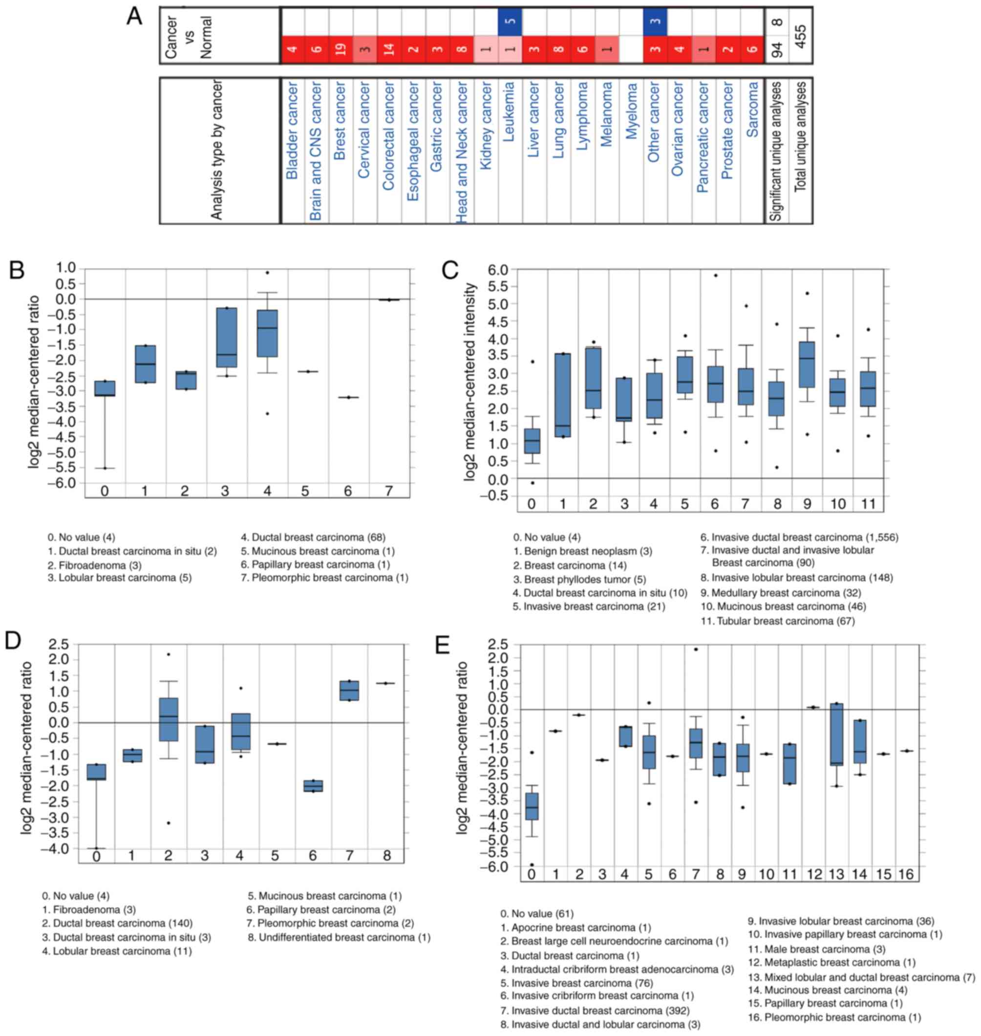
Analysis of CKS2 expression based on
Oncomine and the Human Protein Atlas database
First, CKS2 expression in 20 types of cancer was
examined using the Oncomine database. The results demonstrated that
CKS2 mRNA expression was notably increased in BC samples (Fig. 1A). According to the Sørlie Breast
(13), Curtis Breast (14), Sørlie Breast 2 (15) and TCGA Breast datasets, it was
observed that CKS2 expression was notably increased in BC tissues
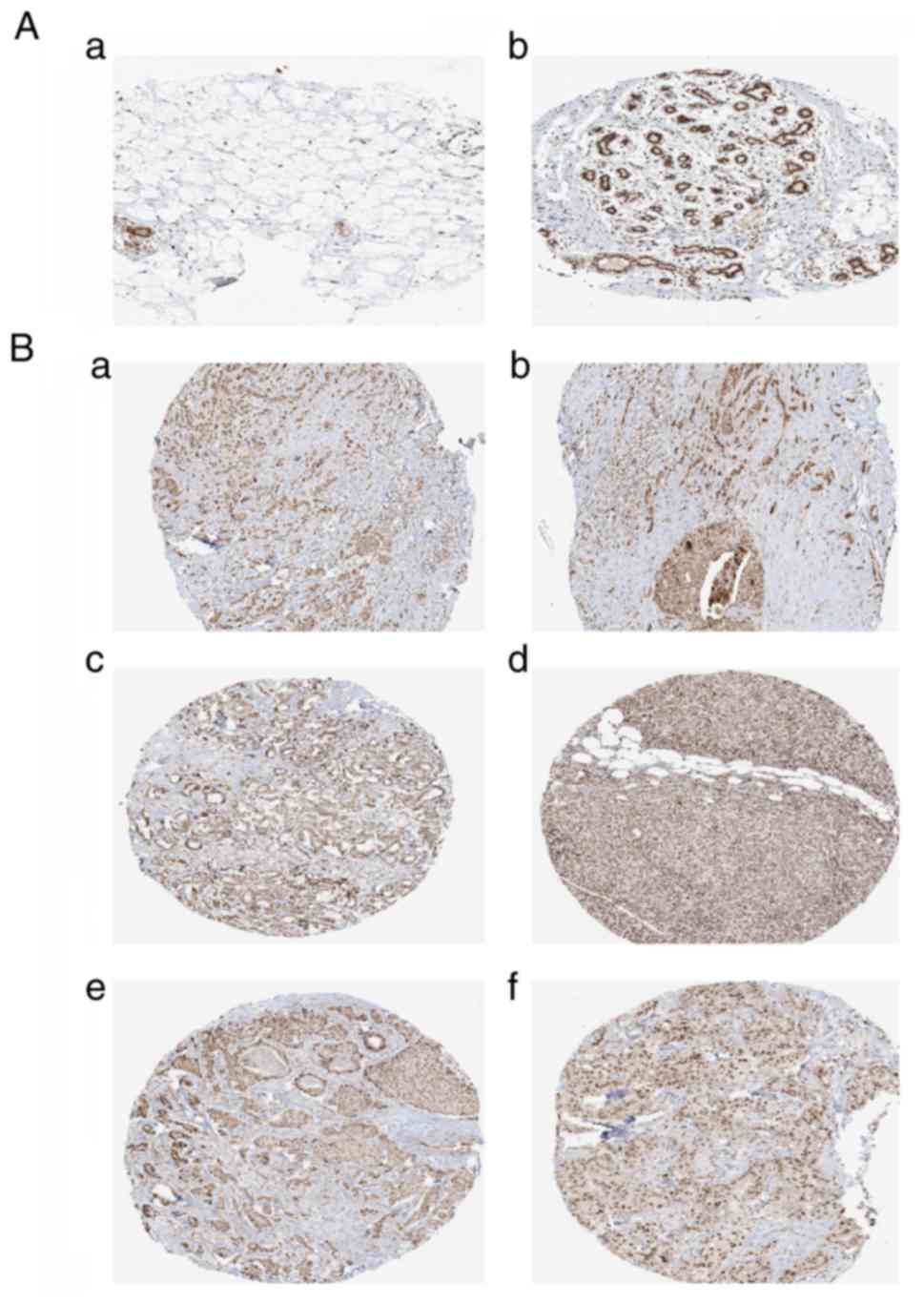
(Fig. 1B-E). Moreover, the Human
Protein Atlas database demonstrated that CKS2 protein expression
was decreased in normal breast samples (Fig. 2A), and increased in BC tissues
(Fig. 2B).
Correlation between CKS2 expression
and clinical features in patients with BC
Next, the bc-GenExMiner database was used to
determine the association between CKS2 expression and clinical
features in patients with BC. The results demonstrated that CKS2
mRNA expression was significantly decreased in estrogen receptor
(ER)-positive and progesterone receptor (PR)-positive BC patients
(Fig. 3A and B). However, there
was no significant difference between the erb-b2 receptor tyrosine
kinase 2 (HER2)-positive group and the HER2-negative group
(Fig. 3C). In addition, it was
demonstrated that CKS2 mRNA expression was significantly increased
patients with BC with positive nodal status, basal-like status and
triple-negative status (Fig.
3D-F).
Correlation between CKS2 expression
and prognosis in patients with BC
Kaplan-Meier Plotter results illustrated that high
CKS2 mRNA expression was correlated with a poor OS, RFS, and DMFS
in patients with BC (Fig. 4). The
bc-GenExMiner results indicated that high CKS2 expression was
associated with an increased risk of MR [Fig. 5A; hazard ratio (HR)=1.32, 95%
confidence interval (CI): 1.25–1.40; P<0.0001] and AE (Fig. 5B; HR=1.26; 95% CI: 1.21–1.32;
P<0.0001). The survival curve demonstrated that high CKS2
expression was associated with poor MR-free survival (Fig. 5C; HR=1.73; 95% CI: 1.53–1.97;
P<0.0001) and AE-free survival (Fig. 5D; HR=1.58; 95% CI: 1.43–1.74;
P<0.0001).
CKS2 inhibition decreased BC cells
progression
To examine the effects of CKS2 on BC progression,
CKS2 expression was first determined in BC cells (Fig. 6A; P<0.05). MCF-7 and MDA-MB-231
cells were selected for the functional experiments. Following
si-CKS2 transfection, the expression of CKS2 was determined by
RT-qPCR and western blot analyses (Fig. 6B and C; P<0.05). The CCK-8 assay
revealed that CKS2 inhibition significantly decreased BC cell
proliferation capacity (Fig. 6D;
P<0.05). The Transwell invasion assay demonstrated that the
invasive capability of BC cells transfected with si-CKS2 was
significantly decreased (Fig. 6E;
P<0.05). Furthermore, the influence of CKS2 on BC cell growth
was assessed in vivo. The results indicated that the average
weights and volumes of the tumors were significantly decreased in
the CKS2 knockdown group (Fig. 6F and
G; P<0.05).
Discussion
CKS2 belongs to the mammalian cyclin kinase subunit
family, which has two members: CKS1 and CKS2 (21). Previous studies indicated that CKS1
was increased in various cancer types, including prostate cancer,
esophageal squamous cell carcinoma, nasopharyngeal carcinoma and
glioma (22–25). The effects of CKS1 have been
studied thoroughly; however, the roles of CKS2 remain unclear.
A previous study reported that the expression of
CKS2 was increased in bladder cancer and associated with cancer
progression (9). In the present
study, the Oncomine database revealed that CKS2 mRNA expression was
markedly increased in invasive ductal breast carcinoma, apocrine
breast carcinoma, invasive breast carcinoma, and mixed lobular and
ductal breast carcinoma. The Human Protein Atlas database indicated
that CKS2 protein expression was significantly increased in BC
tissues. Additionally, the bc-GenExMiner database revealed that
high CKS2 expression was correlated with ER-positive, PR-positive,
positive nodal and positive basal-like status, indicative of
fast-growing invasive tumors. These data indicated that CKS2 may
serve critical roles in BC tumorigenesis.
Kaplan-Meier Plotter analysis demonstrated that high
CKS2 mRNA expression was associated with poor OS, RFS and DMFS in
patients with BC. In addition, bc-GenExMiner analysis revealed that
high CKS2 expression was associated with a higher risk of MR and
AE. Survival curve analysis revealed that high CKS2 expression was
associated with poor MR-free survival and AE-free survival in
patients with BC. These data suggested that CKS2 may act as a
potential prognostic biomarker in BC.
Next, the roles of CKS2 in BC tumorigenesis were
investigated. In vitro functional assays revealed that CKS2
inhibition significantly decreased BC cell proliferation and
invasion capacity compared with the si-NC group. Moreover, the
in vivo assay revealed that the average weights and volumes
of the tumors were significantly decreased in the CKS2 knockdown
group. Therefore, the present data suggested that CKS2 may act as
an oncogene in BC. Kang et al (26) reported that CKS2 was significantly
upregulated in gastric cancers and promoted cell growth by
decreasing p53 and p21cip1 expression. Additionally, Ji
et al (27) revealed that
increased expression of CKS2 was associated with hepatocellular
carcinoma cell proliferation and downregulated the expression of
phosphatase and tensin homolog. Future experiments should
investigate whether CKS2 promotes the proliferation of BC cells via
similar mechanisms.
In conclusion, the present study identified that
CKS2 may act as a novel potential oncogene in BC, serving important
roles in cell proliferation and invasion; however, the research had
the following limitations. Firstly, the data on the expression of
CKS2 in BC and its association with clinical features were obtained
from bioinformatics, and require further analysis in BC samples.
Secondly, the underlying mechanisms of the association between CKS2
and cell proliferation and invasion remain to be investigated.
Future studies will aim to determine these underlying mechanisms to
identify approaches by which CKS2 may be targeted in the treatment
of BC.
Acknowledgements
Not applicable.
Funding
This study was supported by the National Youth
Science Foundation (grant no. 81502426).
Availability of data and materials
All data generated or analyzed during the present
study are available from the corresponding author upon reasonable
request.
Authors' contributions
NQH, ZLW and HGL performed the majority of the
experiments, analyzed the data and drafted the manuscript. HH, XMW,
and FQY contributed to the acquisition of data and revised the
manuscript. HGL designed the study and revised the manuscript.
Ethics approval and consent to
participate
The animal experiments performed in the present
study were approved by the Animal Ethics Committee Review Board at
Tongji University School of Medicine (Shanghai, Chin; no.
T-2018-01-1197).
Patient consent for publication
Not applicable.
Competing interests
The authors declare that they have no competing
interests.
References
|
1
|
Torre LA, Bray F, Siegel RL, Ferlay J,
Lortet-Tieulent J and Jemal A: Global cancer statistics, 2012. CA
Cancer J Clin. 65:87–108. 2015. View Article : Google Scholar : PubMed/NCBI
|
|
2
|
Tao Z, Shi A, Lu C, Song T, Zhang Z and
Zhao J: Breast cancer: Epidemiology and etiology. Cell Biochem
Biophys. 72:333–338. 2015. View Article : Google Scholar : PubMed/NCBI
|
|
3
|
DeSantis CE, Fedewa SA, Goding Sauer A,
Kramer JL, Smith RA and Jemal A: Breast cancer statistics, 2015:
Convergence of incidence rates between black and white women. CA
Cancer J Clin. 66:31–42. 2016. View Article : Google Scholar : PubMed/NCBI
|
|
4
|
Qian C, Guan M, Si C, Shen H, Jin T and
Zhang T: Identification of differentially expressed profiles of
lncRNAs and mRNAs in ER-negative and HER-2 positive breast cancer.
Arch Med Sci Civil Dis. 2:148–160. 2017.
|
|
5
|
Hortobagyi GN, Stemmer SM, Burris HA, Yap
YS, Sonke GS, Paluch-Shimon S, Campone M, Blackwell KL, André F,
Winer EP, et al: Ribociclib as first-line therapy for HR-positive,
advanced breast cancer. N Engl J Med. 375:1738–1748. 2016.
View Article : Google Scholar : PubMed/NCBI
|
|
6
|
You H, Lin H and Zhang Z: CKS2 in human
cancers: Clinical roles and current perspectives. Mol Clin Oncol.
3:459–463. 2015. View Article : Google Scholar : PubMed/NCBI
|
|
7
|
Liberal V, Martinsson-Ahlzén HS, Liberal
J, Spruck CH, Widschwendter M, McGowan CH and Reed SI:
Cyclin-dependent kinase subunit (Cks) 1 or Cks2 overexpression
overrides the DNA damage response barrier triggered by activated
oncoproteins. Proc Natl Acad Sci USA. 109:2754–2759. 2012.
View Article : Google Scholar : PubMed/NCBI
|
|
8
|
Litchfield DW: Protein kinase CK2:
Structure, regulation and role in cellular decisions of life and
death. Biochem J. 369:1–15. 2003. View Article : Google Scholar : PubMed/NCBI
|
|
9
|
Martinsson-Ahlzén HS, Liberal V,
Grünenfelder B, Chaves SR, Spruck CH and Reed SI: Cyclin-dependent
kinase-associated proteins Cks1 and Cks2 are essential during early
embryogenesis and for cell cycle progression in somatic cells. Mol
Cell Biol. 28:5698–5709. 2008. View Article : Google Scholar : PubMed/NCBI
|
|
10
|
Chen R, Feng C and Xu Y: Cyclin-dependent
kinase-associated protein Cks2 is associated with bladder cancer
progression. J Int Med Res. 39:533–540. 2011. View Article : Google Scholar : PubMed/NCBI
|
|
11
|
Lin L, Fang Z, Lin H, You H, Wang J, Su Y,
Wang F and Zhang ZY: Depletion of Cks1 and Cks2 expression
compromises cell proliferation and enhance chemotherapy-induced
apoptosis in HepG2 cells. Oncol Rep. 35:26–32. 2016. View Article : Google Scholar : PubMed/NCBI
|
|
12
|
Wang J, Xu L, Liu Y, Chen J, Jiang H, Yang
S and Tan H: Expression of cyclin kinase subunit 2 in human breast
cancer and its prognostic significance. Int J Clin Exp Pathol.
7:8593–8601. 2014.PubMed/NCBI
|
|
13
|
Sørlie T, Perou CM, Tibshirani R, Aas T,
Geisler S, Johnsen H, Hastie T, Eisen MB, van de Rijn M, Jeffrey
SS, et al: Gene expression patterns of breast carcinomas
distinguish tumor subclasses with clinical implications. Proc Natl
Acad Sci USA. 98:10869–10874. 2001. View Article : Google Scholar : PubMed/NCBI
|
|
14
|
Curtis C, Shah SP, Chin SF, Turashvili G,
Rueda OM, Dunning MJ, Speed D, Lynch AG, Samarajiwa S, Yuan Y, et
al: The genomic and transcriptomic architecture of 2,000 breast
tumours reveals novel subgroups. Nature. 486:346–352. 2012.
View Article : Google Scholar : PubMed/NCBI
|
|
15
|
Sørlie T, Tibshirani R, Parker J, Hastie
T, Marron JS, Nobel A, Deng S, Johnsen H, Pesich R, Geisler S, et
al: Repeated observation of breast tumor subtypes in independent
gene expression data sets. Proc Natl Acad Sci USA. 100:8418–8423.
2003. View Article : Google Scholar : PubMed/NCBI
|
|
16
|
Pontén F, Jirström K and Uhlen M: The
Human Protein Atlas-a tool for pathology. J Pathol. 216:387–393.
2008. View Article : Google Scholar : PubMed/NCBI
|
|
17
|
Lu XF, Zeng D, Liang WQ, Chen CF, Sun SM
and Lin HY: FoxM1 is a promising candidate target in the treatment
of breast cancer. Oncotarget. 9:842–852. 2018. View Article : Google Scholar : PubMed/NCBI
|
|
18
|
Jézéquel P, Frénel JS, Campion L,
Guérin-Charbonnel C, Gouraud W, Ricolleau G and Campone M:
bc-GenExMiner 3.0: New mining module computes breast cancer gene
expression correlation analyses. Database (Oxford).
2013:bas0602013. View Article : Google Scholar : PubMed/NCBI
|
|
19
|
Györffy B, Lanczky A, Eklund AC, Denkert
C, Budczies J, Li Q and Szallasi Z: An online survival analysis
tool to rapidly assess the effect of 22,277 genes on breast cancer
prognosis using microarray data of 1,809 patients. Breast Cancer
Res Treat. 123:725–731. 2010. View Article : Google Scholar : PubMed/NCBI
|
|
20
|
Livak KJ and Schmittgen TD: Analysis of
relative gene expression data using real-time quantitative PCR and
the 2(-Delta Delta C(T)) method. Methods. 25:402–408. 2001.
View Article : Google Scholar : PubMed/NCBI
|
|
21
|
Malumbres M and Barbacid M: Mammalian
cyclin-dependent kinases. Trends Biochem Sci. 30:630–641. 2005.
View Article : Google Scholar : PubMed/NCBI
|
|
22
|
Zhao H, Lu Z, Bauzon F, Fu H, Cui J,
Locker J and Zhu L: p27T187A knockin identifies Skp2/Cks1 pocket
inhibitors for advanced prostate cancer. Oncogene. 36:60–70. 2017.
View Article : Google Scholar : PubMed/NCBI
|
|
23
|
Li Z, Zhou Y, Tu B, Bu Y, Liu A and Kong
J: Long noncoding RNA MALAT1 affects the efficacy of radiotherapy
for esophageal squamous cell carcinoma by regulating Cks1
expression. J Oral Pathol Med. 46:583–590. 2017. View Article : Google Scholar : PubMed/NCBI
|
|
24
|
Xu L, Fan S, Zhao J, Zhou P, Chu S, Luo J,
Wen Q, Chen L, Wen S, Wang L and Shi L: Increased expression of
Cks1 protein is associated with lymph node metastasis and poor
prognosis in nasopharyngeal carcinoma. Diagn Pathol. 12:22017.
View Article : Google Scholar : PubMed/NCBI
|
|
25
|
Wang D, Zhi T, Xu X, Bao Z, Fan L, Li Z,
Ji J and Liu N: MicroRNA-936 induces cell cycle arrest and inhibits
glioma cell proliferation by targeting CKS1. Am J Cancer Res.
7:2131–2143. 2017.PubMed/NCBI
|
|
26
|
Kang MA, Kim JT, Kim JH, Kim SY, Kim YH,
Yeom YI, Lee Y and Lee HG: Upregulation of the cycline kinase
subunit CKS2 increases cell proliferation rate in gastric cancer. J
Cancer Res Clin Oncol. 135:761–769. 2009. View Article : Google Scholar : PubMed/NCBI
|
|
27
|
Ji X, Xue Y, Wu Y, Feng F and Gao X:
High-expressed CKS2 is associated with hepatocellular carcinoma
cell proliferation through down-regulating PTEN. Pathol Res Pract.
214:436–441. 2018. View Article : Google Scholar : PubMed/NCBI
|