Introduction
Hyperglycemia is an important factor involved in the
development of cardiovascular disease, kidney failure and cataract
(1). Patients with diabetes are
more likely to develop cataracts compared with healthy subjects
(2). Diabetic retinopathy is
characterized by a dysfunctional local microcirculation, which
causes structural changes in the microvasculature, leading to the
morphological alterations of large vessels, including arteries and
veins (3). Furthermore,
morphological changes can induce vascular lesions in the retina
(3). Therefore, since the number
of diabetic patients is increasing worldwide, the occurrence rate
of diabetic cataract is also increasing (4).
Multiple factors contribute to cataract development,
including aging, smoking (5),
exposure to ultraviolet-B and ionizing radiations and oxidative
stress (6). Diabetic cataracts are
induced by a glutathione (GSH) deficiency that impairs the
antioxidative mechanisms in the lens of the eye (7). Additionally, deficiency of nutrients,
including vitamins E (8), A
(9) and C (10), and selenium, can also lead to
cataract development.
Puerarin is an isoflavone found at high
concentrations in the dry root extract of Pueraria lobata
(Willd) Ohwi, and was first described in the 1950s as the main
active ingredient in Radix Pueraria lobata (11). Puerarin is commonly used in the
treatment of cerebrovascular disorders, cardiovascular disease
(12), cancer (13), diabetes and diabetes-associated
complications (14). Puerarin
eyedrops have been used to treat eye diseases in China for years
(15). Several previous studies
have demonstrated that puerarin protects against oxidative damage,
inflammation and hyperlipidemia (16,17).
The beneficial effects of puerarin may be due to its ability to
decrease blood glucose levels, reduce insulin resistance and
scavenge oxygen free radicals in diabetic rats (18,19).
Puerarin toxicity is limited, and this safe natural
compound has been shown to inhibit diabetic ocular complications,
such as cataract (20,21). Therefore, over the past decade, an
increasing number of studies have investigated the pharmacological
effects of puerarin (22). A
previous study demonstrated that puerarin ameliorates streptozocin
(STZ)-induced diabetic retinopathy in rats by decreasing the mRNA
or protein expression levels of various factors in the rat retina,
including Fas cell surface death receptor, Fas ligand,
nitrotyrosine, inducible nitric oxide synthase (23), advanced glycation end product
receptors and vascular endothelial growth factor (VEGF) (19,20).
Additionally, puerarin decreases the apoptotic rate of retinal
pigment epithelial cells in diabetic rats induced by STZ injection
(24,25). The aim of the present study was to
investigate the pharmacological mechanism underlying puerarin
function in inhibiting cataract development. In particular, the
present study examined the nuclear factor erythroid 2 like 2
(Nrf2)/heme oxygenase (HO-1) signaling pathway.
Materials and methods
Animal experiment
A total of 90 Male Wistar rats (6–8 weeks, 180–220
g) were purchased from The Shanghai Animal Center, and kept under
specific pathogen-free conditions. The rats were housed at 22±2°C,
under a 12-h light/dark cycle, at 50±10% relative humidity, and
with free access to water and food. Experiments were performed
according to the guidelines for the care and use of experimental
animals established by the Ministry of Science and Technology of
the People's Republic of China (26), and the study was approved by the
Laboratory Animal Management Committee of Linyi Central Hospital
(approval no. 2006-398). After 2 weeks of acclimatization, animals
were administered 65 mg/kg STZ (Sigma-Aldrich; Merck KGaA) in 0.1 M
citrate buffer (pH 4.5), as previously described (27–29).
Rats in the control group were administered 0.1 M citrate buffer
(vehicle control). Blood glucose was measured every week using a
GlucoLeader automatic analyzer. In the present study, animals
exhibiting blood glucose levels ≥16 mmol/l were considered diabetic
(30).
Subsequently, animals treated with STZ were randomly
divided into four experimental groups. Diabetic rats (18 rats in
each group) were administered with 0 (DM group; untreated diabetic
rats), 25, 50 or 100 mg/kg puerarin (Aladdin Reagent) by
intraperitoneal injection, as previously described (31,32).
Animals were treated with puerarin for 12 weeks daily, and they
were provided a standard rodent diet with free access to water.
Untreated non-diabetic rats were also used as the control group
with an equal volume of 0.1 M citrate buffer. Blood was collected
from the tail vein, and blood glucose levels were monitored in all
rats.
Evaluation of cataract
development
After 12 weeks, the development of cataract was
assessed using an ophthalmoscope. Eye inspection was preceded by
topical administration of 1% tropicamide drops. Cataract formation
was graded based on the classification described by Varma (33): Grade 0, clear normal lens; grade I,
peripheral vesicles; grade II, peripheral vesicles and cortical
opacities; grade III, diffuse central opacities; and grade IV,
mature cataract. Cataract formation was considered complete (grade
IV) when the red fundus reflex was not visible through any part of
the lens and the lens appeared completely opaque to the naked
eye.
Lens preparation
After 12 weeks, the rats were sacrificed and the eye
lenses were collected. Each pair of lenses was homogenized in
prechilled 0.2 M potassium phosphate buffer (pH 7.0). This
homogenate was used to assess the activity of glutathione
peroxidase (GPx), the concentrations of GSH and malondialdehyde
(MDA), and the protein expression levels of various factors.
Samples were stored at −80°C prior to use in biochemical
assays.
Measurement of oxidative stress
markers
Antioxidant capacity (AOC) was measured using total
Antioxidant capacity assay kit (A015-1-2, Nanjing Jiancheng
Bioengineering Institute) according to the manufacturer's protocol.
The assay is based on the ability of the sample to inhibit
oxidation of 2,2′-azino-di(3-ethylbenzthiazoline-6-sulphonate)
(ABTS) by metmyoglobin. The antioxidants in the sample cause a
decrease in absorbance at 750 nm, corresponding to the
concentration of ABTS (34). Each
sample was measured in duplicate.
MDA levels were measured using a thiobarbituric acid
(TBA) assay kit (A003-1-2, Nanjing Jiancheng Bioengineering
Institute) according to the manufacturer's protocol. Briefly, 0.1
ml sample was mixed with 1,1,3,3-tetramethoxypropane, 0.75 ml TBA
working solution (0.37%) and perchloric acid. The resulting
solution was incubated at 95°C for 45 min. After cooling (10 min in
ice water bath), the flocculent precipitate was removed by
centrifugation (4,000 × g 10 min at room temperature). The
supernatant was analyzed at 532 nm by multi-scan spectrum
microplate spectrophotometer at room temperature.
GPx activity was assayed using an hydrogen peroxide
assay (A005-1-2, Nanjing Jiancheng Bioengineering Institute), as
previously described (35). The
decrease in NADPH was assessed spectrophotometrically at 340 nm.
The reaction mixture consisted of 240 mU/ml glutathione disulfide
reductase, 1 mM GSH, 0.15 mM NADPH in 0.1 M potassium phosphate
buffer (pH 7.0) containing 1 mM EDTA. In total, 50 µl sample was
added to this mixture and incubated at 37°C for 3 min.
Subsequently, 1.5 mM hydrogen peroxide was added to adjust the
final volume of the assay mixture to 1 ml.
GSH content of lens homogenate was assayed by
spectrophotometric method, using the reduced GSH assay kit
(A006-1-1, Nanjing Jiancheng Bioengineering Institute) according to
the manufacturer's protocol. The detection was performed at 420 nm
by multi-scan spectrum microplate spectrophotometer at room
temperature.
Inflammation biochemical assays
Interleukin (IL)-1β levels were quantified in the
retina using a rat IL-1β ELISA kit (H002, Nanjing Jiancheng
Bioengineering Institute, Nanjing, China) according to the
manufacturer's protocol. The concentration of IL-1β was calculated
as pg IL-1β/mg total retina protein in each sample according to the
manufacturer's protocol. VEGF levels were quantified in the retina
using a rat VEGF ELISA kit (H044, Nanjing Jiancheng Bioengineering
Institute) according to the manufacturer's protocol. The standard
solution or the samples were added in a 96-well plate that was
coated with a monoclonal antibody from the kit. The samples were
incubated for 2 h 37°C. After washing, the samples were detected at
the wavelength of 450 nm using a VERSA max tunable microplate
reader (Molecular Devices, LLC). The ELISA assay could detect
concentrations of VEGF ≥15 ng/ml.
Reverse transcription-quantitative PCR
(RT-qPCR) analysis
The mRNA expression levels of Nrf2 and HO-1 were
determined by RT-qPCR. Total RNA was extracted from rat retinas
using TRIzol reagent (Invitrogen; Thermo Fisher Scientific, Inc.).
RT was performed using 1 µg RNA. Moloney Murine Leukemia Virus
reverse transcriptase kit (cat. no. 28025013, Thermo Fisher
Scientific, Inc.) was used to synthesize the cDNA. The synthesized
cDNA was diluted by adding 75 µl DNase-free water and stored at
−20°C. qPCR was performed using the SYBR-Green PCR kit (Takara
Biotechnology Co., Ltd.) and gene-specific primers were synthesized
by Nanjing Sunshine Biotechnology Co., Ltd. Primer sequences are
listed in Table I. The
thermocycling conditions were: 95°C for 10 min, 95°C for 10 sec and
55°C for 10 sec, with a final step of 72°C for 30 sec. Then steps
2–4 were repeated for 40 cycles followed by a melt curve program
for 60 min. GAPDH served as an internal control. Relative gene
expression levels of Nrf2 and HO-1 were measured using the
2−ΔΔCq method (36).
The expression levels of Nrf2 and HO-1 were normalized to the
control group.
 | Table I.Gene-specific PCR primer
sequences. |
Table I.
Gene-specific PCR primer
sequences.
| Gene symbol | Sequence
(5′-3′) |
|---|
| Nrf2 | F:
TTCCTCTGCTGCCATTAGTCAGTC |
|
| R:
GCTCTTCCATTTCCGAGTCACTG |
| HO-1 | F:
CTGGAAGAGGAGATAGAGCGAA |
|
| R:
TCTTAGCCTCTTCTGTCACCCT |
| GAPDH | F:
TGATGACATCAAGAAGGTGGTGA |
|
| R:
TCCTTGGAGGCCATGTAGGCCAT |
| LMNB1 | F:
CCGGATGGTGGGGCTTTGTT |
|
| R:
CCGGGCCTCTCGATGGATAAGC |
Western blot analysis
For immunoblot analysis, total protein was extracted
using radioimmunoprecipitation buffer (P0013D, Beyotime Institute
of Biotechnology) and nuclear extracts were isolated with a Nuclear
and Cytoplasmic Protein Extraction kit (BioTeke Corporation),
according to the manufacturer's instructions. Equal amounts of
protein samples (~30 µg) were separated by 10% SDS-PAGE. The
protein concentration was determined using a bicinchoninic protein
assay according to the manufacturer's protocol (Thermo Fisher
Scientific, Inc.). The proteins were electro-blotted onto a PVDF
membrane (thickness, 0.45 µm; EMD Millipore). The membranes were
incubated with blocking solution (5% dried milk) for 1 h at room
temperature and incubated overnight at 4°C with specific primary
antibodies anti-Nrf2 (1:1,000; cat. no. ab137550; Abcam), HO-1
(1:1,000; cat. no. ab13248; Abcam), GAPDH (1:5,000, cat. no. A0208;
Beyotime Institute of Biotechnology) and LaminB (1:5,000, cat. no.
AF1408; Beyotime Institute of Biotechnology). After washing with
TBS/0.05% Tween 20 three times, the blots were incubated with
horseradish peroxidase-conjugated immunoglobulin G, horseradish
peroxidase-labeled mouse anti-rabbit IgG (1:5,000; cat. no. 5127,
Cell Signaling Technology, Inc.) and horseradish peroxidase-labeled
rabbit anti-mouse IgG (1:5,000; cat. no. 58802, Cell Signaling
Technology, Inc.), for 1 h at room temperature. The signals were
developed using the Pierce ECL western blotting substrate (Thermo
Fisher Scientific, Inc.). Immunoreactive protein bands were
visualized using the ChemiDoc MP Imaging system and quantified
using Image Lab 3.0 software (Bio-Rad Laboratories, Inc.).
Statistical analysis
Data are presented as the mean ± standard error of
the mean. Comparisons were performed using one-way ANOVA followed
by Tukey's post hoc test using GraphPad Prism software 6.0
(GraphPad Software, Inc.). P<0.05 was considered to indicate a
statistically significant difference.
Results
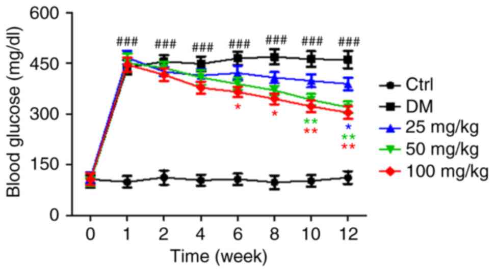
Blood glucose measurement
Blood glucose levels in all experimental animal
groups were measured every week. Puerarin administration
significantly decreased blood glucose levels in diabetic rats,
between 6 and 12 weeks of STZ administration (Fig. 1).
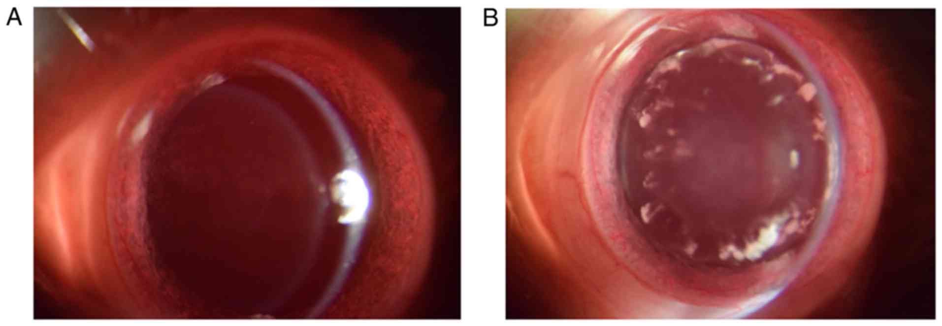
Cataract development and
progression
The effects of puerarin on cataract development in
hyperglycemic rats were examined (Fig.
2). First, the incidence of cataract in diabetic rats following
treatment with puerarin was investigated (Table II). Each group consisted of 18
rats. A total of 15 rats developed cataract in the diabetic group
(rats induced withSTZ and not treated with puerarin). Following
treatment with puerarin at 25, 50 and 100 mg/kg, the number of rats
with cataracts was 13, 11 and 7, respectively (Table II). All animals presented clear
lenses (baseline cataract score, 0) at week 0. However, after 12
weeks, the cataract scores varied among groups (Table III). The average cataract scores
at the end of the experiment were decreased in the diabetic rats
treated with puerarin compared with the untreated group,
particularly in the 100 mg/kg group (P<0.01).
| Table II.Effect of puerarin on the incidence
of cataract in streptozocin-induced diabetic rats. |
Table II.
Effect of puerarin on the incidence
of cataract in streptozocin-induced diabetic rats.
| Groups | Cataract incidence
(number of animals with cataract) |
|---|
| Diabetic | 15/18 |
| Diabetic + 25 mg/kg
puerarin | 13/18 |
| Diabetic + 50 mg/kg
puerarin | 11/18 |
| Diabetic + 100
mg/kg puerarin | 7/18 |
| Table III.Effect of puerarin on the cataract
score in streptozocin-induced diabetic rats. |
Table III.
Effect of puerarin on the cataract
score in streptozocin-induced diabetic rats.
| Cataract score | Diabetic | Diabetic + 25 mg/kg
puerarin | Diabetic + 50 mg/kg
puerarin | Diabetic + 100
mg/kg puerarin |
|---|
| 0 | 3 | 5 | 7 | 10 |
| I | 1 | 1 | 2 | 3 |
| II | 3 | 4 | 4 | 2 |
| III | 5 | 4 | 4 | 3 |
| IV | 6 | 4 | 1 | 0 |
Effects of puerarin on the levels of
MDA, GSH, AOC and GPx in the lens of diabetic rats
The untreated diabetic group exhibited significantly
different levels of AOC, GSH, GPx and MDA compared with the control
group (Fig. 3). Moreover, in the
groups treated with 50 and 100 mg/kg puerarin, AOC and GSH levels
were significantly higher compared with the untreated diabetic
group (Fig. 3A and B). The levels
of GPx in groups treated with 50 and 100 mg/kg puerarin were
significantly higher compared with the untreated diabetic group.
(Fig. 3C). Finally, MDA levels
were significantly decreased by treatment with 50 and 100 mg/kg
puerarin, compared with the untreated diabetic group (Fig. 3D).
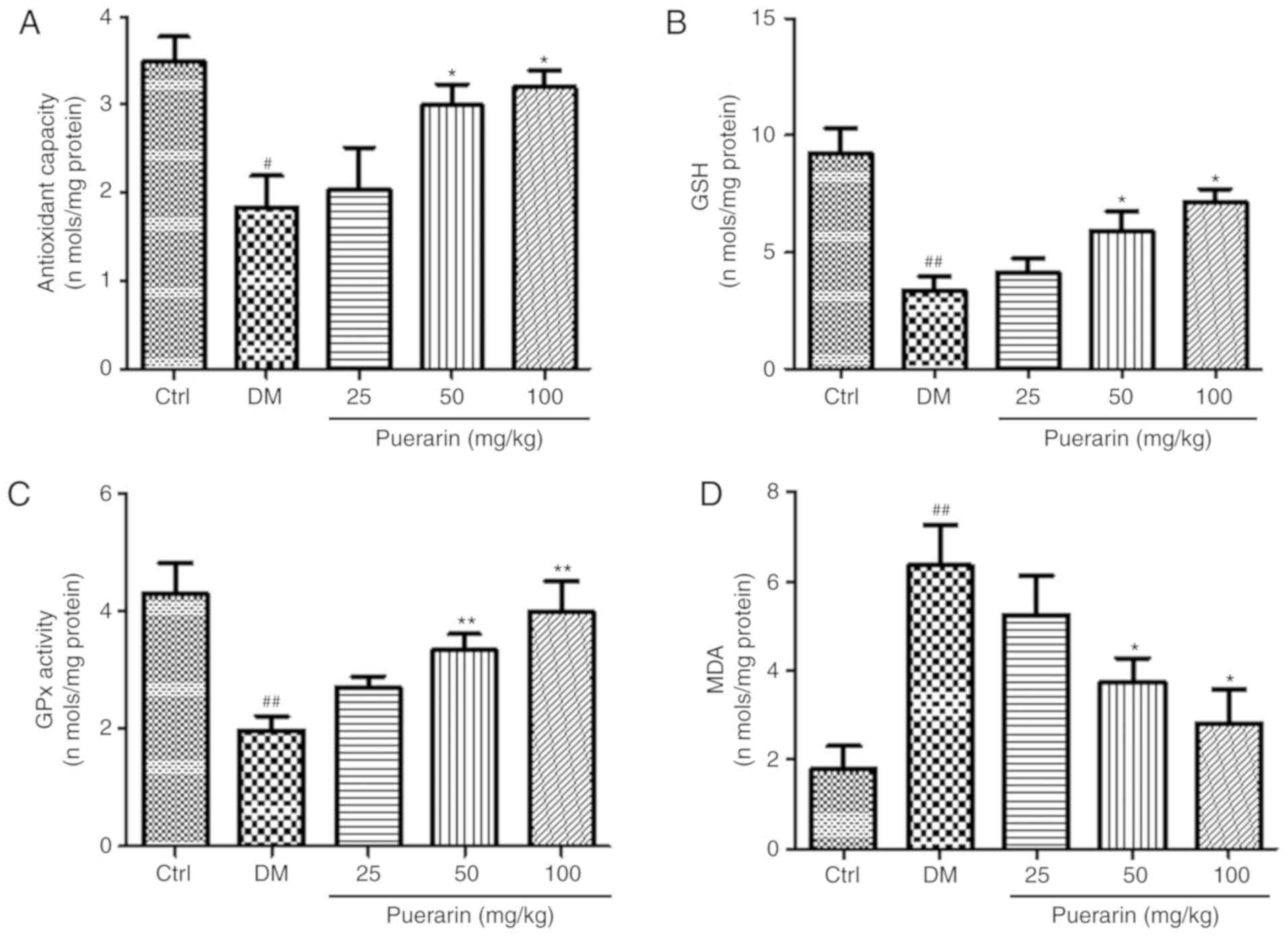 | Figure 3.Effects of puerarin on
diabetes-induced oxidative stress in the lens. (A) Antioxidant
capacity, (B) GSH levels, (C) GPx activity and (D) MDA levels were
detected using specific kits, analyzing protein homogenates of
lenses collected from the different experimental groups (n=18
rats/group). #P<0.05, ##P<0.01 vs.
Ctrl; *P<0.05, **P<0.01 vs. DM. Ctrl, untreated control rats;
DM, untreated diabetic rats; GPx, glutathione peroxidase; GSH,
glutathione; MDA, malondialdehyde. |
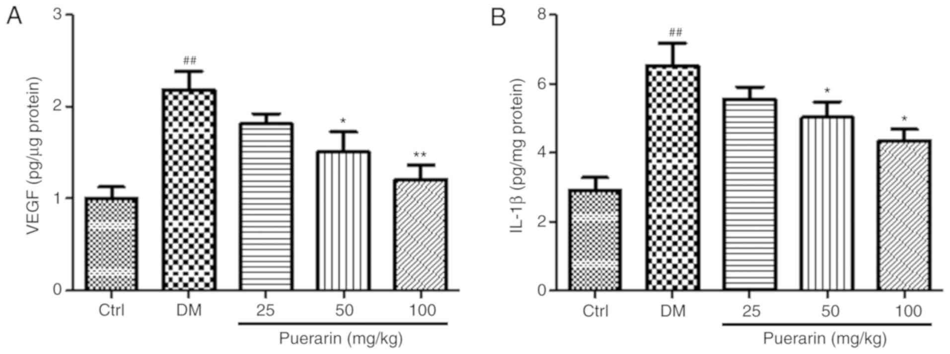
ELISA results of IL-1β and VEGF
levels
To assess the protein levels of IL-1β and VEGF in
the retina, retinas were collected from the different experimental
groups at the end of the experiment and ELISA was performed. The
untreated diabetic group exhibited significantly higher levels of
VEGF and IL-1β levels compared with the control group (Fig. 4). Notably, the protein levels of
IL-1β were significantly decreased in the 50 or 100 mg/kg puerarin
group compared with the untreated diabetic group (Fig. 4B). Similarly, treatment with
puerarin at 50 and 100 mg/kg decreased the VEGF levels compared
with the untreated diabetic group (Fig. 4A).
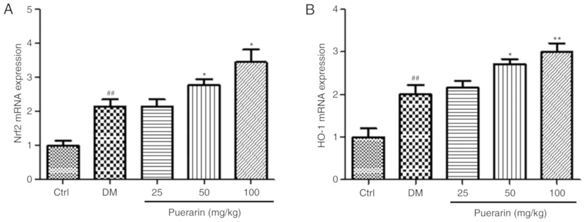
Puerarin upregulates the activity of
the Nrf2/HO-1 signaling pathway in the retinas of diabetic
rats
The effects of puerarin on the mRNA expression
levels of Nrf2 and HO-1 were determined in the retinas in the
different groups (Fig. 5). The
mRNA expression levels of Nrf2 and HO-1 were significantly
increased in the diabetic rats compared with the control group. In
addition, puerarin treatment upregulated the mRNA expression levels
of Nrf2 and HO-1; however, the higher doses of puerarin (50 or 100
mg/kg) significantly increased the mRNA expression levels of Nrf2.
Treatment with puerarin at 50 and 100 mg/kg upregulated the mRNA
expression levels of HO-1 in diabetic rats.
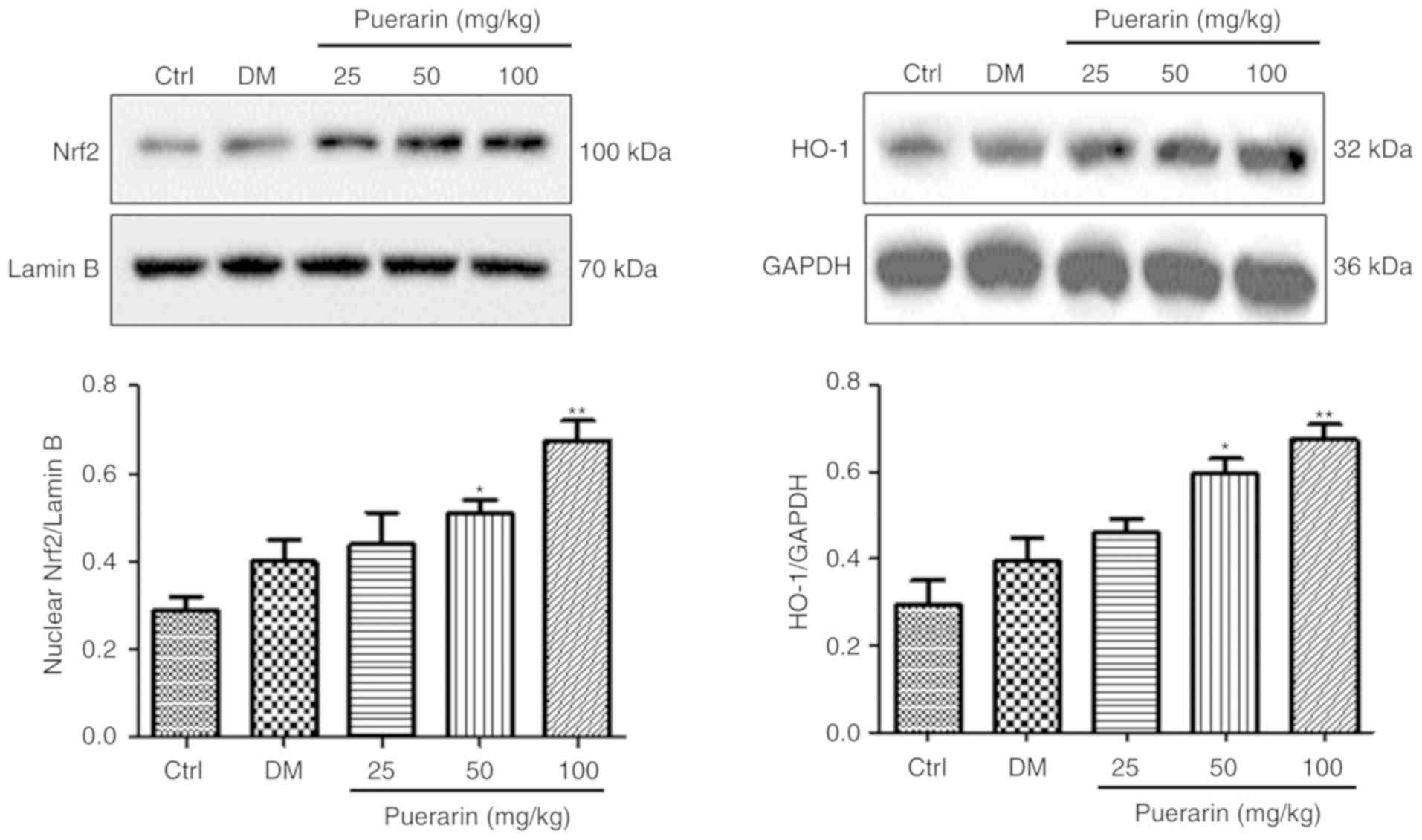
Additionally, the protein expression levels of Nrf2
in the nuclei and the protein expression levels of HO-1 were
examined by western blotting. The protein expression levels of Nrf2
and HO-1 were higher in the untreated diabetic group compared with
the control group, but not statistically significant (Fig. 6). Treatment with puerarin
significantly increased the protein expression levels of these
factors. In conclusion, treatment with puerarin could activate the
Nrf2/HO-1 signaling pathway in retinal tissues from STZ-induced
diabetic rats.
Discussion
Puerarin has been used as a traditional Chinese
medicine to treat diabetes mellitus (37). Many previous studies have reported
the preventative effects of puerarin on cataract development
(38,39); however, the mechanism underlying
the effects of puerarin on cataract development and progression
remains unclear. In the present study, puerarin was identified to
prevent cataract development and progression through the Nrf2/HO-1
signaling pathway in diabetic rats. Puerarin also decreased the
blood glucose levels in STZ-induced diabetic rats. The present
findings are consistent with previous studies showing that puerarin
treatment exhibits hypoglycemic and retinal protective effects in
diabetic rats (40,41).
It is widely accepted that oxidative stress and free
radicals are significant factors in cataract development.
Specifically, oxidative stress induces protein and lipid
peroxidation (42). Peroxidized
lipids and proteins can form insoluble aggregates in the lens,
causing the development of cataract. Lipid peroxidation has also
been associated to cataract development via perturbation of the
cytoplasm and cell membrane (43).
Puerarin is an isoflavone that exhibits a wide range
of biological activities, including antioxidant activity (44). MDA is a marker of oxidative stress
(45). In the present study, the
levels of MDA in the groups treated with puerarin were decreased
compared with the untreated diabetic group. Additionally, the
groups treated with puerarin exhibited significantly higher AOC
compared with the untreated diabetic group. GSH is an important
antioxidant agent in the retina and serves a role similar to
vitamins A, E and C, and selenium in the defense against cataract
formation (46). In the present
study, the levels of GSH in puerarin-treated rats were higher
compared with untreated diabetic rats. Retinal inflammatory
factors, such as IL-1β and VEGF, have been previously associated
with cataract development (47);
the present ELISA results suggested that puerarin induced a
decrease in the protein expression levels of IL-1β and VEGF.
Cataract is a common complication of diabetes. In
addition to oxidative stress, diabetic cataracts are caused by the
accumulation of polyols within the lenses of patients with diabetes
(48). In the lens, insulin is not
involved in the regulation of the levels of glucose or other sugars
(49). The glucose present at high
levels in the aqueous humor can diffuse passively into the lens,
where aldose reductase converts glucose into sorbitol and galactose
into galactitol. These resulting polyols cannot diffuse from the
lens to the aqueous humor (50).
Therefore, the uptake of sodium in the aqueous humor increases,
leading to swelling, electrolyte imbalance (51) and cataract formation. Puerarin is
an isoflavone, and a potent inhibitor of aldose reductase (52). Therefore, in the present study, the
antioxidative effects of puerarin were investigated.
Nrf2 is a member of the ‘cap‘n’collar’ family of
transcription factors, and serves an important role in protecting
cells via antioxidative mechanisms (53). A previous study demonstrated that
the Kelch-like ECH-associating protein 1/Nrf2/antioxidant response
element (ARE) pathway is one of the most important pathways in the
intracellular antioxidant and cytotoxic defense system (54). Nrf2 is an important transcription
factor involved in the regulation of cell responses to oxidative
stress (55,56) and interacts with AREs to induce the
expression of detoxifying enzymes, such as HO-1, a phase-II
detoxifying enzyme (57). A
previous study has demonstrated that HO-1 catalyzes the conversion
of heme into biliverdin, carbon monoxide (CO) and ferric iron
(58). In addition, biliverdin has
antioxidant properties and is an effective free radical scavenger.
Ferric iron facilitates the binding between ferritin and free iron,
thus reducing oxidative stress (59). Therefore, ferric iron is able to
attenuate oxidative toxicity (59). Endogenous CO is a gaseous cellular
messenger molecule that is involved in many biological processes
(60). In addition, HO-1 is
related to reductions in lipid peroxidation and caspase-3 activity,
as well as apoptosis resistance and tumor necrosis factor-α
inhibition (61). Nrf2/HO-1
pathway is activated when damaged and when a drug acts on the
animal, the drug can activate Nrf2/HO-1 signaling as a protective
measure (62). Although many
previous studies have reported that the Nrf2/HO-1 pathway is
involved in the development of various diseases (63,64),
to the best of our knowledge, the present study is the first to
suggest that puerarin served protective effects on cataract
development partly through the Nrf2/HO-1 pathway.
Collectively, the present study demonstrated that
puerarin could prevent cataract development and progression partly
through the Nrf2/mHO-1 signaling pathway in the retina, exhibiting
antioxidative effects in STZ-induced diabetic rats. The present
findings provide insight into the antioxidative mechanisms
underlying puerarin action in the treatment of cataract.
Acknowledgements
Not applicable.
Funding
No funding was received.
Availability of data and materials
The datasets used and/or analyzed during the current
study are available from the corresponding author on reasonable
request.
Authors' contributions
DZ and ML collaborated to the conception and design
of the study. DZ and ML contributed to acquisition, analysis, and
interpretation of the data in writing the manuscript. Both authors
read and approved the final manuscript.
Ethics approval and consent to
participate
The present study was approved by the Laboratory
Animal Management Committee of Linyi Central Hospital (approval no.
2006-398).
Patient consent for publication
Not applicable.
Competing interests
The authors declare that they have no competing
interests.
References
|
1
|
Sen S and Chakraborty R: Treatment and
diagnosis of diabetes mellitus and its complication: Advanced
approaches. Mini Rev Med Chem. 15:1132–1133. 2015. View Article : Google Scholar : PubMed/NCBI
|
|
2
|
Li L, Wan XH and Zhao GH: Meta-analysis of
the risk of cataract in type 2 diabetes. BMC Ophthalmol. 14:942014.
View Article : Google Scholar : PubMed/NCBI
|
|
3
|
Gardiner TA, Archer DB, Curtis TM and
Stitt AW: Arteriolar involvement in the microvascular lesions of
diabetic retinopathy: Implications for pathogenesis.
Microcirculation. 14:25–38. 2007. View Article : Google Scholar : PubMed/NCBI
|
|
4
|
Javadi MA and Zareighanavati S: Cataracts
in diabetic patients: A review article. J Ophthalmic Vis Res.
3:52–65. 2008.PubMed/NCBI
|
|
5
|
Murphy P, Kabir MH, Srivastava T, Mason
ME, Dewi CU, Lim S, Yang A, Djordjevic D, Killingsworth MC, Ho JWK,
et al: Light-focusing human micro-lenses derived from
zebrafish-like lens cell masses model lens development and
drug-induced cataract in vitro. Development. 145(pii):
dev1558382018.doi: 10.1242/dev.155838. View Article : Google Scholar : PubMed/NCBI
|
|
6
|
Chua J, Koh JY, Tan AG, Zhao W, Lamoureux
E, Mitchell P, Wang JJ, Wong TY and Cheng CY: Ancestry,
socioeconomic status, and age-related cataract in asians: The
Singapore epidemiology of eye diseases study. Ophthalmology.
122:2169–2178. 2015. View Article : Google Scholar : PubMed/NCBI
|
|
7
|
Head KA: Natural therapies for ocular
disorders, part two: Cataracts and glaucoma. Altern Med Rev.
6:141–166. 2001.PubMed/NCBI
|
|
8
|
Ross WM, Creighton MO and Trevithick JR:
Radiation cataractogenesis induced by neutron or gamma irradiation
in the rat lens is reduced by vitamin E. Scanning Microsc.
4:641–650. 1990.PubMed/NCBI
|
|
9
|
Brown L, Rimm EB, Seddon JM, Giovannucci
EL, Chasan-Taber L, Spiegelman D, Willett WC and Hankinson SE: A
prospective study of carotenoid intake and risk of cataract
extraction in US men. Am J Clin Nutr. 70:517–524. 1999. View Article : Google Scholar : PubMed/NCBI
|
|
10
|
Varma SD, Kumar S and Richards RD:
Light-induced damage to ocular lens cation pump: Prevention by
vitamin C. Proc Natl Acad Sci USA. 76:3504–3506. 1979. View Article : Google Scholar : PubMed/NCBI
|
|
11
|
Zhou YX, Zhang H and Peng C: Puerarin: A
review of pharmacological effects. Phytother Res. 28:961–975. 2014.
View Article : Google Scholar : PubMed/NCBI
|
|
12
|
Xie RQ, Du J and Hao YM: Myocardial
protection and mechanism of puerarin injection on patients of
coronary heart disease with ischemia/reperfusion. Zhongguo Zhong Xi
Yi Jie He Za Zhi. 23:895–897. 2003.(In Chinese). PubMed/NCBI
|
|
13
|
Jiang K, Chen H, Tang K, Guan W, Zhou H,
Guo X, Chen Z, Ye Z and Xu H: Puerarin inhibits bladder cancer cell
proliferation through the mTOR/p70S6K signaling pathway. Oncol
Lett. 15:167–174. 2018.PubMed/NCBI
|
|
14
|
Lorenzen J, Kumarswamy R, Dangwal S and
Thum T: MicroRNAs in diabetes and diabetes-associated
complications. RNA Biol. 9:820–827. 2012. View Article : Google Scholar : PubMed/NCBI
|
|
15
|
Zhao CY, Hou LH and Che HX: Influence of
puerarin eye drops for the optic disc parameters and antioxidant
capacity of patients with glaucoma. Int Eye Sci. 15:1332–1334.
2015.
|
|
16
|
Liu CM, Ma JQ, Liu SS, Feng ZJ and Wang
AM: Puerarin protects mouse liver against nickel-induced oxidative
stress and inflammation associated with the TLR4/p38/CREB pathway.
Chem Biol Interact. 243:29–34. 2016. View Article : Google Scholar : PubMed/NCBI
|
|
17
|
Ma JQ, Ding J, Xiao ZH and Liu CM:
Puerarin ameliorates carbon tetrachloride-induced oxidative DNA
damage and inflammation in mouse kidney through ERK/Nrf2/ARE
pathway. Food Chem Toxicol. 71:264–271. 2014. View Article : Google Scholar : PubMed/NCBI
|
|
18
|
Jun-Hua L, Zhang SP and Cong-Rong YU:
Effect of puerarin on blood pressure and serum lipid in a rat model
of insulin resistance. Chin J Pathophysiol. 22:997–1000. 2006.(In
Chinese).
|
|
19
|
Chen WC, Hayakawa S, Yamamoto T, Su HC,
Liu IM and Cheng JT: Mediation of beta-endorphin by the isoflavone
puerarin to lower plasma glucose in streptozotocin-induced diabetic
rats. Planta Med. 70:113–116. 2004. View Article : Google Scholar : PubMed/NCBI
|
|
20
|
Hao LN, Ling YL, Zhen-Yong GU, Huang XL
and Shou-Zhi HE: Effects of puerarin on inducible nitric oxide
synthase in lens during diabetic cataract in rats. Chin J
Pathophysiol. 4:620–626. 2004.
|
|
21
|
Qu L, Chen H, Liu X, Bi L, Xiong J, Mao Z
and Li Y: Protective effects of flavonoids against oxidative stress
induced by simulated microgravity in SH-SY5Y cells. Neurochem Res.
35:1445–1454. 2010. View Article : Google Scholar : PubMed/NCBI
|
|
22
|
Rong L, Yang XD, Chen LJ, Wang AQ and He
LF: New research progresses on and mechanism pharmacological
effects of puerarin. Popular Sci Technol. 16:138–142. 2014.
|
|
23
|
Hao LN, Wang M, Ma JL and Yang T: Puerarin
decreases apoptosis of retinal pigment epithelial cells in diabetic
rats by reducing peroxynitrite level and iNOS expression. Sheng Li
Xue Bao. 64:199–206. 2012.PubMed/NCBI
|
|
24
|
Hao LN, He SZ, Shen YH, Zhang YQ, Wang ZY
and Wang YH: Protective effects of puerarin on lens epithelial
cells in rat diabetic cataract. Zhonghua Yan Ke Za Zhi. 47:320–326.
2011.(In Chinese). PubMed/NCBI
|
|
25
|
Teng Y, Cui H, Yang M, Song H, Zhang Q, Su
Y and Zheng J: Protective effect of puerarin on diabetic
retinopathy in rats. Mol Biol Rep. 36:1129–1133. 2009. View Article : Google Scholar : PubMed/NCBI
|
|
26
|
Yuan W, Pan QI, Chen G, Yan J, Xia J and
Chen Y: E-cadherin expression in a rat model of acute pancreatitis.
Exp Ther Med. 10:2088–2092. 2015. View Article : Google Scholar : PubMed/NCBI
|
|
27
|
Dang M, Zeng X, Wang H, Li H, Du F, Chen B
and Guo C: GW28-e0833 Inhibition of myocardial ischemia/reperfusion
apoptosis by soluble receptor for advanced glycation end-product
(sRAGE) via interferon-induced immunoproteasome activity. J Am Coll
Cardiol. 70:C31–C32. 2017. View Article : Google Scholar
|
|
28
|
Park JH, Lee YJ, Kim JJ, Shin YC and Kim
JC: The effect of pinitol on cataractogenesis and anti-oxidative
effect in streptozotocin induced diabetic rats. J Korean Ophthalmol
Soc. 46:1886–1893. 2005.
|
|
29
|
Xiaojian G, Qiuyan Z and Shuhua T:
Inhibitory effect of r-hirudin variant III on
streptozotocin-induced diabetic cataracts in rats. Scientific World
J. 2013:6306512013.
|
|
30
|
Arnal E, Miranda M, Almansa I, Muriach M,
Barcia JM, Romero FJ, Diaz-Llopis M and Bosch-Morell F: Lutein
prevents cataract development and progression in diabetic rats.
Graefes Arch Clin Exp Ophthalmol. 247:115–120. 2009. View Article : Google Scholar : PubMed/NCBI
|
|
31
|
Yao XJ, Yin JA, Xia YF, Xia YF, Wei ZF,
Luo YB, Liu M, Feleder C and Dai Y: Puerarin exerts antipyretic
effect on lipopolysaccharide-induced fever in rats involving
inhibition of pyrogen production from macrophages. J
Ethnopharmacol. 141:322–330. 2012. View Article : Google Scholar : PubMed/NCBI
|
|
32
|
Zhao SS, Yang WN, Jin H, Ma KG and Feng
GF: Puerarin attenuates learning and memory impairments and
inhibits oxidative stress in STZ-induced SAD mice. Neurotoxicology.
51:166–171. 2015. View Article : Google Scholar : PubMed/NCBI
|
|
33
|
Varma SD: Scientific basis for medical
therapy of cataracts by antioxidants. Am J Clin Nutr. 53 (Suppl
1):S335–S345. 1991. View Article : Google Scholar
|
|
34
|
Kowluru RA and Kanwar M: Effects of
curcumin on retinal oxidative stress and inflammation in diabetes.
Nutr Metab (Lond). 4:82007. View Article : Google Scholar : PubMed/NCBI
|
|
35
|
Zelinová V, Mistrík I, Pavlovkin J and
Tamás L: Glutathione peroxidase expression and activity in barley
root tip after short-term treatment with cadmium, hydrogen peroxide
and t-butyl hydroperoxide. Protoplasma. 250:1057–1065. 2013.
View Article : Google Scholar : PubMed/NCBI
|
|
36
|
Livak KJ and Schmittgen TD: Analysis of
relative gene expression data using real-time quantitative PCR and
the 2(-Delta Delta C(T)) method. Methods. 25:402–408. 2001.
View Article : Google Scholar : PubMed/NCBI
|
|
37
|
Sun W, Zheng XZ, Xu QL, Nian H and Liu GL:
Effects of puerarin on ADRP gene expression in fatty tissue of type
2 diabetes mellitus rats. Zhongguo Zhong Yao Za Zhi. 33:2026–2028.
2008.(In Chinese). PubMed/NCBI
|
|
38
|
Hao LN, Ling YQ, Luo XM, Mao YX, Mao QY,
He SZ and Ling YL: Puerarin decreases lens epithelium cell
apoptosis induced partly by peroxynitrite in diabetic rats. Sheng
Li Xue Bao. 58:584–592. 2006.PubMed/NCBI
|
|
39
|
Wan L, Liu WB, Shen YY, Yu QL and Zhang
JJ: Oxidative stress-apoptosis mediated STZ-induced diabetic
cataract and the interventions of puerarin. Int J Ophthalmol.
14:1773–1775. 2014.
|
|
40
|
Xu X, Zheng N, Chen Z, Huang W, Liang T
and Kuang H: Puerarin, isolated from Pueraria lobata (Willd.),
protects against diabetic nephropathy by attenuating oxidative
stress. Gene. 591:411–416. 2016. View Article : Google Scholar : PubMed/NCBI
|
|
41
|
Cai Y, Zhang X, Xu X and Yu Y: Effects of
puerarin on the retina and STAT3 expression in diabetic rats. Exp
Ther Med. 14:5480–5484. 2017.PubMed/NCBI
|
|
42
|
Babizhayev MA, Vishnyakova KS and Yegorov
YE: Telomere-dependent senescent phenotype of lens epithelial cells
as a biological marker of aging and cataractogenesis: The role of
oxidative stress intensity and specific mechanism of phospholipid
hydroperoxide toxicity in lens and aqueous. Fundam Clin Pharmacol.
25:139–162. 2011. View Article : Google Scholar : PubMed/NCBI
|
|
43
|
Katta AV, Katkam RV and Geetha H: Lipid
peroxidation and the total antioxidant status in the pathogenesis
of age related and diabetic cataracts: A study on the lens and
blood. J Clin Diagn Res. 7:978–981. 2013.PubMed/NCBI
|
|
44
|
Li S, Yue J, Zhou W and Li L: An
investigation into the preparation, characterization and
antioxidant activity of puerarin/cyclodextrin inclusion complexes.
J Inclusion Phenomena Macrocyclic Chem. 82:1–8. 2015. View Article : Google Scholar
|
|
45
|
Cui X, Gong J, Han H, He L, Teng Y, Tetley
T, Sinharay R, Chung KF, Islam T, Gilliland F, et al: Relationship
between free and total malondialdehyde, a well-established marker
of oxidative stress, in various types of human biospecimens. J
Thorac Dis. 10:3088–3097. 2018. View Article : Google Scholar : PubMed/NCBI
|
|
46
|
Kamegawa M, Nakanishi-Ueda T, Iwai S, Ueda
T, Kosuge S, Ogura H, Sasuga K, Inagaki M, Watanabe M, Oguchi K, et
al: Effect of lipid-hydroperoxide-induced oxidative stress on
vitamin E, ascorbate and glutathione in the rabbit retina.
Ophthalmic Res. 39:49–54. 2007. View Article : Google Scholar : PubMed/NCBI
|
|
47
|
Gologorsky D, Thanos A and Vavvas D:
Therapeutic interventions against inflammatory and angiogenic
mediators in proliferative diabetic retinopathy. Mediators Inflamm.
2012:6294522012. View Article : Google Scholar : PubMed/NCBI
|
|
48
|
Abdul Nasir NA, Agarwal R, Sheikh Abdul
Kadir SH, Vasudevan S, Tripathy M, Iezhitsa I, Mohammad Daher A,
Ibrahim MI and Mohd Ismail N: Reduction of oxidative-nitrosative
stress underlies anticataract effect of topically applied
tocotrienol in streptozotocin-induced diabetic rats. PLoS One.
12:e01745422017. View Article : Google Scholar : PubMed/NCBI
|
|
49
|
Levari R, Wertheimer E, Berman ER and
Kornblueth W: Effect of insulin on pathways of glucose oxidation in
the rat lens. Nature. 192:1075–1076. 1961. View Article : Google Scholar : PubMed/NCBI
|
|
50
|
Sabah JR, Davidson H, Mcconkey EN and
Takemoto L: In vivo passage of albumin from the aqueous humor into
the lens. Mol Vis. 10:254–259. 2004.PubMed/NCBI
|
|
51
|
Drozdov AD and deClaville Christiansen J:
Modeling the effects of pH and ionic strength on swelling of
anionic polyelectrolyte gels. J Chem Phys. 142:1149042015.
View Article : Google Scholar : PubMed/NCBI
|
|
52
|
Feng CG, Zhang LX and Liu X: Progress in
research of aldose reductase inhibitors in traditional medicinal
herbs. Zhongguo Zhong Yao Za Zhi. 30:1496–1500. 2005.(In Chinese).
PubMed/NCBI
|
|
53
|
Kobayashi A, Ohta T and Yamamoto M: Unique
function of the Nrf2-Keap1 pathway in the inducible expression of
antioxidant and detoxifying enzymes. Methods Enzymol. 378:273–286.
2004. View Article : Google Scholar : PubMed/NCBI
|
|
54
|
Magesh S, Chen Y and Hu L: Small molecule
modulators of Keap1-Nrf2-ARE pathway as potential preventive and
therapeutic agents. Med Res Rev. 32:687–726. 2012. View Article : Google Scholar : PubMed/NCBI
|
|
55
|
Nakagami Y: Nrf2 is an attractive
therapeutic target for retinal diseases. Oxid Med Cell Longev.
2016:74693262016. View Article : Google Scholar : PubMed/NCBI
|
|
56
|
Foresti R, Bucolo C, Platania CM, Drago F,
Dubois-Randé JL and Motterlini R: Nrf2 activators modulate
oxidative stress responses and bioenergetic profiles of human
retinal epithelial cells cultured in normal or high glucose
conditions. Pharmacol Res. 99:296–307. 2015. View Article : Google Scholar : PubMed/NCBI
|
|
57
|
Chen CY, Jang JH, Li MH and Surh YJ:
Resveratrol upregulates heme oxygenase-1 expression via activation
of NF-E2-related factor 2 in PC12 cells. Biochem Biophys Res
Commun. 331:993–1000. 2005. View Article : Google Scholar : PubMed/NCBI
|
|
58
|
Kim KC, Kang KA, Rui Z, Piao MJ, Kim GY,
Kang MY, Lee SJ, Lee NH, Surh YJ and Hyun JW: Up-regulation of
Nrf2-mediated heme oxygenase-1 expression by eckol, a phlorotannin
compound, through activation of Erk and PI3K/Akt. Int J Biochem
Cell Biol. 42:297–305. 2010. View Article : Google Scholar : PubMed/NCBI
|
|
59
|
Finazzi D and Arosio P: Biology of
ferritin in mammals: An update on iron storage, oxidative damage
and neurodegeneration. Arch Toxicol. 88:1787–1802. 2014. View Article : Google Scholar : PubMed/NCBI
|
|
60
|
Bucolo C and Drago F: Carbon monoxide and
the eye: Implications for glaucoma therapy. Pharmacol Ther.
130:191–201. 2011. View Article : Google Scholar : PubMed/NCBI
|
|
61
|
Zhou W, Yuan X, Zhang L, Su B, Tian D, Li
Y, Zhao J, Wang Y and Peng S: Overexpression of HO-1 assisted
PM2.5-induced apoptosis failure and autophagy-related cell
necrosis. Ecotoxicol Environ Saf. 145:605–614. 2017. View Article : Google Scholar : PubMed/NCBI
|
|
62
|
Lou J, Cao G, Li R, Liu J, Dong Z and Xu
L: β-caryophyllene attenuates focal cerebral ischemia-reperfusion
injury by Nrf2/HO-1 pathway in rats. Neurochem Res. 41:1291–1304.
2016. View Article : Google Scholar : PubMed/NCBI
|
|
63
|
He M, Pan H, Chang RC, So KF, Brecha NC
and Pu M: Activation of the Nrf2/HO-1 antioxidant pathway
contributes to the protective effects of Lycium barbarum
polysaccharides in the rodent retina after
ischemia-reperfusion-induced damage. PLoS One. 9:e848002014.
View Article : Google Scholar : PubMed/NCBI
|
|
64
|
Yalniz M, Demirel U, Orhan C, Bahcecioglu
IH, Ozercan IH, Aygun C, Tuzcu M and Sahin K: Nadroparin sodium
activates Nrf2/HO-1 pathway in acetic acid-induced colitis in rats.
Inflammation. 35:1213–1221. 2012. View Article : Google Scholar : PubMed/NCBI
|