Introduction
Total hip arthroplasty is considered to be one of
the most effective therapeutic approaches for hip joint diseases
(1). It can effectively treat
late/severe arthritis, relieve pain and restore joint function.
However, with increasing life expectancy, hip joint replacements
must last longer, and the problem of prosthetic loosening is
becoming increasingly prominent (2). As a result, revision arthroplasty
procedures are increasingly required. Revision surgery is
expensive, time-consuming and dangerous for the patient, and the
results are not as good as for primary arthroplasties (3). Studies have indicated that 5–20% of
prostheses are affected by aseptic loosening, which is now widely
believed to be induced by wear debris generated from prosthetic
implant materials (4–6).
Subsequent to implantation of hip prosthesis, wear
debris may be produced between prosthetic implants and the bone
interface due to micro-movement and friction. The wear debris
activates numerous cell types, including fibroblasts, monocytes,
macrophages, giant cells, osteocytes, osteoblasts and osteoclasts
(7,8), which then induce chronic inflammatory
responses and the release of cytokines and chemokines in the
periprosthetic soft tissue (9,10).
These inflammatory factors and reactions hamper bone formation by
decreasing the synthesis of type I collagen, the major component of
bone matrix, and elevating the levels of matrix-degrading enzymes
(11,12). In addition, bone resorption is
enhanced via activation of the adhesion and differentiation of
osteoclasts (13). Overall, the
phagocytosis of wear particles by various cells disrupts the
balance between bone formation and resorption in the bone
remodeling process, potentially leading to periprosthetic
osteolysis (14).
In addition to the inflammation responsible for wear
debris-induced osteolysis, the apoptosis of several associated cell
types has recently been demonstrated to be one of the main events
involved in wear particle-associated bone resorption (4,8,15,16).
For instance, Wang et al (17) reported that direct exposure to
commercially pure Ti and ZrO2 particles compromises the
viability of human mesenchymal stem cells through the induction of
apoptosis in a manner dependent on material composition, particle
dosage and time. Landgraeber et al (18) also detected apoptosis of
T-lymphocytes in the capsules and interface membranes of patients
with aseptic hip implant loosening. Furthermore, Zhang et al
(8) demonstrated that the
β-tricalcium phosphate wear particles caused osteocyte dysfunction
by mediating apoptotic death and protein kinase B inactivation of
osteocytes. In our previous study, the apoptosis of macrophages was
identified in the interface membrane of patients undergoing
revision surgery for aseptic loosening of hip joint prostheses
(4). As the main cells involved in
bone formation, osteoblasts have also been reported to be
associated with improper bone remodeling around the prosthesis site
due to a decrease in their proliferation and an increase in their
apoptosis (13,14). However, the specific mechanism
underlying the interaction of wear debris and osteoblasts has not
been closely investigated (19).
Depending on different stimuli, apoptosis can be
executed via extrinsic and intrinsic pathways, which both involve
mitochondrial injury and endoplasmic reticulum (ER) stress
(20). Certain apoptotic stimuli
induce reactive oxygen species (ROS) release, resulting in a drop
in MMP, which causes pro-apoptotic factors to move from the
intermembrane space into the cytosol and activates apoptosis
(21). In a number of recent
studies, ROS release has also been demonstrated to induce ER
stress, which leads to unfolded protein response (UPR), ER overload
response and caspase-12-dependent apoptosis (22,23).
Our previous study further demonstrated that the
mitochondria-caspase-dependent pathway and ER stress pathway were
involved in the process of macrophage apoptosis in the capsules of
patients with aseptic loosening (4).
However, the specific mechanisms of ROS generation,
mitochondrial injury and ER stress, as well as the roles that they
serve in wear debris-induced osteoblast apoptosis and aseptic
loosening, are not well understood. Therefore, in the current
study, the direct biological effects of metallic wear debris from
loosened implants on osteoblasts were investigated, including
apoptosis, ROS release, mitochondrial injury and ER stress. The aim
of the current study was to expand our understanding of the
mechanism underlying metallic wear debris-induced aseptic
loosening.
Patients and methods
Patients
The current study was approved by the Ethics
Committee of Shanghai Ninth People's Hospital affiliated to
Shanghai Jiao Tong University School of Medicine (Shanghai, China).
Written informed consents were obtained from three patients for
inclusion and participation prior to commencing the present study.
In total, 3 patients with aseptic loosening of hip joint prostheses
were included in the study, and none of the included patients had
any infections. The patients included 1 male and 2 females, and all
exhibited definite aseptic loosening signs under X-ray scanning.
The male patient was 63 years old, had an initial diagnosis of
traumatic arthritis of the hip joint, and the implantation was
conducted 27 years before the revision surgery. One female patient
was 60 years old with an initial diagnosis of femoral neck
fracture, and underwent implantation 8 years before the revision
surgery. The other female patient was 72 years old with an initial
diagnosis of developmental dislocation of the hip, and underwent
implantation 15 years before the revision surgery. In each case,
the articulations and coupling of the loosened prostheses both
comprised a metal femoral head and a polyethylene cup. The tissue
around the osteolytic region was obtained from the three patients
at the time of revision surgery and used to extract metallic
particles.
Particle extraction and
characterization
Approximately 5 g tissue was cut into pieces of ~1
mm diameter, and repeatedly washed with deionized water to remove
blood clots. Next, the tissue was placed into a clean glass
container, and 30 ml degreasing mixture (chloroform to methanol,
2:1) was added. Subsequent to shaking at room temperature
overnight, 12 ml of 5 M NaOH solution was added for 48-h digestion
at 65°C (24). The digestion
solution was subjected to sonication for 10 min after cooling to
room temperature. A 50% sucrose solution was then added to the
digested sample and centrifuged at 3,792 × g for 1 h at room
temperature. The lower particles were harvested, and deionized
water was added for sonication for 5 min at 80°C in order to remove
residual sucrose. Subsequently, the metal particles were further
separated by density gradient centrifugation at 3,792 × g for 1 h
at room temperature (25). The
pellet suspension was then centrifuged at 474 × g for 4 min at room
temperature, and the supernatant was collected and resuspended
again. This process was repeated three times to collect all
supernatants. The collected liquid was centrifuged twice at 948 × g
for 40 min at room temperature, and the supernatant was discarded.
Sonication was used to prevent the aggregation of particles prior
to each centrifugation step, and the final collection of particles
was stored at 4°C.
The collected metallic particles were subjected to
quantitative analysis of elemental composition using inductively
coupled plasma spectroscopy (Agilent 725 ICP-OES; Agilent
Technologies, Inc., Santa Clara, CA, USA). The size and
distribution of the metallic particles was analyzed using a laser
particle size analyzer (Mastersizer 2000; Malvern Panalytical Ltd.,
Malvern, UK). The final collected metallic particles were
sterilized at 180°C for 6 h, followed by treatment with 70% ethanol
for 48 h (26). Subsequently, the
metallic wear debris was sonicated for 30 min using a SB3200
Ultrasonic Generator (Shanghai Branson Ultrasonic Co., Ltd.,
Shanghai, China), prior to incubation with cells. The
concentrations of metallic particles used for cell incubation were
0.05 and 0.1 mg/ml.
Cell culture and treatment
The study was approved by the Ethics Committee of
Shanghai Ninth People's Hospital affiliated to Shanghai Jiao Tong
University School of Medicine. A total of 10 newborn (<24 h)
Sprague-Dawley rats (5 male and 5 female, weighing 5–6 g) were
purchased from the Shanghai Model Organisms Center, Inc. (Shanghai,
China). Animals were housed under specific pathogen-free conditions
at 26°C, under a 12-h light/dark cycle, and with free access to
sterile food and water. Primary osteoblasts were isolated from the
calvarias, as previously described with modifications (27,28).
Briefly, neonatal rat calvarias were dissected from adherent soft
tissue and washed in a PBS solution. The tissue was cut into pieces
using ophthalmic scissors and then sequentially digested with 1%
trypsin for 10 min, followed by addition of 0.2% collagenase
type≈II for 60 min, in order to release the cells. The cells from
the final digestion were resuspended in culture medium and plated
in flasks. All the cells were routinely cultured in high-glucose
Dulbecco's modified Eagle's medium (DMEM; Hyclone; GE Healthcare
Life Sciences, Logan, UT, USA), supplemented with 10% FBS (Gibco;
Thermo Fisher Scientific, Inc., Waltham, MA, USA) and 1%
penicillin-streptomycin in a humidified atmosphere with 5%
CO2 and 95% humidity at 37°C in an incubator. The medium
was changed every other day, and cells in the third passage were
used for all experiments. In order to evaluate the role of wear
debris-induced ROS on apoptosis, cells were treated with 0.1 mg/ml
wear debris for 24 h in the absence or presence of 50 mM
antioxidant N-acetyl-L-cysteine (NAC; Sigma-Aldrich; Merck KGaA,
Darmstadt, Germany) dissolved in deionized water. Cells cultured in
medium alone were used as the control group.
Analysis of apoptosis by flow
cytometry
Annexin V and propidium iodide (PI) double staining
was used to determine the rate of apoptosis, using the Annexin
V/Dead Cell Apoptosis kit (Invitrogen; Thermo Fisher Scientific,
Inc.). Briefly, following treatment with wear debris at 0.05 and
0.1 mg/ml for 12, 24 and 48 h, the primary osteoblasts were
harvested and centrifuged at 948 × g for 5 min at room temperature.
Subsequent to discarding the supernatant, the cell pellets were
washed twice with cold PBS and resuspended in 100 µl Annexin V
binding buffer. Next, 5 µl Alexa Fluor® 488 Annexin V
and 1 µl PI (100 µg/ml) staining of the suspension was conducted at
room temperature for 15 min, followed by addition and gentle mixing
with 400 µl Annexin V binding buffer. Flow cytometry (FACSAria; BD
Biosciences, Franklin Lakes, NJ, USA) was finally conducted at a
wavelength of 488 nm to analyze the cell apoptosis.
ROS measurement
2′,7′-dichlorofluorescein diacetate (DCFH-DA;
Sigma-Aldrich; Merck KGaA) can be deacetylated by intracellular
esterase to form non-fluorescent DCFH, which can be oxidized by
ROS, resulting in the formation of the fluorescent compound
2′,7′-dichloroflorescein (DCF). The fluorescence intensity of DCF
is proportional to the amount of ROS produced by the cells
(29,30). In the present study, osteoblasts
treated with 0.1 mg/ml wear debris for 6, 12 and 24 h in the
absence or presence of the antioxidant NAC were collected, washed
once with ice-cold PBS and incubated with DCFH-DA (50 µM final
concentration) at 37°C for 30 min in the dark. Next, the cells were
washed twice and maintained in 1 ml of PBS. ROS generation was then
assessed using a fluorescence microscope (Axio Imager A2; Zeiss AG,
Oberkochen, Germany) or a fluorescence plate reader (Tecan Infinite
M200; Tecan Group Ltd., Mannedorf, Switzerland) at excitation and
emission wavelengths of 485 and 530 nm, respectively.
Determination of mitochondrial
membrane potential (MMP) with JC-1 staining
The primary osteoblasts were treated with wear
debris for 12 and 24 h. Next, the medium was removed, and cells
were washed with PBS and incubated with JC-1 staining working
solution (MitoProbe JC-1 assay kit; Thermo Fisher Scientific, Inc.)
at 37°C for 20 min. Following washing with staining buffer and PBS,
the changes in MMP were visualized using a confocal laser scanning
microscope (LSM510; Zeiss AG), and images were captured.
Analysis of extracellular activated
caspases
Following incubation with wear debris for 24 and 48
h in the absence or presence of the antioxidant NAC, the amount of
caspase-3/9/12 proteins secreted into the culture medium was
determined using the corresponding caspase-3/9/12 fluorometric
assay kits (BioVision, Inc., Milpitas, CA, USA), according to the
manufacturer's protocol. The absorbance was measured at 400 nm
using a microplate reader (Tecan Infinite M200), and activated
caspase-3/9/12 levels were quantified from the relevant standard
curve. All samples were measured in triplicate, and the experiment
was repeated three times independently.
Western blot analysis
Following treatment under different conditions, the
cells were harvested and the medium was removed. A nuclear/cytosol
fractionation kit and a mitochondria isolation kit (both purchased
from BioVision, Inc.) were used to isolate the nuclei and
mitochondria, respectively, according to the manufacturer's
protocols. Cells lysis was then performed for 30 min on ice using
radioimmunoprecipitation assay lysis buffer and protease inhibitor
cocktail (both Beyotime Institute of Biotechnology, Shanghai,
China). Following centrifugation of the lysate at 12,000 × g for 10
min at 4°C, the protein in the supernatant was collected, and a
bicinchoninic acid assay was performed to measure protein
concentrations. Protein lysate samples (20 mg) were resolved by
sodium dodecyl sulfate-polyacrylamide gel electrophoresis using 10%
gels, and proteins were then transferred to polyvinylidene
difluoride membranes (EMD Millipore, Billerica, MA, USA).
Subsequent to blocking nonspecific interactions by incubation with
5% skim milk for 1 h at room temperature, the membranes were probed
overnight at 4°C with specific primary antibodies against
cytochrome c (Cyto-c; 1:1,000; cat. no. 11940), glucose-regulated
protein 78 (GRP78; 1:1,000; cat. no. 3177), growth arrest DNA
damage 153 (GADD153; 1:1,000; cat. no. 2895) and porin (1:1,000;
cat. no. 4866; all purchased from Cell Signaling Technology, Inc.,
Danvers, MA, USA), as well as with GAPDH antibody (1:5,000; cat.
no. KC-5G4; Kangcheng, Shanghai, China). Then, membranes were
incubated with goat anti-rabbit or goat anti-mouse horseradish
peroxidase-conjugated secondary antibodies (1:5,000; cat. no. A0208
or A0216; Beyotime Institute of Biotechnology) for 1 h at room
temperature. The bound antibodies were then visualized using an
enhanced chemiluminescence detection system (EMD Millipore).
Statistical analysis
All data were obtained from three or more
experiments, and values are presented as the mean ± standard
deviation. Statistical analysis was conducted using SPSS software
version 20 (IBM Corp., Armonk, NY, USA). Statistical comparisons
were performed using one-way analysis of variance, followed by the
Student-Newman-Keul post hoc test. P<0.01 was considered to
indicate a statistically significant difference.
Results
Patients
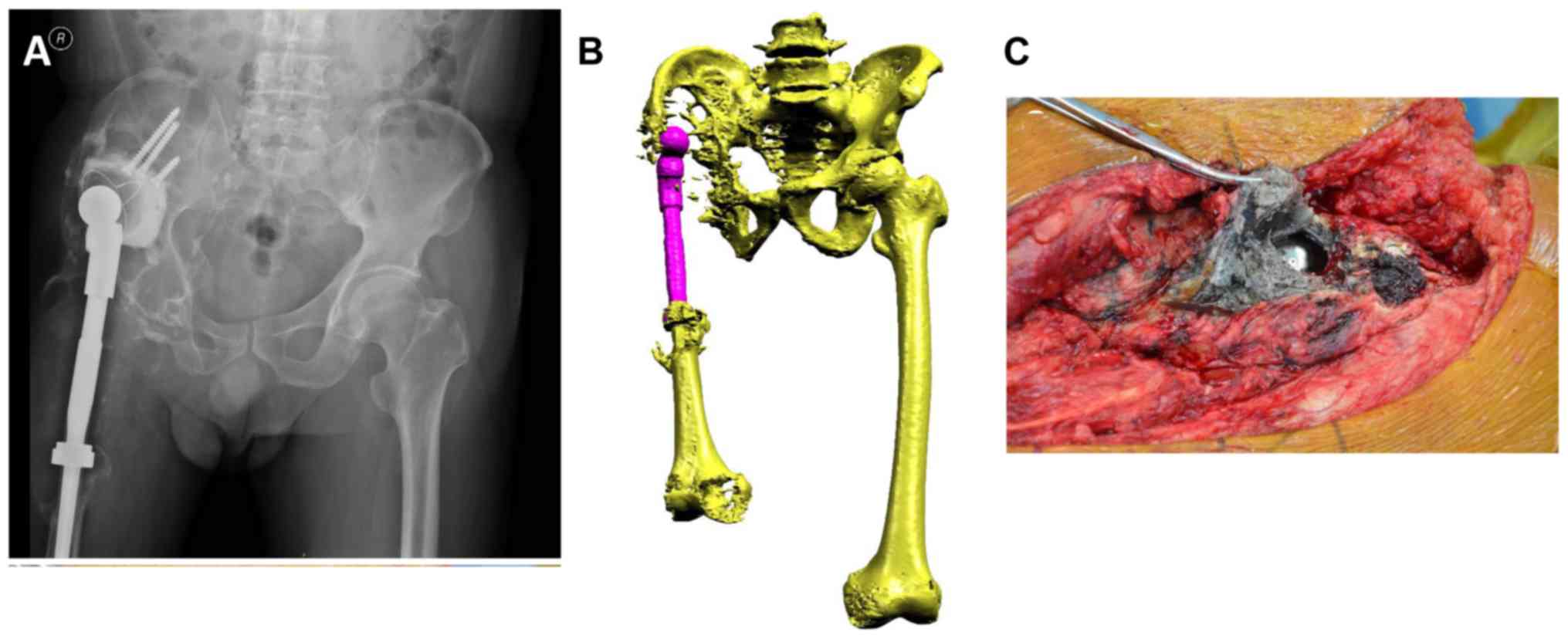
The representative case of an aseptic loosening
patient (63-year-old male) is depicted in Fig. 1. Pre-operative X-ray (Fig. 1A) and three-dimensional computed
tomography (Fig. 1B) scans
indicated the migration of the acetabular component and massive
periacetabular osteolysis. During surgery, the tissue around the
osteolytic region appeared black due to the accumulation of a large
number of metallic wear particles (Fig. 1C).
Characterization of wear debris
Inductively coupled plasma analysis indicated that
the collected metallic wear debris from all three patients were
primarily composed of Ti, Co, Cr, Mo, Al and V, which was
consistent with the prosthesis alloy composition (data not shown).
The size of the lower particles following the initial
centrifugation ranged between 0.05 and 47.30 µm, with a mean size
of 19.06 µm. Subsequent to density gradient centrifugation, the
final collected metallic particles ranged between 0.45 and 1.67 µm
in size, with a mean size of 1.22 µm, and >90% of the particles
were 0.90–1.50 µm in size.
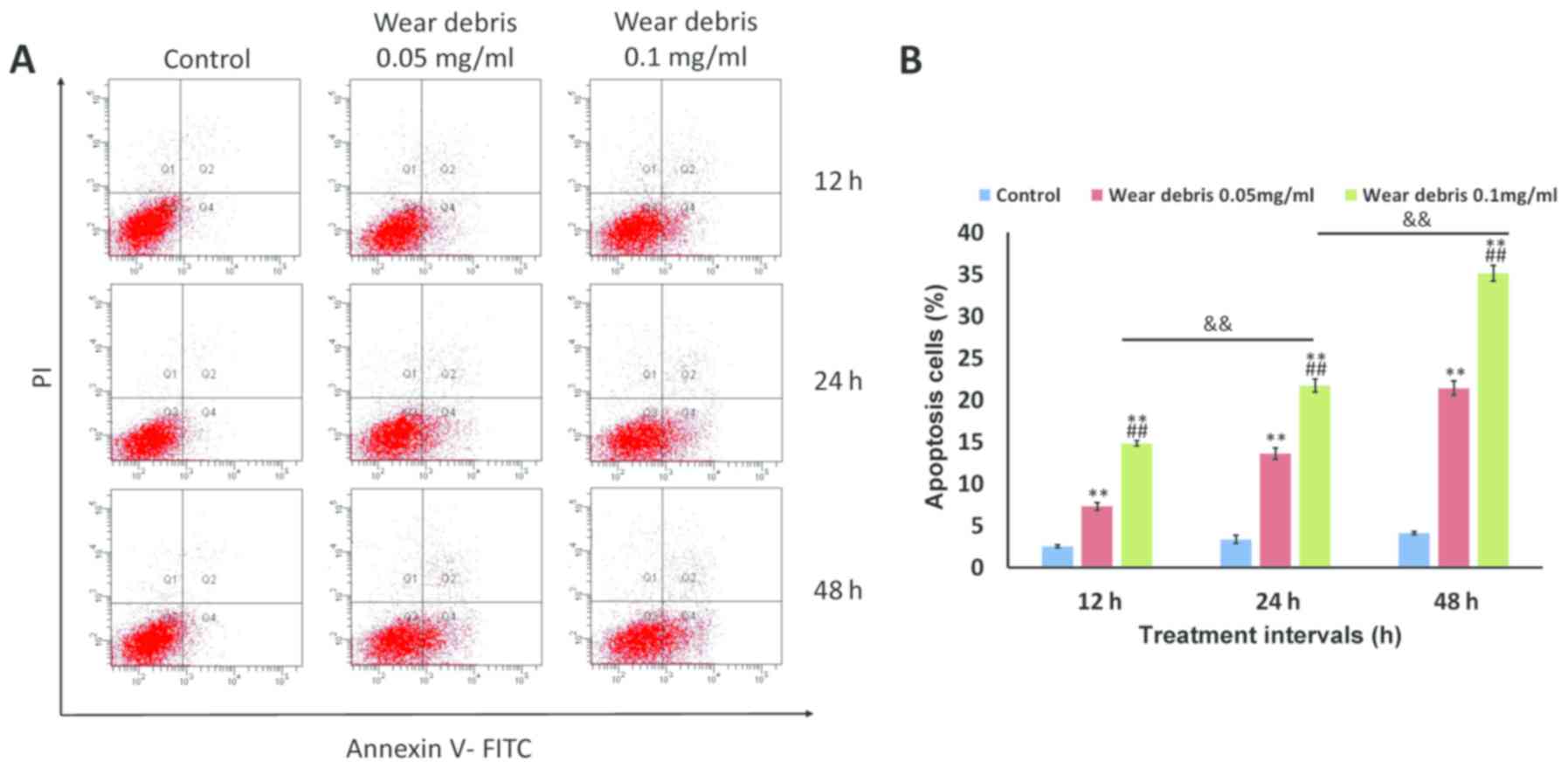
Induction of apoptosis by metallic
wear debris at different concentrations in rat primary
osteoblasts
The results of flow cytometry analysis indicated
that significant apoptosis was induced in rat primary osteoblasts
following exposure to metallic wear debris in a concentration- and
time-dependent manner (Fig. 2).
The strongest induction of apoptosis was observed at 0.1 mg/ml wear
debris; therefore, this concentration was selected for use in
subsequent experiments.
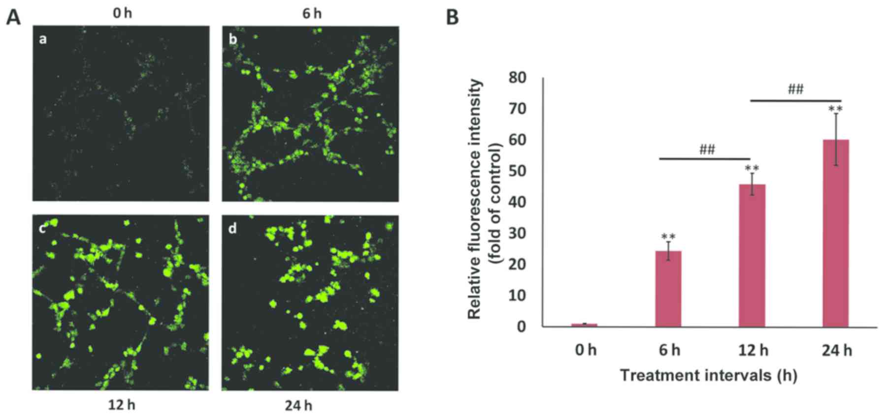
ROS generation and MMP reduction in
metallic wear debris-induced apoptosis
ROS is known to serve an essential role in the
regulation of cellular apoptosis (31). As depicted in Fig. 3, ROS release was highly induced by
wear debris treatment in a time-dependent manner in rat primary
osteoblasts, indicating that the metallic wear debris-induced
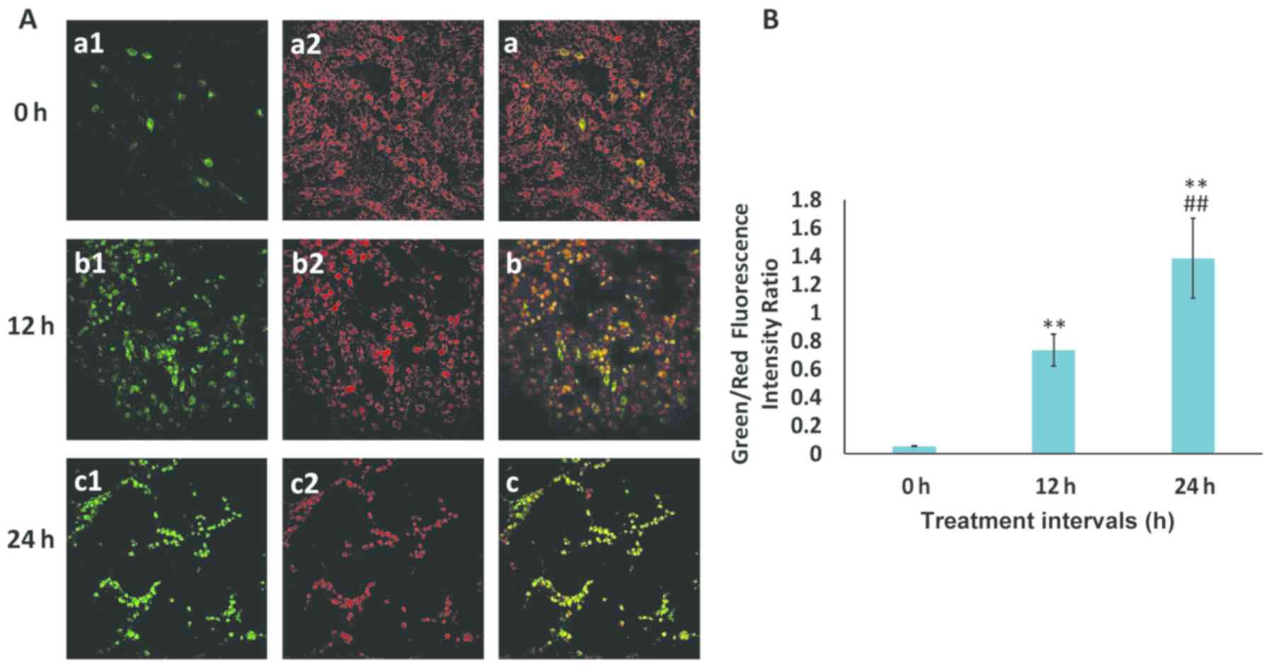
apoptosis was associated with ROS production. Furthermore, to
detect MMP, the JC-1 probe was used, which is a marker of
mitochondrial depolarization in the early apoptotic process. During
early apoptosis, as a result of the drop in MMP, JC-1 aggregates
(red fluorescence) cannot accumulate within the mitochondria and
dissipate into JC-1 monomers (green fluorescence), leading to a
loss of red fluorescence. Therefore, a decrease in MMP is signified
by an increase in the ratio of green to red fluorescence. In the
current study, the MMP was reduced in a time-dependent manner
following wear debris treatment (Fig.
4), implying that metallic wear debris may mediate the
intrinsic apoptotic signaling pathway in osteoblasts.
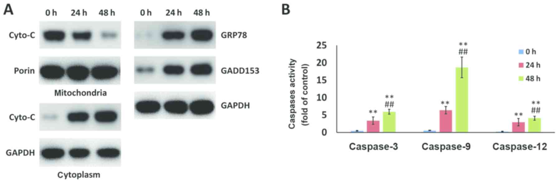
Metallic wear debris induces apoptosis
through the mitochondria-caspase-dependent and ER stress
pathways
In addition to mitochondria-associated apoptosis, ER
stress-induced apoptosis also serves a pivotal role in cell death.
Western blot analysis indicated that mitochondrial Cyto-c was
released into the cytoplasm following wear debris treatment. In
addition, the markers of the ER stress apoptotic pathway, namely
GRP78 and GADD153, were enhanced by wear debris treatment in a
time-dependent manner (Fig. 5A).
In addition, activated caspase-3, caspase-9 and caspase-12 levels
were examined, and these were observed to be significantly
upregulated following treatment for 48 h as compared with the value
at 24 h (Fig. 5B). These results
suggested that the metallic wear debris induced apoptosis in rat
primary osteoblasts by both the mitochondria-caspase-dependent
pathway and the ER stress pathway.
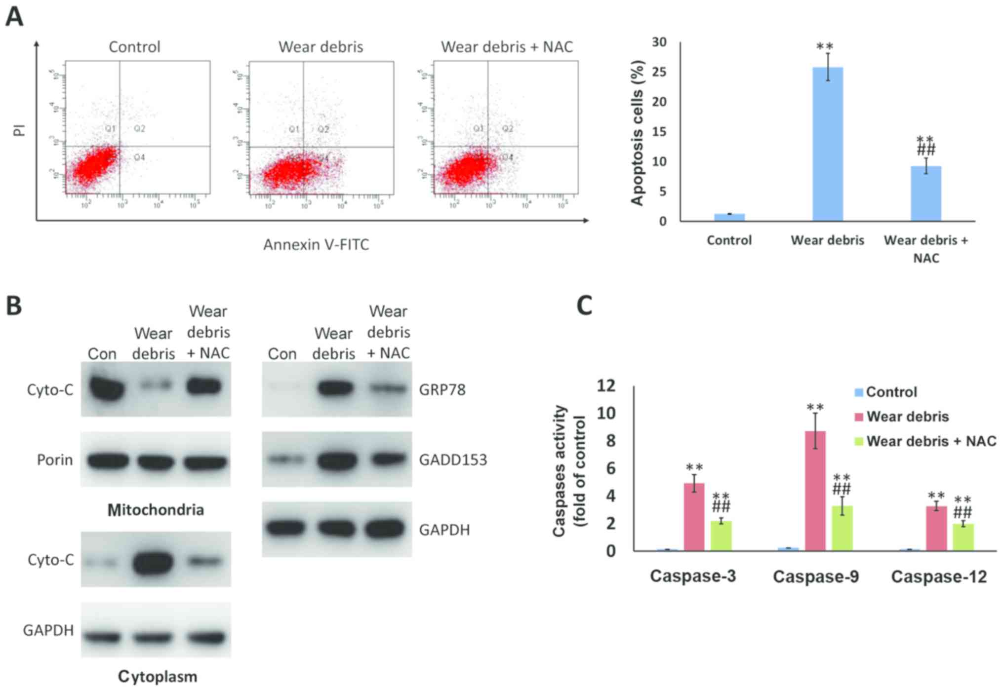
Inhibition of ROS attenuates metallic
wear debris-induced apoptosis
Antioxidant NAC, as an inhibitor of ROS, was
observed to markedly downregulate the metallic wear debris-induced
apoptosis (Fig. 6A). In addition,
treatment with NAC hindered the increase of Cyto-c in the
cytoplasm, and of GRP78 and GADD153 expression levels, as compared
with those in cells treated with metallic wear debris alone
(Fig. 6B). NAC treatment also
significantly suppressed the activation of caspase-3, caspase-9 and
caspase-12 (Fig. 6C). These
findings provided further evidence that the ROS release caused by
metallic wear debris resulted in the mitochondria- and ER
stress-regulated apoptosis of rat primary osteoblasts.
Discussion
As a successful treatment for end-stage arthritis,
artificial joint replacement surgery restores mobility, reduces
pain and improves the quality of life for millions of patients.
However, wear of the implants over time generates debris, which may
cause a series of biological responses in the tissues surrounding
the joint space, finally leading to aseptic loosening and limiting
the longevity of the prosthesis. The wear debris around the
prosthesis mainly includes polyethylene (PE), metal and bone cement
particles. These particles cause multiple biological reactions,
among which metallic particle-induced osteoblast apoptosis has been
proposed to be one of the main causes of prosthesis osteolysis in
recent years (14,32,33).
However, owing to the complexity of the factors involved, the
specific regulatory mechanism has not yet been revealed. The
current study aimed to investigate the direct biological effects of
metallic wear particles on osteoblasts and to elucidate the
possible signaling pathways that result in cell dysfunction.
In the majority of studies, the wear debris used for
evaluation has been mainly derived from commercial particles
(2,34,35).
However, due to the various types of wear and the in vivo
environment, there are several differences between commercial
particles and the wear debris extracted from the interfacial
membrane in vivo, including the morphology, size and
biological behavior of particles. Therefore, in the present study,
metallic wear debris obtained from patients with aseptic joint
loosening was used to ensure that the experiments more closely
resembled the biological response in vivo and to provide
more relevant insights into clinical treatment.
It has been demonstrated that the cellular response
to wear debris varies with the size, shape, composition, charge and
concentration of particles (36,37).
Furthermore, particle phagocytosis represents an important
component of the cellular response to implants; thus, the size of
wear particles may be a significant factor affecting osteolysis.
Studies have estimated that small particles ranging between 0.2 and
10 µm in diameter undergo phagocytosis by macrophages (38), and particles <20 µm elicit a
significantly greater inflammatory cytokine response (39). In the present study, the wear
debris obtained by preliminary extraction included various sizes of
particles, which suggested that there may be a variety of wear
mechanisms involved. Therefore, density gradient centrifugation was
performed to isolate metallic particles of a similar size, which
were <2 µm in diameter and within the reported range of
phagocytosis.
Wear debris from prostheses has been demonstrated to
serve an important role in the initiation of aseptic loosening.
Beside the direct effect on bone homeostasis by stimulating
osteoclastogenesis, particle exposure also has adverse effects on
osteoblastogenesis. Osteoblasts, derived from mesenchymal stem
cells, are the main cells in bone tissue and bone formation, and
are considered to be critical for prosthesis stability. Continuous
exposure to wear debris gives rise to the phagocytosis of mature
osteoblasts, and compromises osteoblast function and survival, such
as by inducing apoptosis, which finally suppresses bone formation
around the prosthesis.
The mitochondrial pathway is considered to be an
important apoptotic pathway. Landgraeber et al (40) reported strong expression of
Bcl-2-antagonist/killer in macrophages and giant cells in interface
tissue, and proposed that the intrinsic apoptotic pathway
(mitochondrial pathway) was activated in these cells. As the ER
regulates apoptosis, the ER stress pathway has been proposed as
another apoptotic pathway that differs from mitochondrial pathways.
In our previous study, ER stress pathway-associated markers,
including GRP78, GADD153 and caspase-4, were demonstrated to be
activated in the aseptic loosening of prostheses using
immunohistochemistry (4). It has
been demonstrated that ROS are formed as a natural byproduct of the
normal metabolism of oxygen, and serve an important role in a
series of biological behaviors, such as lipid peroxidation, DNA
chain fragmentation and protein modification (41). However, during periods of
environmental stress, ROS levels can increase dramatically,
resulting in significant damage to cell structures. In the present
study, the phagocytosis of metallic wear particles on rat primary
osteoblasts directly led to an increase in ROS, which then
triggered downstream cellular changes against oxidative stress and
finally induced apoptosis. This was also confirmed by the finding
that apoptosis was evidently attenuated subsequent to treatment
with the antioxidant NAC to inhibit ROS generation. Notably, it has
been indicated that ROS can be produced in the ER as a byproduct of
oxidative protein folding (42)
and in the mitochondria as a byproduct of mitochondrial respiration
(43). Furthermore, the ER,
mitochondria and oxidative stress interact with each other to
interfere with cell function and activate apoptosis signaling in
vitro and in vivo (44,45).
Hence, in the current study, it was speculated that the stimulation
of metallic wear debris induced cellular stress through the
activation of ER stress and mitochondrial oxidative stress by ROS
generation. In the early stages of ER stress, ROS generation is
also associated with the induction of UPR genes, such as GRP78,
suggesting the functional propagation of an ER stress response.
However, when the UPR process fails to restore adequate ER
function, cell death signaling pathways will be activated, as
indicated by the activation of GADD153 and caspase-12 (46). By contrast, oxidative stress
induces excessive ROS production, activating apoptosis signaling,
such as Cyto-c release and caspase cascades, which will contribute
to cell apoptosis. In the current study, the aforementioned
proteins increased upon metallic wear debris treatment, and their
levels were restored by addition of the antioxidant NAC to inhibit
ROS production, indicating that wear debris induced-ROS generation
was one of the initiating factors of apoptosis.
In conclusion, in the present study, metallic wear
debris particles of a similar size were isolated and collected from
the tissue around the loosened implants of patients, and these
particles were found to directly cause the apoptosis of rat primary
osteoblasts. The results indicated that ROS generation induced by
metallic wear debris triggered ER stress, mitochondrial dysfunction
and downstream caspase cascades, ultimately leading to cell
apoptosis. This was further verified by the finding that apoptosis
was attenuated following antioxidant treatment. These findings
improved our knowledge on the mechanism of metallic wear
debris-induced osteoblast apoptosis and may provide a basis for
controlling bone destruction during the treatment of implant
loosening.
Acknowledgements
Not applicable.
Funding
The present study was supported by grants from the
National Nature Science Foundation of China (no. 81301548), the
Shanghai Municipal Natural Science Foundation (no. 13ZR1424100),
the Natural Science Foundation of Shanghai (no. 16ZR1441700), the
Shanghai Jiaotong University ‘Cross Research Fund of Medical
Engineering’ (no. YG2013MS57), and the Fund for Key Disciplines of
Shanghai Municipal Education Commission (no. J50206).
Availability of data and materials
All data generated or analyzed during this study are
included in this published article.
Authors' contributions
FY and YH designed the study and performed the
experiments. JT obtained patient tissue for particle extraction,
and provided X-ray and three-dimensional CT images. KD contributed
to method design and revised the manuscript critically for
important intellectual content. All authors read and approved the
manuscript.
Ethics approval and consent to
participate
The current study was approved by the Ethics
Committee of Shanghai Ninth People's Hospital affiliated to
Shanghai Jiao Tong University School of Medicine. Written informed
consent was obtained from all patients.
Patient consent for publication
Not applicable.
Competing interests
The authors declare that they have no competing
interests.
References
|
1
|
Meermans G, Konan S, Das R, Volpin A and
Haddad FS: The direct anterior approach in total hip arthroplasty:
A systematic review of the literature. Bone Joint J. 99-B:732–740.
2017. View Article : Google Scholar : PubMed/NCBI
|
|
2
|
Petis S, Howard JL, Lanting BL and
Vasarhelyi EM: Surgical approach in primary total hip arthroplasty:
Anatomy, technique and clinical outcomes. Can J Surg. 58:128–139.
2015. View Article : Google Scholar : PubMed/NCBI
|
|
3
|
Sundfeldt M, Carlsson LV, Johansson CB,
Thomsen P and Gretzer C: Aseptic loosening, not only a question of
wear: A review of different theories. Acta Orthop. 77:177–197.
2006. View Article : Google Scholar : PubMed/NCBI
|
|
4
|
Yang F, Wu W, Cao L, Huang Y, Zhu Z, Tang
T and Dai K: Pathways of macrophage apoptosis within the interface
membrane in aseptic loosening of prostheses. Biomaterials.
32:9159–9167. 2011. View Article : Google Scholar : PubMed/NCBI
|
|
5
|
Wooley PH and Schwarz EM: Aseptic
loosening. Gene Ther. 11:402–407. 2004. View Article : Google Scholar : PubMed/NCBI
|
|
6
|
Cherian JJ, Jauregui JJ, Banerjee S,
Pierce T and Mont MA: What host factors affect aseptic loosening
after THA and TKA? Clin Orthop Relat Res. 473:2700–2709. 2015.
View Article : Google Scholar : PubMed/NCBI
|
|
7
|
Shimizu S, Okuda N, Kato N, Rittling SR,
Okawa A, Shinomiya K, Muneta T, Denhardt DT, Noda M, Tsuji K and
Asou Y: Osteopontin deficiency impairs wear debris-induced
osteolysis via regulation of cytokine secretion from murine
macrophages. Arthritis Rheum. 62:1329–1337. 2010. View Article : Google Scholar : PubMed/NCBI
|
|
8
|
Zhang Y, Yan M, Yu A, Mao H and Zhang J:
Inhibitory effects of β-tricalciumphosphate wear particles on
osteocytes via apoptotic response and Akt inactivation. Toxicology.
297:57–67. 2012. View Article : Google Scholar : PubMed/NCBI
|
|
9
|
Hallab NJ and Jacobs JJ: Biologic effects
of implant debris. Bull NYU Hosp Jt Dis. 67:182–188.
2009.PubMed/NCBI
|
|
10
|
Goodman SB and Ma T: Cellular chemotaxis
induced by wear particles from joint replacements. Biomaterials.
31:5045–5050. 2010. View Article : Google Scholar : PubMed/NCBI
|
|
11
|
Vidovszky TJ, Cabanela ME, Rock MG, Berry
DJ, Morrey BF and Bolander ME: Histologic and biochemical
differences between osteolytic and nonosteolytic membranes around
femoral components of an uncemented total hip arthroplasty. J
Arthroplasty. 13:320–330. 1998. View Article : Google Scholar : PubMed/NCBI
|
|
12
|
Yao J, Cs-Szabó G, Jacobs JJ, Kuettner KE
and Glant TT: Suppression of osteoblast function by titanium
particles. J Bone Joint Surg Am. 79:107–112. 1997. View Article : Google Scholar : PubMed/NCBI
|
|
13
|
Vermes C, Chandrasekaran R, Jacobs JJ,
Galante JO, Roebuck KA and Glant TT: The effects of particulate
wear debris, cytokines, and growth factors on the functions of
MG-63 osteoblasts. J Bone Joint Surg Am. 83-A:201–211. 2001.
View Article : Google Scholar : PubMed/NCBI
|
|
14
|
Lochner K, Fritsche A, Jonitz A, Hansmann
D, Mueller P, Mueller-Hilke B and Bader R: The potential role of
human osteoblasts for periprosthetic osteolysis following exposure
to wear particles. Int J Mol Med. 28:1055–1063. 2011.PubMed/NCBI
|
|
15
|
Posada OM, Gilmour D, Tate RJ and Grant
MH: CoCr wear particles generated from CoCr alloy metal-on-metal
hip replacements, and cobalt ions stimulate apoptosis and
expression of general toxicology-related genes in monocyte-like
U937 cells. Toxicol Appl Pharmacol. 281:125–135. 2014. View Article : Google Scholar : PubMed/NCBI
|
|
16
|
Thomas V, Halloran BA, Ambalavanan N,
Catledge SA and Vohra YK: In vitro studies on the effect of
particle size on macrophage responses to nanodiamond wear debris.
Acta Biomater. 8:1939–1947. 2012. View Article : Google Scholar : PubMed/NCBI
|
|
17
|
Wang ML, Tuli R, Manner PA, Sharkey PF,
Hall DJ and Tuan RS: Direct and indirect induction of apoptosis in
human mesenchymal stem cells in response to titanium particles. J
Orthop Res. 21:697–707. 2003. View Article : Google Scholar : PubMed/NCBI
|
|
18
|
Landgraeber S, von Knoch M, Löer F,
Brankamp J, Tsokos M, Grabellus F, Schmid KW and Totsch M:
Association between apoptotis and CD4(+)/CD8(+) T-lymphocyte ratio
in aseptic loosening after total hip replacement. Int J Biol Sci.
5:182–191. 2009. View Article : Google Scholar : PubMed/NCBI
|
|
19
|
O'Neill SC, Queally JM, Devitt BM, Doran
PP and O'Byrne JM: The role of osteoblasts in peri-prosthetic
osteolysis. Bone Joint J. 95-B:1022–1026. 2013. View Article : Google Scholar : PubMed/NCBI
|
|
20
|
Orrenius S, Gogvadze V and Zhivotovsky B:
Mitochondrial oxidative stress: Implications for cell death. Annu
Rev Pharmacol Toxicol. 47:143–183. 2007. View Article : Google Scholar : PubMed/NCBI
|
|
21
|
Kroemer G, Galluzzi L and Brenner C:
Mitochondrial membrane permeabilization in cell death. Physiol Rev.
87:99–163. 2007. View Article : Google Scholar : PubMed/NCBI
|
|
22
|
Sano R and Reed JC: ER stress-induced cell
death mechanisms. Biochim Biophys Acta 1833. 3460–3470. 2013.
|
|
23
|
Gorman AM, Healy SJ, Jäger R and Samali A:
Stress management at the ER: Regulators of ER stress-induced
apoptosis. Pharmacol Ther. 134:306–316. 2012. View Article : Google Scholar : PubMed/NCBI
|
|
24
|
Minovic A, Milowsev I, Pisot V, Cör A and
Antolic V: Isolation of polyacetal wear particles from
periprosthetic tissue of isoelastic femoral stems. J Bone Joint
Surg Br. 83:1182–1190. 2001. View Article : Google Scholar : PubMed/NCBI
|
|
25
|
Maloney WJ, Smith RL, Schmalzried TP,
Chiba J, Huene D and Rubash H: Isolation and characterization of
wear particles generated in patients who have had failure of a hip
arthroplasty without cement. J Bone Joint Surg Am. 77:1301–1310.
1995. View Article : Google Scholar : PubMed/NCBI
|
|
26
|
Liu F, Zhu Z, Mao Y, Liu M, Tang T and Qiu
S: Inhibition of titanium particle-induced osteoclastogenesis
through inactivation of NFATc1 by VIVIT peptide. Biomaterials.
30:1756–1762. 2009. View Article : Google Scholar : PubMed/NCBI
|
|
27
|
Wornham DP, Hajjawi MO, Orriss IR and
Arnett TR: Strontium potently inhibits mineralisation in
bone-forming primary rat osteoblast cultures and reduces numbers of
osteoclasts in mouse marrow cultures. Osteoporos Int. 25:2477–2484.
2014. View Article : Google Scholar : PubMed/NCBI
|
|
28
|
Hou JM, Chen EY, Wei SC, Lin F, Lin QM,
Lan XH, Xue Y and Wu M: Lactoferrin inhibits apoptosis through
insulin-like growth factor I in primary rat osteoblasts. Acta
Pharmacol Sin. 35:523–530. 2014. View Article : Google Scholar : PubMed/NCBI
|
|
29
|
Zhu WB, Tian FJ and Liu LQ: Chikusetsu
(CHI) triggers mitochondria-regulated apoptosis in human prostate
cancer via reactive oxygen species (ROS) production. Biomed
Pharmacother. 90:446–454. 2017. View Article : Google Scholar : PubMed/NCBI
|
|
30
|
Erl W, Weber C and Hansson GK: Pyrrolidine
dithiocarbamate-induced apoptosis depends on cell type, density,
and the presence of Cu(2+) and Zn(2+). Am J Physiol Cell Physiol.
278:C1116–C1125. 2000. View Article : Google Scholar : PubMed/NCBI
|
|
31
|
Sinha K, Das J, Pal PB and Sil PC:
Oxidative stress: The mitochondria-dependent and
mitochondria-independent pathways of apoptosis. Arch Toxicol.
87:1157–1180. 2013. View Article : Google Scholar : PubMed/NCBI
|
|
32
|
Haleem-Smith H, Argintar E, Bush C,
Hampton D, Postma WF, Chen FH, Rimington T, Lamb J and Tuan RS:
Biological responses of human mesenchymal stem cells to titanium
wear debris particles. J Orthop Res. 30:853–863. 2012. View Article : Google Scholar : PubMed/NCBI
|
|
33
|
Pioletti DP, Takei H, Kwon SY, Wood D and
Sung KL: The cytotoxic effect of titanium particles phagocytosed by
osteoblasts. J Biomed Mater Res. 46:399–407. 1999. View Article : Google Scholar : PubMed/NCBI
|
|
34
|
Yang X and Hutchinson CR: Corrosion-wear
of β-Ti alloy TMZF (Ti-12Mo-6Zr-2Fe) in simulated body fluid. Acta
Biomater. 42:429–439. 2016. View Article : Google Scholar : PubMed/NCBI
|
|
35
|
Cheng T, Zhao Y, Li B, Cheng M, Wang J and
Zhang X: Curcumin attenuation of wear particle-induced osteolysis
via RANKL signaling pathway suppression in mouse calvarial model.
Mediators Inflamm 2017. 57843742017.
|
|
36
|
González O, Smith RL and Goodman SB:
Effect of size, concentration, surface area, and volume of
polymethylmethacrylate particles on human macrophages in vitro. J
Biomed Mater Res. 30:463–473. 1996. View Article : Google Scholar : PubMed/NCBI
|
|
37
|
Sabokbar A, Pandey R and Athanasou NA: The
effect of particle size and electrical charge on
macrophage-osteoclast differentiation and bone resorption. J Mater
Sci Mater Med. 14:731–738. 2003. View Article : Google Scholar : PubMed/NCBI
|
|
38
|
Gelb H, Schumacher HR, Cuckler J, Ducheyne
P and Baker DG: In vivo inflammatory response to
polymethylmethacrylate particulate debris: Effect of size,
morphology, and surface area. J Orthop Res. 12:83–92. 1994.
View Article : Google Scholar : PubMed/NCBI
|
|
39
|
Abu-Amer Y, Darwech I and Clohisy JC:
Aseptic loosening of total joint replacements: Mechanisms
underlying osteolysis and potential therapies. Arthritis Res Ther.
9 Suppl 1:S62007. View
Article : Google Scholar : PubMed/NCBI
|
|
40
|
Landgraeber S, von Knoch M, Löer F, Wegner
A, Tsokos M, Hussmann B and Totsch M: Extrinsic and intrinsic
pathways of apoptosis in aseptic loosening after total hip
replacement. Biomaterials. 29:3444–3450. 2008. View Article : Google Scholar : PubMed/NCBI
|
|
41
|
Devasagayam TP, Tilak JC, Boloor KK, Sane
KS, Ghaskadbi SS and Lele RD: Free radicals and antioxidants in
human health: Current status and future prospects. J Assoc
Physicians India. 52:794–804. 2004.PubMed/NCBI
|
|
42
|
Santos CX, Tanaka LY, Wosniak J and
Laurindo FR: Mechanisms and implications of reactive oxygen species
generation during the unfolded protein response: Roles of
endoplasmic reticulum oxidoreductases, mitochondrial electron
transport, and NADPH oxidase. Antioxid Redox Signal. 11:2409–2427.
2009. View Article : Google Scholar : PubMed/NCBI
|
|
43
|
Brand MD: The sites and topology of
mitochondrial superoxide production. Exp Gerontol. 45:466–472.
2010. View Article : Google Scholar : PubMed/NCBI
|
|
44
|
Malhotra JD and Kaufman RJ: Endoplasmic
reticulum stress and oxidative stress: A vicious cycle or a
double-edged sword? Antioxid Redox Signal. 9:2277–2293. 2007.
View Article : Google Scholar : PubMed/NCBI
|
|
45
|
Wang J, Yang X and Zhang J: Bridges
between mitochondrial oxidative stress, ER stress and mTOR
signaling in pancreatic β cells. Cell Signal. 28:1099–1104. 2016.
View Article : Google Scholar : PubMed/NCBI
|
|
46
|
Buytaert E, Matroule JY, Durinck S, Close
P, Kocanova S, Vandenheede JR, de Witte PA, Piette J and Agostinis
P: Molecular effectors and modulators of hypericin-mediated cell
death in bladder cancer cells. Oncogene. 27:1916–1929. 2008.
View Article : Google Scholar : PubMed/NCBI
|