Introduction
Osteosarcoma (OS) is the most common type of primary
bone malignancy, which occurs during adolescence, as well as
sporadically in adulthood (1,2).
Although modern combination therapies have markedly improved, in
the past 30 years, the five-year survival rate for patients with OS
remains at ~60–70% (3,4). Mortality as a result of OS is usually
due to pulmonary metastases, as in the majority of cases the local
disease may be surgically treated. At presentation of OS, ~20% of
patients already exhibit pulmonary metastases and almost all
patients with recurrent OS demonstrate metastatic disease (5,6).
Patients exhibiting metastasis or disease recurrence are associated
with a long-term survival rate of <20% (7,8). Over
the past 20 years, the understanding of OS metastasis has improved
significantly, however, this has not been translated into
substantial therapeutic advances or clinical outcomes. Therefore,
there is a marked requirement for the development of a clinically
relevant animal model to further investigate the mechanism(s)
involved in OS metastasis.
Survivin is a unique member of the inhibitor of
apoptosis protein family, which interferes with post-mitochondrial
events, including the activation of caspases. Furthermore, survivin
regulates the cell cycle (9), and
functions to inhibit apoptosis, promote cell proliferation and
enhance angiogenesis (10,11). Notably, while it is expressed in
almost all malignancies, survivin is rarely detected in healthy
differentiated adult tissues (12).
In OS, survivin is overexpressed and is considered to be important
in protecting cells from apoptosis (13), however, the correlation between
survivin expression levels and lung metastasis of OS remains
unknown.
Vascular endothelial growth factor (VEGF) is a
well-known angiogenic factor, which is important for vascular
development and maintenance in all mammalian organs (14), and is also involved in tumor
angiogenesis and metastasis (15).
VEGF expression has been found to correlate with the prognosis and
metastasis of OS (16,17). Furthermore, VEGF expression is often
detected in tumoral tissues; however, its involvement in
spontaneous metastatic lung cancer is poorly understood.
In the present study, an orthotopic mouse model was
developed to investigate the mechanisms associated with OS
metastasis. Using this model, the protein expression levels of
survivin and VEGF of in situ tumors and metastatic lungs of
mice were analyzed. The aim of the present study was to establish a
novel model within which to test the efficacy of treatment options
for metastatic OS and to identify a potential tool to determine
factors which promote or inhibit the growth and metastasis.
Materials and methods
Cell culture and animal maintenance
The UMR106-01 rat OS cell line was cultured in
high-glucose Dulbecco’s modified Eagle’s medium supplemented with
10% fetal bovine serum (HyClone; Thermo Fisher Scientific,
Rockford, IL, USA) and antibiotics at 37°C in an atmosphere of 5%
CO2. A total of 30 four-week-old male Balb/c nu/nu mice
(Shanghai Laboratory Animal Co., Ltd., Shanghai, China) were
purchased and housed in the animal center at Fujian University of
Traditional Chinese Medicine (Fuzhou, China). The mice were
acclimatized for one week prior to experimental manipulation. The
care and use of the mice followed the approved guidelines of the
Institutional Animal Care and Use Committee and was approved by the
ethics committee of Fujian University of Traditional Chinese
Medicine (Fuzhou, China).
Orthotopic intratibial injection
OS cells were injected into the right tibiae of
mice, as described previously (3,18).
Specifically, 25 mice were anesthetized with 4% chloral hydrate
(0.1 ml/10 g), and the right leg was cleaned using 75% ethanol.
Cultured OS cells were harvested, counted and re-suspended in
phosphate-buffered saline (PBS) at a final concentration of
5×10⁶ cells/ml. The cortical layer of the bone was
pierced using a 25-gauge needle (Zhejiang KangKang Medical-Devices
Co., Ltd., Zhejiang, China), which was then inserted ~3–5 mm into
the diaphyseal shaft of the tibiae for the delivery of
5×10⁵ OS cells in 100 μl PBS. The left limb of each
mouse was inoculated with PBS and served as a control.
Primary tumor growth and pulmonary
metastases
Tibial tumor growth was measured weekly using a
micrometer for five weeks, beginning one week following
inoculation. The tumors grew to be almost spherical ellipsoids. The
tumor diameter (skin to skin) was measured in two perpendicular
dimensions (D1 and D2), and the tumor volume was calculated
according to the following formula: Tumor volume = 4/3π [1/4 (D1 +
D2)]2. Five mice were sacrificed by cervical dislocation
at one, two, three, four and five weeks following inoculation.
Tumor size and pulmonary metastases were recorded weekly. The
primary tumors in the tibia were excised and stored in 4%
paraformaldehyde. In addition, the lungs were excised and stored in
4% paraformaldehyde following inspection with a microscope (BX51,
Olympus Corporation, Tokyo, Japan).
Histology
The fixed tumor masses from the tibiae were embedded
in paraffin and sectioned at 5 μm. The sections were stained with
Harris’ hematoxylin and eosin (H&E) for histological analysis,
according to general laboratory instructions. The lung specimens
were also embedded and sectioned to 5 μm, whereby every fifth
section was stained with hematoxylin and eosin for tumor
screening.
Immunohistochemistry (IHC)
A standard IHC method was adopted to stain the tumor
and lung sections. Polyclonal rabbit anti-rat survivin (Santa Cruz
Biotechnology, Inc., Santa Cruz, CA, USA) and polyclonal mouse
anti-rat VEGF antibodies (Santa Cruz Biotechnology, Inc.) were
used. The samples were then incubated with horseradish
peroxidase-conjugated secondary goat anti-rabbit and goat
anti-mouse polyclonal antibodies (Envision; Dakocytomation,
Shanghai, China) and liquid 3,3’-diaminobenzidine+ (Dako
NorthAmerica, Inc., Carpinteria, CA, USA) was used for detection.
The sections were subsequently counterstained with H&E. The
right tibiae of the control group were fixed and decalcified in 10%
EDTA for two weeks, and the solution was replaced every second day
(3). The tibiae from mice in the
treatment group did not require decalcification, as the tibia was
almost completely replaced by the tumor. Decalcified right tibiae
and lungs of a healthy mouse were also stained for survivin and
VEGF using the aforementioned antibodies and served as a negative
control, with the PBS-inoculated left limbs of each mouse serving
as non-specific controls.
Results
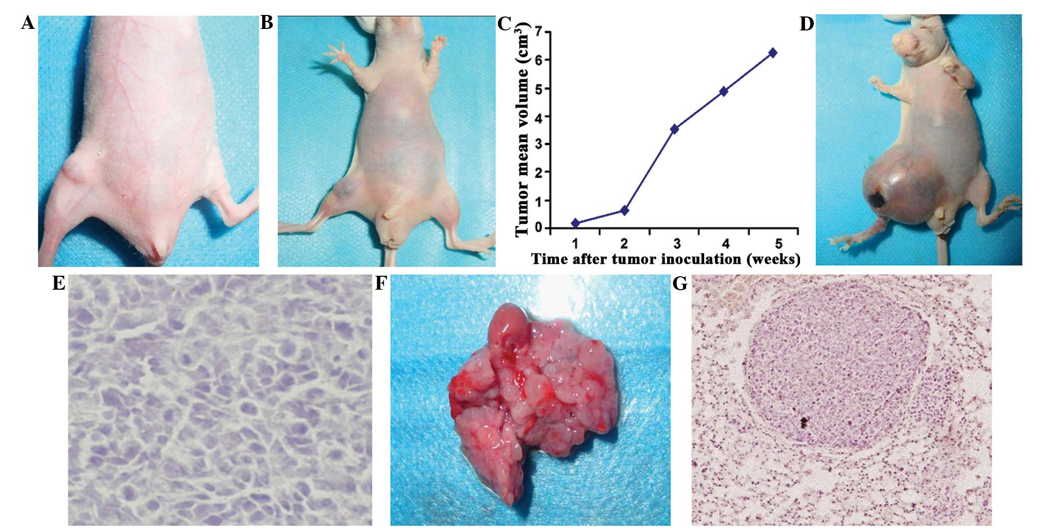
Characterization of the orthotopic mouse
model of OS
Tibial tumors were observed macroscopically at the
first week following inoculation (Fig.
1A; Table I). The tumor size
had increased rapidly two weeks following inoculation (mean tumor
volume, 0.76 cm3) and the skin overlying the tumor was
red (Fig. 1B and C). After five
weeks, a large, local tumor mass (mean tumor volume, 6.3
cm3) was observed (Fig.
1D). Microscopically, the tumor exhibited numerous pleomorphic
matrix components consisting of osteoblasts and pleomorphic tumor
cells (Fig. 1E). No lung metastases
were identified macroscopically after one or two weeks. However,
macroscopic nodules were visible in the lungs at weeks three (4/5
mice), four (5/5 mice) and five (5/5 mice) (Fig. 1F and G; Table I).
 | Table ITransplantation and spontaneous
metastasis of UMR106-01 cells orthotopically injected into nude
mice. |
Table I
Transplantation and spontaneous
metastasis of UMR106-01 cells orthotopically injected into nude
mice.
| Time after tumor
inoculation, weeks | Tumor-take rate at
inoculation site | Mice with
microscopically visible lung metastases, n | Mice with
macroscopically visible lung metastases, n |
|---|
| 1 | 5/5 | 0/5 | 0/5 |
| 2 | 5/5 | 4/5 | 0/5 |
| 3 | 5/5 | 5/5 | 4/5 |
| 4 | 5/5 | 5/5 | 5/5 |
| 5 | 5/5 | 5/5 | 5/5 |
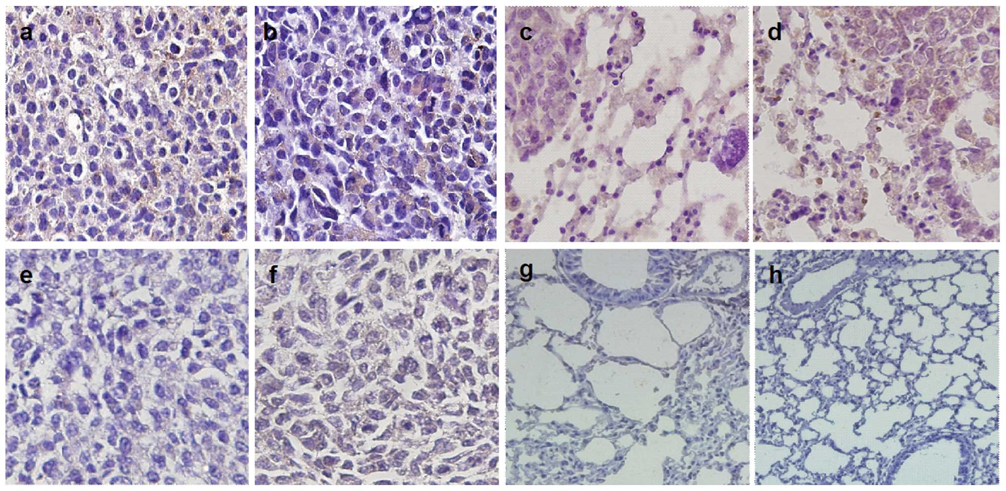
Survivin and VEGF expression in tumors
and metastatic lungs
IHC revealed positive survivin and VEGF expression
in the tibial tumors (Fig. 2A and
B), and pulmonary metastases (Fig.
2C and D), which was predominantly observed in the cytoplasm of
the tumor cells. The healthy mouse control samples exhibited no
survivin and VEGF expression in the tibiae (Fig. 2E and F) or the lungs (Fig. 2G and H). The experiments were
repeated three times and similar results were obtained.
Discussion
The high rate of pulmonary metastases of OS and the
intolerance of patients to the toxicity levels experienced during
current treatment regimes presents a therapeutic challenge in the
field of orthopedics. Consequently, molecular targeted therapies
are becoming a focus of cancer research with the aim of identifying
future therapeutic treatment strategies (19). Therefore, the identification of
proteins that are associated with the progression and metastatic
nature of OS is critical. In the present study, a spontaneous
pulmonary metastases orthotopic model of OS was developed using
UMR106-01 cells. In contrast to previous studies (3), a high rate of in situ tumor
formation and pulmonary metastases was observed following
inoculation (Table I). The
difference between the results of the present study and those of
previous studies may be attributed to the method, as the cells were
re-suspended in PBS and an increased concentration of cells was
injected (5×10⁶ cells/ml) during the present study. The
orthotopic model used in the current study presents a valuable tool
for investigating the biomolecules that promote or inhibit OS
growth and/or metastasis, and may also be used to assess the effect
of anti-target agents, which are specific to OS.
Survivin inhibits apoptosis, and promotes
proliferation and angiogenesis, which are key components of tumor
growth and metastasis. While previous studies have shown that
survivin is involved in the inhibition of apoptosis in OS (20,21),
few studies have investigated the correlation between survivin and
pulmonary metastasis. In the present study, survivin was observed
to be expressed in the metastatic lungs, indicating that survivin
may be associated with spontaneous pulmonary metastases. Previous
studies have identified a positive correlation between angiogenic
stimulation and vascular endothelial cell growth, and survivin
expression. Furthermore, survivin, in addition to AKT, is known to
activate anti-apoptotic signaling pathways that lead to vascular
endothelial cell growth and tumor angiogenesis (22). These results facilitate the
elucidation of the possible role of survivin in pulmonary
metastases of OS. An elevation in the levels of VEGF expression was
also identified in the metastatic lungs. VEGF is involved in
pulmonary metastases of OS and its elevated expression may also be
associated with spontaneous pulmonary metastases of OS. As survivin
promotes the growth of vascular endothelial cells, it is
hypothesized in the present study that survivin may affect the
secretion of VEGF, however, further study is required to
investigate this theory.
In conclusion, the present study successfully
established a novel spontaneous pulmonary metastases orthotopic
mouse model of OS. Using this model, the expression levels of
survivin and VEGF were identified in the metastatic lungs,
indicating that these two factors may be important in OS
metastases. These results have a potential clinical implication in
the prediction of metastasis and in the development of novel
combined targeted therapies for the treatment of OS.
Acknowledgements
The present study was supported by the key project
of Fujian provincial social development funds (grant no.
2012Y0038).
Abbreviations:
|
OS
|
osteosarcoma
|
|
VEGF
|
vascular endothelial growth factor
|
References
|
1
|
Assi H, Missenard G, Terrier P, et al:
Intensive induction chemotherapy without methotrexate in adult
patients with localized osteosarcoma: results of the Institut
Gustave-Roussy phase II trial. Curr Oncol. 17:23–31. 2010.
|
|
2
|
Yu Y, Luk F, Yang JL and Walsh WR:
Ras/Raf/MEK/ERK pathway is associated with lung metastasis of
osteosarcoma in an orthotopic mouse model. Anticancer Res.
31:1147–1152. 2011.
|
|
3
|
Fisher JL, Mackie PS, Howard ML, et al:
The expression of the urokinase plasminogen activator system in
metastatic murine osteosarcoma: an in vivo mouse model. Clin Cancer
Res. 7:1654–1660. 2001.
|
|
4
|
Hingorani P, Zhang W, Gorlick R and Kolb
EA: Inhibition of Src phosphorylation alters metastatic potential
of osteosarcoma in vitro but not in vivo. Clin Cancer Res.
15:3416–3422. 2009.
|
|
5
|
Sueyoshi T, Jono H, Shinriki S, et al:
Therapeutic approaches targeting midkine suppress tumor growth and
lung metastasis in osteosarcoma. Cancer Lett. 316:23–30. 2012.
|
|
6
|
Janeway KA and Walkley CR: Modeling human
osteosarcoma in the mouse: From bedside to bench. Bone. 47:859–865.
2010.
|
|
7
|
Sandberg AA and Bridge JA: Updates on the
cytogenetics and molecular genetics of bone and soft tissue tumors:
osteosarcoma and related tumors. Cancer Genet Cytogenet. 145:1–30.
2003.
|
|
8
|
Meyers PA, Schwartz CL, Krailo M, et al:
Osteosarcoma: a randomized, prospective trial of the addition of
ifosfamide and/or muramyl tripeptide to cisplatin, doxorubicin, and
high-dose methotrexate. J Clin Oncol. 23:2004–2011. 2005.
|
|
9
|
Sah NK, Khan Z, Khan GJ and Bisen PS:
Structural, functional and therapeutic biology of survivin. Cancer
Lett. 244:164–171. 2006.
|
|
10
|
Duffy MJ, O’Donovan N, Brennan DJ, et al:
Survivin: a promising tumor biomarker. Cancer Lett. 249:49–60.
2007.
|
|
11
|
Ryan BM, O’Donovan N and Duffy MJ:
Survivin: a new target for anti-cancer therapy. Cancer Treat Rev.
35:553–562. 2009.
|
|
12
|
Johnson ME and Howerth EW: Survivin: a
bifunctional inhibitor of apoptosis protein. Vet Pathol.
41:599–607. 2004.
|
|
13
|
Zou J, Gan M, Mao N, et al: Sensitization
of osteosarcoma cell line SaOS-2 to chemotherapy by downregulating
survivin. Arch Med Res. 41:162–169. 2010.
|
|
14
|
Breen EC: VEGF in biological control. J
Cell Biochem. 102:1358–1367. 2007.
|
|
15
|
Saharinen P, Eklund L, Pulkki K, et al:
VEGF and angiopoietin signaling in tumor angiogenesis and
metastasis. Trends Mol Med. 17:347–362. 2011.
|
|
16
|
Lin F, Zheng SE, Shen Z, et al:
Relationships between levels of CXCR4 and VEGF and blood-borne
metastasis and survival in patients with osteosarcoma. Med Oncol.
28:649–653. 2011.
|
|
17
|
Bajpai J, Sharma M, Sreenivas V, et al:
VEGF expression as a prognostic marker in osteosarcoma. Pediatr
Blood Cancer. 53:1035–1039. 2009.
|
|
18
|
Berlin O, Samid D, Donthineni-Rao R, et
al: Development of a novel spontaneous metastasis model of human
osteosarcoma transplanted orthotopically into bone of athymic mice.
Cancer Res. 53:4890–4895. 1993.
|
|
19
|
Thompson PA and Chintagumpala M: Targeted
therapy in bone and soft tissue sarcoma in children and
adolescents. Curr Oncol Rep. 14:197–205. 2012.
|
|
20
|
Zou J, Gan M, Mao N, et al: Sensitization
of osteosarcoma cell line SaOS-2 to chemotherapy by downregulating
survivin. Arch Med Res. 41:162–169. 2010.
|
|
21
|
Wu YF, Liang XJ, Liu YY, et al: +Antisense
oligonucleotide targeting survivin inhibits growth by inducing
apoptosis in human osteosarcoma cells MG-63. Neoplasma. 57:501–506.
2010.
|
|
22
|
Kanwar JR, Kamalapuram SK and Kanwar RK:
Targeting survivin in cancer: patent review. Expert Opin Ther Pat.
20:1723–1737. 2010.
|