Introduction
Hepatocellular carcinoma (HCC) is responsible for
numerous clinical therapeutic problems and is ranked as the third
leading cause of cancer-associated mortality worldwide (1). As only patients with early-stage HCC are
able to achieve optimal survival by radical hepatectomy or liver
transplantation, the majority of patients are beyond curative
intervention upon HCC diagnosis (1,2). Notably,
existing alternative strategies, including chemotherapy and
immunotherapy, have seldom elicited satisfactory responses in HCC
(2,3).
Therefore, the development of novel therapeutic agents against HCC
is urgently required.
Survivin belongs to the family of apoptosis
inhibitors (IAPs). Accumulating evidence has revealed that survivin
overexpression is a critical contributor to HCC pathogenesis via
its anti-apoptotic and pro-mitotic activities (4,5). In
addition, survivin possesses a distinct feature of high expression
specificity in cancer, in contrast to that of normally
differentiated tissues (6). Thereby,
survivin represents an attractive therapeutic target for the
development of antitumor agents, although progression in this field
has remained static.
YM155 is a recently developed survivin inhibitor
(6). To date, this novel, small,
imidazolium-based chemical has exhibited broad antitumor effects on
various tumors, including Wilm's tumor, glioblastoma and breast
cancer (6–9). In addition, ongoing studies have
demonstrated that YM155 may also enhance the chemosensitivity of
cells to existing anticancer agents (4,10,11). Furthermore, phase II clinical trials
of YM155 for the treatment of non-small cell lung carcinoma are
currently underway (7). However, the
mechanisms underlying YM155 antitumor activity have not yet been
fully elucidated, and the potent effect of YM155 in HCC remains
unclear. Therefore, the present study aimed to investigate how and
via what mechanism YM155 functions in HCC.
Materials and methods
Cells and reagents
Human HCC cell lines HepG2 and Huh7 were purchased
from Shanghai Laboratory Animal Centre (SLRC; Shanghai, China).
YM155 was obtained from Selleck Chemicals (Houston, TX, USA).
Dulbecco's modified Eagle's medium (DMEM), fetal bovine serum (FBS)
and MTT were purchased from Sigma-Aldrich (St. Louis, MO, USA).
Annexin V/fluorescein isothiocyanate (FITC) apoptosis detection kit
(cat. no. 556547) was purchased from BD Pharmingen (San Diego, CA,
USA). Bicinchoninic Acid (BCA) Protein Assay and Protein Lysis
kits, phosphate-buffered saline (PBS), streptomycin and penicillin
were purchased from the Beyotime Institute of Biotechnology
(Nanjing, China). The primary rabbit monoclonal anti-survivin (cat.
no. 76424), anti-procaspase 3 (cat. no. 32499), anti-β-actin (cat.
no. 8227) and anti-phosphorylated retinoblastoma tumor suppressor
protein (p-Rb; Ser807/811; cat. no. D20B12) antibodies, as well as
secondary goat anti-rabbit horseradish peroxidase-conjugated
antibody (cat. no. 97080) were purchased from Abcam (Cambridge, MA,
USA).
Immunohistochemistry
To explore the protein expression profiles of
survivin and p-Rb, eight paraffin-embedded HCC specimens (T group)
and matched adjacent normal tissues (N group) were retrieved from
the Department of Pathology (Taizhou People's Hospital, Taizhou,
China) for immunohistochemical assessment. All patient samples
recruited to the present study were approved by the ethical review
committee (Institutional Ethical Board of Taizhou People's
Hospital).
Consecutive sections (4 µm) of paraffin-embedded
normal and tumor specimens were prepared as described previously
(12). Survivin and p-Rb protein
expression in these sections was detected by incubation with
respective antibodies against survivin (1:150) and p-Rb (1:150) at
37°C for 1 h. Two individual pathologists, blinded to patient
characteristics, scored the sections using an Olympus CX32
microscope (Olympus Corp., Tokyo, Japan). Protein expression was
classified according to the staining intensity as follows: 0,
absence of staining; 1, mild expression; 2, moderate expression and
3, high expression. A mean percentage of positive tumor cell was
determined in at least five areas at x400 magnification. The
percentage of positive tumor cells and the staining intensity were
multiplied to produce a weighted score for each case.
Theoretically, the total scores ranged from 0 (0% of cells stained)
to 3 (100×3/100).
Cell culture and viability
measurement
Human HCC cell lines HepG2 and Huh7 were cultured in
DMEM supplemented with 10% FBS and 1% streptomycin and penicillin,
in an atmosphere of 5% CO2 at 37°C. Cells were allowed
to adhere overnight. Subsequently, HCC cells incubated with and
without YM155 were designated as the YM155 and control groups,
respectively.
For cell viability assessment, HCC cells were seeded
in 96-well plates (Nunc, Roskilde, Denmark) at 5000 cells/well and
subsequently exposed to YM155 at the IC50 concentration
of 100 nM according to previous literature (13) for up to 72 h. At various time-points
(0, 24, 48 and 72 h), 20 µl MTT (5 mg/ml) was added to each well.
The HCC cells were then incubated at 37°C for a further 4 h. The
culture medium was removed and 150 µl 0.1% dimethylsulfoxide (DMSO)
was added to each well to resolve the MTT. The absorbance of each
well was measured at 450 nm on a Multiskan FC 51119000 photometer
(BioTek Instruments, Inc., Winooski, VT, USA). For apoptosis
measurements and protein sample preparation, cells were cultured in
6-well plates (Nunc) with YM155 or without YM155 (control group)
and harvested 48 h later.
Apoptosis detection
The apoptotic degree of YM155-treated HCC cells was
quantified by flow cytometry using the FITC Annexin V Apoptosis
Detection kit I. Briefly, cell samples were sequentially incubated
for 15 min at 28°C with 5 mg/ml Annexin V-FITC (AV-FITC) and 10
mg/ml propidium iodide (PI) diluted in Annexin V binding buffer.
The cells were then resuspended in PBS and analyzed with a flow
cytometer (FACSCalibur; BD Biosciences, San Jose, CA, USA), using a
530/30 nm signal detector for AV-FITC and a 582/42 nm signal
detector for PI. The data were subsequently analyzed by Flow J
software (version 7.6.5; Tree Star, Inc., San Carlos, CA, USA). The
upper left and lower left quadrants represented late and early
apoptosis, respectively. The total apoptosis ratio was calculated
by adding the late and early apoptosis proportions.
Western blotting
Cell samples grown in 6-well plates were incubated
with ice-cold lysis buffer [0.1% Triton X-100, 50 mM HEPES (pH
7.5), 150 mM NaCl, 10% (v/v) glycerol, 1.5 mM MgCl2, 1
mM dithiothreitol, 1 mM sodium fluoride, 0.1 mM sodium
orthovanadate, 1 mM phenylmethylsulfonyl fluoride and 2 mg/ml
leupeptin and aprotinin]. The total protein sols were centrifuged
at 12,000 × g at 4°C for 10 min and the supernatants were bolted at
100°C by iron heating for a further 10 min. A BCA Protein Assay kit
was used to determine the protein concentration. Equivalent samples
(30 mg protein) were then subjected to 12% SDS-PAGE (Beyotime
Institute of Biotechnology). The proteins were transferred to
nitrocellulose membranes (Beyotime Institute of Biotechnology) and
probed with specific primary antibodies. The antibodies were used
at the following concentrations: survivin (1:1,500), procaspase-3
(1:2,000), p-Rb (1:2,000), β-actin (1:2,000), followed by secondary
antibody (1:2,500). The molecular sizes of the target proteins were
determined by comparison with prestained Bio-Rad protein markers
(Bio-Rad Laboratories, Inc., Hercules, CA, USA). The protein bands
were then scanned using the Western Lightning Chemiluminescent
Reagent Plus (Perkin Elmer, Boston, MA, USA) detection system.
Protein immunoreactivity was quantified by densitometry analysis
using the Gel DocXR System 170–8170 device and (Bio-Rad
Laboratories, Inc., Nazareth, Belgium) and Quantity One software
(version 4.4; Bio-Rad Laboratories, Inc.). β-actin was used as a
loading control.
Statistical analysis
All parametric data are expressed as the mean ±
standard error of the mean, and analyzed by Student's
t-test, whereas non-parametric data analysis was performed
by Mann-Whitney U test. For all tests, analyses were performed
using SPSS 19.0 statistical software (IBM SPSS, Armonk, NY, USA)
and two-sided P<0.05 was considered to indicate a statistically
significant difference.
Results
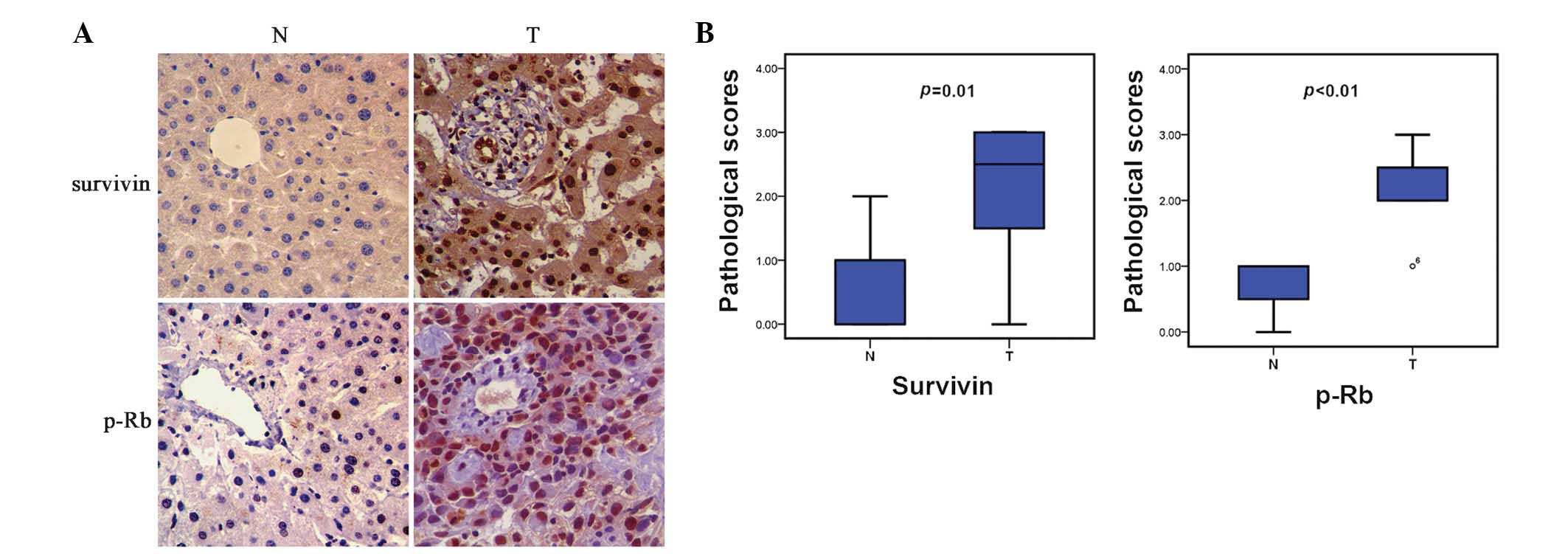
Survivin and p-Rb protein expression
is enhanced in HCC cells
To investigate the variance in protein expression of
survivin between human HCC and normal liver tissues,
immunohistochemistry assays were performed. As shown in Fig. 1, HCC specimens presented with markedly
greater survivin expression than that of noncancerous tissues, and
the distribution was characterized by expression primarily in the
nucleus, but also the cytoplasm.
It is well established that Rb protein functions as
a cell-cycle suppressor, and its functional loss through protein
phosphorylation has been implicated to the tumorigenesis of various
types of neoplasm (14,15). Therefore, the phosphorylation level of
Rb protein in HCC was investigated. Immunohistochemical analysis
revealed that HCC samples displayed elevated p-Rb expression, as
compared with that of normal liver tissues, in a nucleus-exclusive
distribution model (Fig. 1).
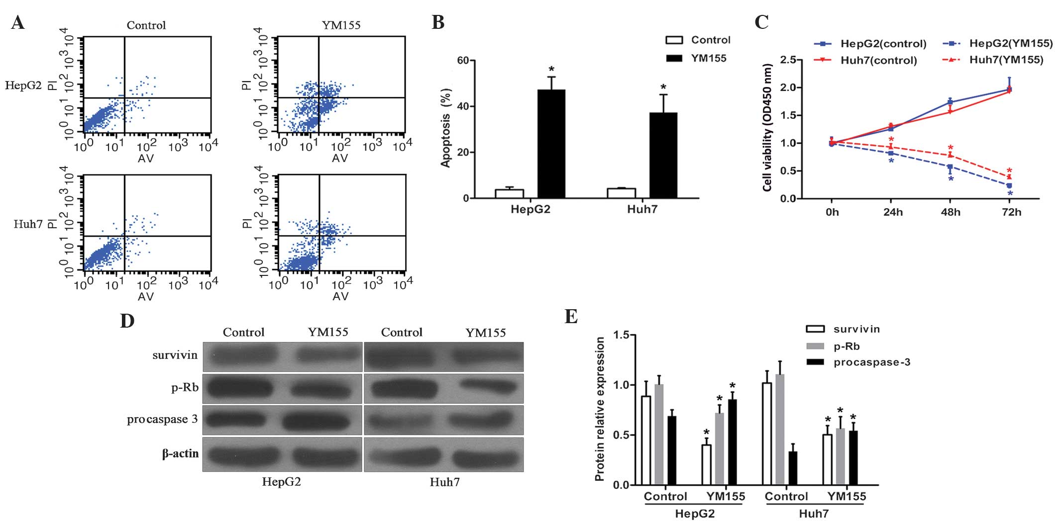
YM155 promotes HCC cell apoptosis
Cell apoptosis status was determined by flow
cytometry. Following 48 h of exposure to YM155, the cell apoptosis
ratios in HepG2 and Huh7 cells were 47.3±5.5 and 37.3±7.8%,
respectively, markedly higher than those of the corresponding
control groups, which were 3.7±1.2 and 4.2±0.4%, respectively
(P<0.05; Fig. 2A and B). These
results indicated a significant pro-apoptotic effect of YM155 on
HCC cell lines.
YM155 induces growth arrest in HCC
cell lines
To verify the antitumor activity of YM155, HCC cell
viabilities were investigated by MTT assay. Following YM155
exposure, HepG2 cell viabilities at 24, 48 and 72 h were 0.81±0.02,
0.55±0.09 and 0.25±0.05 respectively, which were markedly lower
than those at the corresponding times in the control group
(1.00±0.04, 1.74±0.10 and 1.97±0.19) (P<0.05; Fig. 2C). Concurrently, the Huh7 cell
survival at 24, 48 and 72 h in the YM155 group was 0.93±0.06,
0.78±0.06 and 0.38±0.06, respectively. These results were markedly
lower than those at the corresponding time-points in the control
group: 1.30±0.06, 1.56±0.12 and 1.92±0.10 (P<0.05; Fig. 2C). These data demonstrated a
significant anti-proliferative effect of YM155 in HCC cells.
YM155 inhibits survivin and p-Rb
protein expression and enhances procaspase-3
To detect the variations in the protein expression
of survivin, p-Rb and procaspase-3 in HCC cells following YM155
pretreatment, immunoblotting assays were performed (Fig. 2D and E). The relative survivin protein
expression levels of YM155-treated HepG2 and Huh7 cells were
0.40±0.07 and 0.50±0.09, respectively; these were markedly lower
than those of the corresponding control groups (0.89±0.15 and
1.02±0.12, respectively). Relative p-Rb protein levels in HepG2 and
Huh7 cells following YM155 exposure (0.72±0.08 and 0.57±0.12,
respectively) were also lower than those in the associated control
group (1.01±0.09 and 1.11±0.13, respectively). The relative protein
expression levels of procaspase-3 in control HepG2 and Huh7 cells
were 0.69±0.06 and 0.34±0.08, respectively, which were
significantly lower than those of the corresponding YM155-treated
groups (0.86±0.07 and 0.53±0.08, respectively). These results
revealed that YM155-treatment was not only able to directly
suppress survivin protein expression, but also indirectly inhibited
p-Rb and enhanced procaspase-3 expression.
Discussion
In the present study, the protein expression of
survivin and p-Rb in HCC tissues was evaluated, and the apoptosis
and proliferation status of YM155-treated HCC cells was examined.
The results demonstrated that silencing of survivin expression, via
specific inhibitor YM155, induced HCC cell apoptosis and growth
arrest. These results corroborated those of previous studies
regarding other neoplasms (8–11,13,16). In
order to represent a potential therapeutic strategy for HCC, the
impacts of survivin, as well as YM155, on HCC should be
sufficiently understood and validated.
Apoptosis is a natural defense mechanism for the
elimination of unhealthy cells, and the loss of sensitivity to
apoptosis has been identified as a hallmark of cancer (6,17–19). Accumulating evidence has revealed that
survivin overactivation contributes to apoptotic dysfunction via
the intrinsic and extrinsic apoptosis pathways (4,7,17–20). Given
that survivin contains a single BIR domain, similarly to other IAP
family members, it is not surprising that survivin is able to
interfere with the activity of caspases in the mediation of
apoptosis (18–20). In addition, survivin has demonstrated
the ability to prevent the release of apoptosis-induced factor from
the mitochondrial intermembrane space, protecting cells against
apoptotic death (19,21,22).
Futhermore, survivin also suppresses apoptosis via inhibition of
the pathway of death receptors and downstream tumor necrosis
factor-related apoptosis-inducing ligand (13,18,23,24).
The present study was limited to examination of the total apoptosis
profile and the protein expression levels of procaspase-3, a
principle ‘executor’, committing cells to death via the intrinsic
apoptosis pathway (19,25). The results of the present study
revealed that YM155 positively modulated the intrinsic apoptosis
pathway and enhanced the total apoptosis ratios in HCC cells, which
was consistent with the results of previous studies (18–20).
Notably, the survivin distribution identified by
immunohistochemical analysis exhibited evident characteristics of
expression in the nucleus first and cytoplasm second. These
findings were in accordance with an early hypothesis that survivin
is able to shuttle between the nucleus and cytoplasm through active
nucleocytoplasmic transport (6,19). On the
other hand, the presence of cytoplasmic survivin has been further
associated with poor prognosis in HCC, as members of the caspase
protein family occur predominantly in the cytoplasm (5,6,19). Combining these factors with another
generally accepted notion that survivin expression may correlate
negatively with the distal prognosis of cancer patients even
following radical resection, indicates that postoperative
immunohistochemistry may be of interest in the selection of
postoperative therapeutic schedules for patients with HCC, through
assessment of survivin expression levels and distribution (5,6,18).
Apart from its role in apoptosis suppression,
survivin represents a widely recognized mitotic regulator of
various cell division processes (6,18–20). Notably, survivin is able to
participate in the chromosomal passenger complex, and thereby
accelerate chromosomal segregation and cytokinesis (18,19). In
addition, survivin is also able to promote the formation of
microtubules and the spindle. Furthermore, the DNA damage-mediated
activation of checkpoint kinase 2 always results in secondary
survivin release and apoptosis dysfunction, facilitating the
survival of tumorous cells (18,20). To
date, accumulating evidence has exemplified the proliferative
impact of survivin on various tumor cells (6,8,18–19). In
the current study, the results of cell viability analysis supported
this hypothesis. However, the potential anti-proliferative and
pro-apoptotic effects of YM155 were indistinguishable by MTT
assessment, and therefore the phosphorylation level of Rb protein
was evaluated. Rb is another generally acknowledged cell-cycle
regulator, downstream of survivin (20,26,27). Rb
protein primarily presents as a hypophosphorylated form within
quiescent cells, whereas its phosphorylation promotes cell
proliferation and tumorigenesis (14,15,20,26–28).
In the present study, p-Rb was used as an indicator to assess the
proliferation profile of HCC cells. The results revealed that YM155
induced the phosphorylation of Rb protein, which indicated a
proliferative effect of survivin on HCC cell lines.
Survivin is a significant therapeutic target for the
development of anticancer agents, due to its role in tumorigenesis
and exclusive expression in tumor cells (18,19).
However, targeting survivin may be difficult as it is not an enzyme
or a cell surface protein (7,13). Recently, low molecular weight YM155
has emerged as a novel breakthrough drug, associated with existing
survivin inhibitors. In addition to direct potential antitumor
activity, YM155 has also displayed an ability to sensitize HCC
cells to other chemotherapeutic agents (10,11,18–20).
Cancer is essentially a consequence of an imbalance
between cell death and proliferation (18,19).
Aberrant cell proliferation, combined with apoptotic resistance, is
a crucial contributor to tumorigenesis (18–20). In
this context, YM155 may possess potential to be a dual ‘protector’
for patients with HCC, by suppressing survivin. The results of the
present study preliminarily exhibited the potency of YM155 on HCC
cells via blocking cell growth and enhancing apoptosis. Further
studies are required in order to elucidate whether this novel agent
may be utilized as a potential therapeutic strategy for HCC.
References
|
1
|
Guglielmi A, Ruzzenente A, Conci S, et al:
Hepatocellular carcinoma: Surgical perspectives beyond the
barcelona clinic liver cancer recommendations. World J
Gastroenterol. 20:7525–7533. 2014. View Article : Google Scholar : PubMed/NCBI
|
|
2
|
Zhang Y, Shi ZL, Yang X and Yin ZF:
Targeting of circulating hepatocellular carcinoma cells to prevent
postoperative recurrence and metastasis. World J Gastroenterol.
20:142–147. 2014. View Article : Google Scholar : PubMed/NCBI
|
|
3
|
Shindoh J, Kaseb A and Vauthey JN:
Surgical strategy for liver cancers in the era of effective
chemotherapy. Liver Cancer. 2:47–54. 2013. View Article : Google Scholar : PubMed/NCBI
|
|
4
|
Zhao X, Ogunwobi OO and Liu C: Survivin
inhibition is critical for Bcl-2 inhibitor-induced apoptosis in
hepatocellular carcinoma cells. PLoS One. 6:e219802011. View Article : Google Scholar : PubMed/NCBI
|
|
5
|
Liu JL, Zhang XJ, Zhang Z, Zhang AH, Wang
W and Dong JH: Meta-analysis: Prognostic value of survivin in
patients with hepatocellular carcinoma. PLoS One. 8:e833502013.
View Article : Google Scholar : PubMed/NCBI
|
|
6
|
Cheung CH, Huang CC, Tsai FY, et al:
Survivin - biology and potential as a therapeutic target in
oncology. Onco Targets Ther. 6:1453–1462. 2013. View Article : Google Scholar : PubMed/NCBI
|
|
7
|
Yamanaka K, Nakata M, Kaneko N, et al:
YM155, a selective survivin suppressant, inhibits tumor spread and
prolongs survival in a spontaneous metastatic model of human triple
negative breast cancer. Int J Oncol. 39:569–575. 2011.PubMed/NCBI
|
|
8
|
Tao YF, Lu J, Du XJ, et al: Survivin
selective inhibitor YM155 induce apoptosis in SK-NEP-1 Wilms tumor
cells. BMC Cancer. 12:6192012. View Article : Google Scholar : PubMed/NCBI
|
|
9
|
Lai PC, Chen SH, Yang SH, Cheng CC, Chiu
TH and Huang YT: Novel survivin inhibitor YM155 elicits
cytotoxicity in glioblastoma cell lines with normal or deficiency
DNA-dependent protein kinase activity. Pediatr Neonatol.
53:199–204. 2012. View Article : Google Scholar : PubMed/NCBI
|
|
10
|
Liang H, Zhang L, Xu R and Ju XL:
Silencing of survivin using YM155 induces apoptosis and
chemosensitization in neuroblastomas cells. Eur Rev Med Pharmacol
Sci. 17:2909–2915. 2013.PubMed/NCBI
|
|
11
|
Koike H, Nitta T, Sekine Y, et al: YM155
reverses rapamycin resistance in renal cancer by decreasing
survivin. J Cancer Res Clin Oncol. 140:1705–1713. 2014. View Article : Google Scholar : PubMed/NCBI
|
|
12
|
Wang H, Jiang S, Zhang Y, et al: High
expression of thymosin beta 10 predicts poor prognosis for
hepatocellular carcinoma after hepatectomy. World J Surg Oncol.
12:2262014. View Article : Google Scholar : PubMed/NCBI
|
|
13
|
Charette N, De Saeger C, Horsmans Y,
Leclercq I and Stärkel P: Salirasib sensitizes hepatocarcinoma
cells to TRAIL-induced apoptosis through DR5 and survivin-dependent
mechanisms. Cell Death Dis. 4:e4712013. View Article : Google Scholar : PubMed/NCBI
|
|
14
|
Reed CA, Mayhew CN, McClendon AK, Yang X,
Witkiewicz A and Knudsen ES: RB has a critical role in mediating
the in vivo checkpoint response, mitigating secondary DNA damage
and suppressing liver tumorigenesis initiated by aflatoxin B1.
Oncogene. 28:4434–4443. 2009. View Article : Google Scholar : PubMed/NCBI
|
|
15
|
Nojiri S and Joh T: Albumin suppresses
human hepatocellular carcinoma proliferation and the cell cycle.
Int J Mol Sci. 15:5163–5174. 2014. View Article : Google Scholar : PubMed/NCBI
|
|
16
|
Kaneko N, Mitsuoka K, Amino N, et al:
Combination of YM155, a survivin suppressant, with bendamustine and
rituximab: A new combination therapy to treat relapsed/refractory
diffuse large B-cell lymphoma. Clin Cancer Res. 20:1814–1822. 2014.
View Article : Google Scholar : PubMed/NCBI
|
|
17
|
Fabregat I: Dysregulation of apoptosis in
hepatocellular carcinoma cells. World J Gastroenterol. 15:513–520.
2009. View Article : Google Scholar : PubMed/NCBI
|
|
18
|
Mobahat M, Narendran A and Riabowol K:
Survivin as a preferential target for cancer therapy. Int J Mol
Sci. 15:2494–2516. 2014. View Article : Google Scholar : PubMed/NCBI
|
|
19
|
Coumar MS, Tsai FY, Kanwar JR, Sarvagalla
S and Cheung CH: Treat cancers by targeting survivin: Just a dream
or future reality? Cancer Treat Rev. 39:802–811. 2013. View Article : Google Scholar : PubMed/NCBI
|
|
20
|
Rauch A, Hennig D, Schäfer C, et al:
Survivin and YM155: How faithful is the liaison? Biochim Biophys
Acta. 1845:202–220. 2014.PubMed/NCBI
|
|
21
|
Zeng J, Sun Y, Wu K, et al:
Chemopreventive and chemotherapeutic effects of intravesical
silibinin against bladder cancer by acting on mitochondria. Mol
Cancer Ther. 10:104–116. 2011. View Article : Google Scholar : PubMed/NCBI
|
|
22
|
Zhang R, Humphreys I, Sahu RP, Shi Y and
Srivastava SK: In vitro and in vivo induction of apoptosis by
capsaicin in pancreatic cancer cells is mediated through ROS
generation and mitochondrial death pathway. Apoptosis.
13:1465–1478. 2008. View Article : Google Scholar : PubMed/NCBI
|
|
23
|
Taniguchi H, Horinaka M, Yoshida T, et al:
Targeting the glyoxalase pathway enhances TRAIL efficacy in cancer
cells by downregulating the expression of antiapoptotic molecules.
Mol Cancer Ther. 11:2294–2300. 2012. View Article : Google Scholar : PubMed/NCBI
|
|
24
|
Sakai T, Eskander RN, Guo Y, et al:
Flavokawain B, a kava chalcone, induces apoptosis in synovial
sarcoma cell lines. J Orthop Res. 30:1045–1050. 2012. View Article : Google Scholar : PubMed/NCBI
|
|
25
|
Loor G, Kondapalli J, Iwase H, et al:
Mitochondrial oxidant stress triggers cell death in simulated
ischemia-reperfusion. Biochim Biophys Acta. 1813:1382–1394. 2011.
View Article : Google Scholar : PubMed/NCBI
|
|
26
|
Baker SJ and Reddy EP: CDK4: A key player
in the cell cycle, development and cancer. Genes Cancer. 3:658–669.
2012. View Article : Google Scholar : PubMed/NCBI
|
|
27
|
Hu H, Li Z, Chen J, et al: P16
reactivation induces anoikis and exhibits anti tumour potency by
down regulating Akt/survivin signalling in hepatocellular carcinoma
cells. Gut. 60:710–721. 2011. View Article : Google Scholar : PubMed/NCBI
|
|
28
|
Deng L, Lu Y, Zhao X, et al: Ran GTPase
protein promotes human pancreatic cancer proliferation by
deregulating the expression of Survivin and cell cycle proteins.
Biochem Biophys Res Commun. 440:322–329. 2013. View Article : Google Scholar : PubMed/NCBI
|