Introduction
Temozolomide (TMZ) is a second-generation oral
alkylating agent with the ability to penetrate the blood-brain
barrier, and is widely used in the management of high-grade brain
neoplasms, including anaplastic astrocytoma and glioblastoma
multiforme, in addition to brain metastasis from solid tumors
(1,2).
TMZ is a prodrug and imidazotetrazine analog that exerts its action
following spontaneous conversion to its active form,
5-(3-methyltriazen-1-yl)imidazole-4-carboxamide (3). This active drug subsequently exerts its
antitumor effect by methylating the purine bases of chromosomal DNA
including O6-guanine, which results in the failure of DNA
replication, cell cycle arrest and subsequent apoptosis (4,5). TMZ is
generally considered effective and relatively safe (6–8); however,
increased survival rates in certain patients have uncovered
toxicities arising from the long-term use of alkylating agents,
including TMZ (9). Treatment-related
secondary myelodysplastic syndrome (t-MDS) and treatment-related
acute myeloid leukemia (t-AML) have been recorded in patients
following prolonged (5–10 years) exposure to alkylating agents
(10–12).
TMZ is a relatively new drug and glioblastoma is an
aggressive neoplasm with poor prognosis, thus the types of
secondary cancer arising due to TMZ treatment have not yet been
fully characterized. A review of the existing literature
demonstrated that only 7 cases of TMZ-related acute lymphoblastic
leukemia (ALL) have been reported thus far (13–18). The
current case describes an 11-year-old boy with glioblastoma
multiforme, who developed B-cell ALL 6 months after the last dose
of TMZ. To the best of our knowledge, the present case is the
youngest known patient with TMZ-related ALL. In addition, a review
of the available literature regarding TMZ-related ALL was
performed.
Case report
An 11-year-old boy presented at The First Affiliated
Hospital of Jilin University (Changchun, China) in July 2013 with a
3-day history of generalized tonic-clonic seizures. The patient was
otherwise healthy and had no other significant medical or family
history. Magnetic resonance imaging (MRI) of the brain revealed a
contrast-enhanced lesion (4.5×3 cm) in the left temporo-occipital
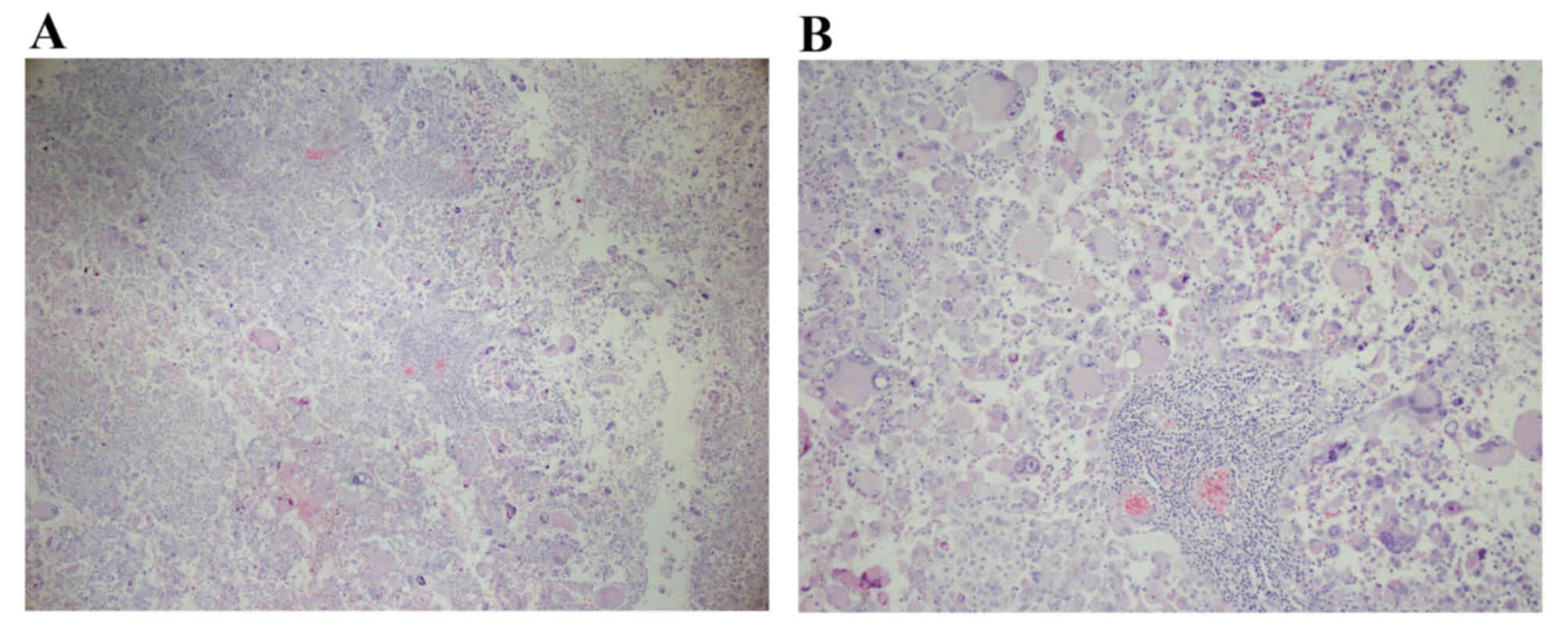
region with focal cystic degeneration (Fig. 1). Craniotomy and gross-total resection
were subsequently performed. A post-operative pathological
examination identified that the tumor cells varied in size, the
nucleus was enlargened, the chromatin was granulated, and
thickened-karyotheca and focal necrosis were observed. (Fig. 2; hematoxylin and eosin staining).
Immunochemistry, performed as previously described (19) revealed positive staining for epidermal
growth factor receptor (anti-EGFR; cat. no. SP111; dilution, 1:100;
Fuzhou Maixin Biotech Co., Ltd., Fuzhou, China), sixty percent
positive staining for Ki-67 (anti-Ki-67; cat. no. MX006; dilution,
1:100), GFAP (anti-GFAP; cat. no. GA-5; dilution, 1:100; both
Fuzhou Maixin Biotech Co., Ltd.). Pathological analysis confirmed
the diagnosis of giant cell glioblastoma with necrosis (Fig. 2) and this was classified as World
Health Organization grade IV based on the 2007 World Health
Organization Classification of Tumors of the Central Nervous System
(20).
Postoperatively, the patient received TMZ-based
concurrent chemoradiation with TMZ at a dose of 75
mg/m2/day for 5 days during radiotherapy in August 2013.
Radiotherapy was administered at a total dose of 60 Gy in 30
fractions for 6 weeks (5 days per week) and limited to the
temporo-occipital region. Following concurrent chemoradiotherapy,
the patient received a total of 5 cycles of maintenance TMZ from
September 2013 to January 2014 (150 mg/m2/day for 5 days
every 28 day course). During this period the patient remained
stable with no evidence of recurrence on surveillance MRI
examinations.
At 6 months after the final dose of TMZ, the patient
presented with increasing fatigue and easy bruising. Physical
examination reported pallor, scattered petechiae and ecchymosis of
the skin, tenderness of the sternum on percussion and
hepatosplenomegaly (the liver was firm and palpable 6 cm below the
costal margin). Blood counts indicated absolute lymphocytosis
(white blood cell count, 16,750/µl, normal range, 3,500–9,500/µl,
including 74% lymphocytes, normal range 20–50%) with anemia
(hemoglobin count, 75 g/l, normal range 130–175g/l) and
thrombocytopenia (platelet count, 24,000/µl; normal range,
125,000–350,000/µl). The serum lactate dehydrogenase (LDH) level
was markedly elevated, at 1,243 U/l. Bone marrow aspiration
exhibited 91.5% lymphocytes with 46% lymphoblasts (Fig. 3), and bone marrow biopsy demonstrated
diffuse infiltration of the peripheral blood by lymphoblasts
(Fig. 4). Peripheral blood flow
cytometry indicated 63.58% phenoty pically abnormal cells, which
were positive for CD10 (anti-CD10 mAb; cat. no. MX002; dilution,
1:100), CD19 (anti-CD19 mAb; cat. no. LE-CD19; dilution, 1:100),
CD20 (anti-CD20 mAb; cat. no. MX003; dilution, 1:100), CD22
(anti-CD22 mAb; cat. no. MS-1087; dilution, 1:100) and CD79a
(anti-CD79a mAb; cat. no. SP18; dilution, 1:100; all Fuzhou Maixin
Biotech Co., Ltd.), and negative for immunoglobulin M
(anti-Immunoglobulin M; cat. no. 20-2786; dilution, 1:100; Dakewe
Biotech Ltd., Shanghai, China) and CD7 (anti-CD7 mAb; cat. no. 272;
dilution, 1:100; Fuzhou Maixin Biotech Co., Ltd.). Staining was
performed as described previously (21). Bone marrow cytogenetics revealed a
normal male karyotype (Fig. 5) and an
MRI scan demonstrated no signs of recurrence. Based on these
results, the patient was diagnosed with B-cell ALL. A four-week
combination chemotherapy induction protocol was administered,
comprising vincristine (1.5 mg/m2, intravenous injection
at days 8, 15, 22 and 29), daunorubicin (30 mg/m2,
intravenous injectionat days 8–10, 22 and 29), L-asparaginase
(6,000–10,000 U/kg, intravenous injectionat days 11, 13, 15, 17,
19, 21, 23, 25, 27 and 29) and prednisone (60 mg/m2/day,
oral administration in days 1–7; 40 mg/m2/day, oral
administration in days 8–28), and the patient achieved complete
hematological remission. The patient remains on chemotherapy and
has been in remission for 8 months, undergoing regular surveillance
MRI three times a month.
Discussion
A large number of randomized clinical trials have
demonstratedthe safety and efficacy of TMZ in the treatment of
aggressive central nervous system tumors, revealing significantly
improved overall survivalcompared with the first-generation oral
alkylating agent (6–8). The United States Food and Drug
Administration approved TMZ as a treatment for glioblastoma in 1999
(22). However, TMZ is a relatively
new alkylating agent and its long-term safety is not fully
characterized.
The present study performed a literature review and
identified only 6 cases of therapy-associated ALL that occurred
subsequent to TMZ administration include the current patient. The
clinical profiles of the six patients are summarized in Table I. Of the patients identified, three
were males and three werefemales and all except onewere adults. A
total of six patients were diagnosed with primary glioblastoma and
received TMZ-based concurrent chemoradiation. The other patient was
diagnosed with oligodendroglioma and was treated with TMZ-based
adjuvant chemotherapy. Bone marrow cytogenetics revealed chromosome
abnormalities in 2 cases: 1 with t(4;11)(q21;q23)(14), and the other with breakpoint cluster
region/Abelson murine leukemia rearrangement, t(9;22) and monosomy
7 (13). During concurrent
chemoradiation, TMZ was administered orally at 75
mg/m2/d 5 consecutive days a week for 42 days.
Subsequent maintenance therapy with TMZ was administered at 150
mg/m2/day for 5 days in a 28 day cycle, with the total
number of cycles ranging from 1–36 prior to the diagnosis of
secondary hematological disorders. A total of 5 patients were
diagnosed with pre-B ALL, and the other two with precursor T-cell
ALL (pre-T ALL).
 | Table I.Cases of TMZ-associated ALL in
patients with glioblastoma. |
Table I.
Cases of TMZ-associated ALL in
patients with glioblastoma.
| Case | Age/sex | Diagnosis | Chemoradiation
strategya | Latency, months | Immunophenotype | Cytogenetic
findings | Outcome | Leukemia-associated
mortality |
|---|
| 1 | 40/M | Glioblastoma
multiforme | 60 cGy with 70
mg/m2/daily; 200 mg/m2/daily X1 cycle | 4 | Precursor B-ALL | 45, XY, −7,
der(9)(p12)t(9;22) | CR | N/A |
| 2 | 12/F | Anaplastic
astrocytoma | 60 cGy with 75
mg/m2/daily; 150 mg/m2/daily X8 cycles | 13 | Precursor B-ALL | Normal | S | 8 months |
| 3 | 26/M | Astrocytoma
andoligodendroglioma (WHO grade II) | 60 cGy with 75
mg/m2/daily; 150 mg/m2/daily X8 cycles | 17 | Precursor
B-ALL | Normal | N/A | N/A |
| 4 | 49/F | Astrocytoma (WHO
grade II) | 80 cGy with 75
mg/m2/daily; 150 mg/m2/daily X6 cycles | 57 | Precursor
T-ALL | Normal | CR | N/A |
| 5 | 54/F | Glioblastoma
multiforme | 60 cGy with 75
mg/m2/daily; 150 mg/m2/daily X11 cycles | 15 | Precursor
B-ALL |
t(4;11)(q21;q23) | S | 1 month |
| Present case | 11/M | Giant-cell
Glioblastoma (WHO grade IV) | 60 cGy with 75
mg/m2/daily; 150 mg/m2/daily X5 cycles | 6 | Precursor
B-ALL | Normal | CR | N/A |
The leukemogenic potential of alkylating agents is
well-known and it usually takes 5–10 years after exposure for
leukemia to develop (10–12). Following prolonged TMZ treatment,
t-MDS and t-AML have been widely reported in patients (22–24).
Results from phase I and II clinical trials suggest that the
primary toxicity associated with TMZ is myelotoxicity (13). Reported TMZ-related secondary
hematological disorders, including t-MDS and t-AML, are usually
secondary to the administration of TMZ in combination with other
alkylating agents (25) and few cases
have been documented following TMZ monotherapy (24,26).
Meanwhile, T-ALL subsequent to TMZ-based concurrent chemoradiation
in patients with glioblastoma is extremely rare (13–18). In
the limited number of patients studied, B-ALL was more common than
T-ALL.
The first case of TMZ therapy-related ALL was
reported by De Vita et al (13) in 2005, documenting a case of Ph+ acute
precursor B-cell lymphoblastic leukemia French-American-British L1
subtype. Subsequently, four other cases were reported with B- or
T-ALL with or without cytogenetic abnormalities (14–17). The
latency period from the first dose of TMZ to the onset of ALL was
an average of 17.7 months (range, 4–57 months), which is notably
shorter than the latency period of therapy-related ALL subsequent
to other conventional alkylating agents (mean, 63 months) (27). The development of secondary
malignancies usually requires the accumulation of multiple genetic
abnormalities, causing a period of latency.
Treatment-related MDS and t-AML are the most common
types of secondary leukemia and secondary ALL accounts for ~10% of
all secondary leukemia cases (28,29).
TMZ-related ALL is rare and the specific mechanisms underlying its
development remain unclear. Previous studies suggest that several
genetic predisposing factors may be involved in the pathogenesis of
secondary ALL. De Vita et al (13) indicated that monosomy 7 and t(9;22)
may be associated with the onset of TMZ-related ALL. In addition,
Chou et al (14) suggested
that t(4;11)(q21;q23) may be a potential genetic predisposing
factor (14). However, the literature
review performed in the current study demonstrated that >50% of
the secondary ALL cases exhibited no associated chromosome
abnormality (15–17). Onset of ALL may thus be an incidental
event following TMZ treatment, and the relatively short latency
period suggests that TMZ administration may be the primary
pathogenic factor contributing to the development of TMZ-related
ALL. Geiger et al (30)
confirmed the mutagenic potential of TMZ for bone marrow cells in
an in vivo murine model and suggested that this may be the
underlying cause of therapy-related leukemia in TMZ-treated
patients.
The optimal treatment and prognosis of TMZ-related
ALL remain undetermined. To the best of our knowledge, the present
case is only the sixth patient reported with TMZ-related ALL, and
also the youngest. Half of all patients have achieved complete
remission following induction chemotherapy. However, the therapy
details and outcomes of these cases are incomplete. In the present
case, a standard combination chemotherapy protocol comprising
vincristine, daunorubicin, L-asparaginase and prednisone was
employed and the outcome was favorable. The implication of
therapy-related leukemia is that the prognosis may be worse than
de novo leukemia; however, it has been consistently
demonstrated in various studies that standard or more intensive
therapies improve patient outcomes (31–33).
A high index of suspicion is required by
practitioners when following up patients with glioblastoma who have
received TMZ as a part of their treatment. Perry et al
(34) suggested that the cumulative
dose of alkylating agents is a major risk factor for the
pathogenesis of secondary leukemia, and concurrent radiotherapy
does not appear to confer additional leukemogenic risk. Pagano
et al (28,35) supported this hypothesis and added that
it is not only exposure to chemotherapy, but also genetic
predisposition that is important in the pathogenesis of secondary
leukemia.
Alkylating agents have been used to treat high-grade
gliomas for decades and their leukemogenicity is well known.
However, TMZ is a second-generation oral alkylating agent and has
only been widely used for the last decade, therefore its
leukemogenicity has not been comprehensively evaluated. TMZ-related
hematological disorders have increasingly been reported in the
literature, suggesting that TMZ has a similar leukemogenic
potential to other alkylating agents (9,25).
However, the latency period from TMZ exposure to the development of
secondary leukemia appears to be considerably shorter than other
alkylating agents.
In conclusion, TMZ is included in the standard
treatment of high-grade gliomas and it is generally safe and
effective; however, its leukemogenic potential should be noted.
Close hematological monitoring and a high index of suspicion by
practitioners is required, and further studies are warranted to
determine the specific pathogenic mechanism of TMZ-related ALL.
Acknowledgements
The authors would like to thank Dr Jinlu Yu (The
First Hospital of Jilin University, Changchun, Jilin, China) for
his contribution to the language editing of the present
manuscript.
Funding
No funding was received.
Availability of data and materials
The datasets used and/or analyzed during the current
study are available from the corresponding author on reasonable
request.
Authors' contributions
HH conceived and designed the study. PLi, TL and LQ
acquired the data, acquired and managed the patients and provided
the radiology images. QM contributed to the study design and PL
analyzed and interpreted the data. HH supervised the study.
Ethics approval and consent to
participate
The present study was approved by the Ethics
Committee of the First Hospital of Jilin University. The patient
provided written informed consent for the present study.
Consent for publication
The patient and his father consented to contribute
his radiology images, hematology and pathological sections to
medical research, for copyright and ethics without controversy.
Competing interests
The authors declare that there are no competing
interests.
References
|
1
|
Macdonald DR: Temozolomide for recurrent
high-grade glioma. Semin Oncol. 28 4 Suppl 13:S3–S12. 2001.
View Article : Google Scholar
|
|
2
|
Yung WK: Temozolomide in malignant
gliomas. Semin Oncol. 27 3 Suppl 6:S27–S34. 2000.
|
|
3
|
Wang T, Pickard AJ and Gallo JM: Histone
methylation by temozolomide; a classic DNA methylating anticancer
drug. Anticancer Res. 36:3289–3299. 2016.PubMed/NCBI
|
|
4
|
Roos WP, Batista LF, Naumann SC, Wick W,
Weller M, Menck CF and Kaina B: Apoptosis in malignant glioma cells
triggered by the temozolomide-induced DNA lesion O6-methylguanine.
Oncogene. 26:186–197. 2007. View Article : Google Scholar : PubMed/NCBI
|
|
5
|
Villano JL, Seery TE and Bressler LR:
Temozolomide in malignant gliomas: Current use and future targets.
Cancer Chemother Pharmacol. 64:647–655. 2009. View Article : Google Scholar : PubMed/NCBI
|
|
6
|
Le Rhun E, Taillibert S and Chamberlain
MC: Anaplastic glioma: Current treatment and management. Expert Rev
Neurother. 15:601–620. 2015. View Article : Google Scholar : PubMed/NCBI
|
|
7
|
Nayak L, Panageas KS, Reiner AS, Huse JT,
Pentsova E, Braunthal SG, Abrey LE, DeAngelis LM and Lassman AB:
Radiotherapy and temozolomide for anaplastic astrocytic gliomas. J
Neurooncol. 123:129–134. 2015. View Article : Google Scholar : PubMed/NCBI
|
|
8
|
Taal W, Bromberg JE and van den Bent MJ:
Chemotherapy in glioma. CNS Oncol. 4:179–192. 2015. View Article : Google Scholar : PubMed/NCBI
|
|
9
|
Momota H, Narita Y, Miyakita Y and Shibui
S: Secondary hematological malignancies associated with
temozolomide in patients with glioma. Neuro Oncol. 15:1445–1450.
2013. View Article : Google Scholar : PubMed/NCBI
|
|
10
|
Björkholm M, Hultcrantz M and Derolf ÅR:
Leukemic transformation in myeloproliferative neoplasms:
Therapy-related or unrelated. Best Pract Res Clin Haematol.
27:141–153. 2014. View Article : Google Scholar : PubMed/NCBI
|
|
11
|
Ishikawa M, Nakayama K, Rahman MT, Rahman
M, Katagiri H, Katagiri A, Ishibashi T, Iida K, Nakayama N and
Miyazaki K: Therapy-related myelodysplastic syndrome and acute
myeloid leukemia following chemotherapy (paclitaxel and
carboplatin) and radiation therapy in ovarian cancer: A case
report. Eur J Gynaecol Oncol. 35:443–448. 2014.PubMed/NCBI
|
|
12
|
Hamano A, Shingaki S, Abe Y, Miyazaki K,
Sekine R, Nakagawa Y, Tsukada N, Hattori Y and Suzuki K: Impacts of
new agents for multiple myeloma on development of secondary
myelodysplastic syndrome and acute myeloid leukemia. Rinsho
Ketsueki. 55:428–435. 2014.(In Japanese). PubMed/NCBI
|
|
13
|
De Vita S, De Matteis S, Laurenti L,
Chiusolo P, Reddiconto G, Fiorini A, Leone G and Sica S: Secondary
Ph+ acute lymphoblastic leukemia after temozolomide. Ann Hematol.
84:760–762. 2005. View Article : Google Scholar : PubMed/NCBI
|
|
14
|
Chou KN, Lin YC, Liu MY and Chang PY:
Temozolomide-related acute lymphoblastic leukemia with
translocation (4;11)(q21;q23) in a glioblastoma patient. J Clin
Neurosci. 21:701–704. 2014. View Article : Google Scholar : PubMed/NCBI
|
|
15
|
Shaikh AJ and Masood N: Acute
lymphoblastic leukemia subsequent to temozolomide use in a
26-year-old man: A case report. J Med Case Rep. 4:2742010.
View Article : Google Scholar : PubMed/NCBI
|
|
16
|
Ogura M, Todo T, Tanaka M, Nannya Y,
Ichikawa M, Nakamura F and Kurokawa M: Temozolomide may induce
therapy-related acute lymphoblastic leukaemia. Br J Haematol.
154:663–665. 2011. View Article : Google Scholar : PubMed/NCBI
|
|
17
|
Momota H, Narita Y, Miyakita Y, Hosono A,
Makimoto A and Shibui S: Acute lymphoblastic leukemia after
temozolomide treatment for anaplastic astrocytoma in a child with a
germline TP53 mutation. Pediatr Blood Cancer. 55:577–579. 2010.
View Article : Google Scholar : PubMed/NCBI
|
|
18
|
Prevost C, Ferrandi D, Corsetti MT,
Mascolo M, Depaoli L, Salvi F, Bottaro R, Melato M, Palermo M and
Ruiz L: p17.72acute lymphoblastic leukemia after temozolomide
treatment for oligodendroglioma: Case report. Neuro Oncol. 16 Suppl
2:ii1042014. View Article : Google Scholar
|
|
19
|
Kiviniemi A, Gardberg M, Frantzén J,
Parkkola R, Vuorinen V, Pesola M and Minn H: Serum levels of GFAP
and EGFR in primary and recurrent high-grade gliomas: Correlation
to tumor volume, molecular markers, and progression-free survival.
J Neurooncol. 124:237–245. 2015. View Article : Google Scholar : PubMed/NCBI
|
|
20
|
Louis DN, Ohgaki H, Wiestler OD, Cavenee
WK, Burger PC, Jouvet A, Scheithauer BW and Kleihues P: The 2007
WHO classification of tumours of the central nervous system. Acta
Neuropathol. 114:97–109. 2007. View Article : Google Scholar : PubMed/NCBI
|
|
21
|
Alvarnas JC, Brown PA, Aoun P, Ballen KK,
Barta SK, Borate U, Boyer MW, Burke PW, Cassaday R, Castro JE, et
al: acute lymphoblastic leukemia, Version 2.2015. J Natl Compr Canc
Netw. 13:1240–1279. 2015. View Article : Google Scholar : PubMed/NCBI
|
|
22
|
Villano JL, Letarte N, Yu JM, Abdur S and
Bressler LR: Hematologic adverse events associated with
temozolomide. Cancer Chemother Pharmacol. 69:107–113. 2012.
View Article : Google Scholar : PubMed/NCBI
|
|
23
|
Scaringi C, De Sanctis V, Minniti G and
Enrici RM: Temozolomide-related hematologic toxicity. Onkologie.
36:444–449. 2013. View Article : Google Scholar : PubMed/NCBI
|
|
24
|
Su YW, Chang MC, Chiang MF and Hsieh RK:
Treatment-related myelodysplastic syndrome after temozolomide for
recurrent high-grade glioma. J Neurooncol. 71:315–318. 2005.
View Article : Google Scholar : PubMed/NCBI
|
|
25
|
Kim SJ, Park TS, Lee ST, Song J, Suh B,
Kim SH, Jang SJ, Lee CH and Choi JR: Therapy-related
myelodysplastic syndrome/acute myeloid leukemia after treatment
with temozolomide in a patient with glioblastoma multiforme. Ann
Clin Lab Sci. 39:392–398. 2009.PubMed/NCBI
|
|
26
|
Chamberlain MC and Raizer J: Extended
exposure to alkylator chemotherapy: Delayed appearance of
myelodysplasia. J Neurooncol. 93:229–232. 2009. View Article : Google Scholar : PubMed/NCBI
|
|
27
|
Mauritzson N, Albin M, Rylander L,
Billström R, Ahlgren T, Mikoczy Z, Björk J, Strömberg U, Nilsson
PG, Mitelman F, et al: Pooled analysis of clinical and cytogenetic
features in treatment-related and de novo adult acute myeloid
leukemia and myelodysplastic syndromes based on a consecutive
series of 761 patients analyzed 1976–1993 and on 5098 unselected
cases reported in the literature 1974–2001. Leukemia. 16:2366–2378.
2002. View Article : Google Scholar : PubMed/NCBI
|
|
28
|
Pagano L, Pulsoni A, Tosti ME, Annino L,
Mele A, Camera A, Martino B, Guglielmi C, Cerri R, Di BE, et al:
Acute lymphoblastic leukaemia occurring as second malignancy:
Report of the GIMEMA archive of adult acute leukaemia. Gruppo
Italiano Malattie Ematologiche Maligne dell' Adulto. Br J Haematol.
106:1037–1040. 1999. View Article : Google Scholar : PubMed/NCBI
|
|
29
|
Hunger SP, Sklar J and Link MP: Acute
lymphoblastic leukemia occurring as a second malignant neoplasm in
childhood: Report of three cases and review of the literature. J
Clin Oncol. 10:156–163. 1992. View Article : Google Scholar : PubMed/NCBI
|
|
30
|
Geiger H, Schleimer D, Nattamai KJ,
Dannenmann SR, Davies SM and Weiss BD: Mutagenic potential of
temozolomide in bone marrow cells in vivo. Blood. 107:3010–3011.
2006. View Article : Google Scholar : PubMed/NCBI
|
|
31
|
Alam N, Atenafu EG, Kuruvilla J, Uhm J,
Lipton JH, Messner HA, Kim DH, Seftel M and Gupta V: Outcomes of
patients with therapy-related AML/myelodysplastic syndrome
(t-AML/MDS) following hematopoietic cell transplantation. Bone
Marrow Transplant. 50:1180–1186. 2015. View Article : Google Scholar : PubMed/NCBI
|
|
32
|
Aldoss I, Dagis A, Palmer J, Forman S and
Pullarkat V: Therapy-related ALL: Cytogenetic features and
hematopoietic cell transplantation outcome. Bone Marrow Transplant.
50:746–748. 2015. View Article : Google Scholar : PubMed/NCBI
|
|
33
|
Ferrero D, Crisà E, Marmont F, Audisio E,
Frairia C, Giai V, Gatti T, Festuccia M, Bruno B, Riera L, et al:
Survival improvement of poor-prognosis AML/MDS patients by
maintenance treatment with low-dose chemotherapy and
differentiating agents. Ann Hematol. 93:1391–1400. 2014. View Article : Google Scholar : PubMed/NCBI
|
|
34
|
Perry JR, Brown MT and Gockerman JP: Acute
leukemia following treatment of malignant glioma. J Neurooncol.
40:39–46. 1998. View Article : Google Scholar : PubMed/NCBI
|
|
35
|
Pagano L, Pulsoni A, Tosti ME, Avvisati G,
Mele L, Mele M, Martino B, Visani G, Cerri R, Di Bona E, et al:
Clinical and biological features of acute myeloid leukaemia
occurring as second malignancy: GIMEMA archive of adult acute
leukaemia. Br J Haematol. 112:109–117. 2001. View Article : Google Scholar : PubMed/NCBI
|