Introduction
Breast cancer is the second leading cause of cancer
death in the US (1). Squamous cell
carcinoma and salivary gland malignancies are common types of head
and neck cancer (2). For advanced or
metastatic disease, there are limited acceptable therapeutics
options (1,2). These types of cancer represent a
significant unmet medical need and the identification of new
molecular targets and targeted therapies are urgently needed.
Glycogen Synthase Kinase-3 (GSK-3), a
serine/threonine protein kinase, has been identified as a regulator
of diverse cellular functions (3).
GSK-3α and GSK-3β are two highly homologous forms of GSK-3 in
mammals (3). Historically, GSK-3β had
been thought to be a potential tumor suppressor, via suppression of
pro-oncogenic molecules such as cyclin D1 (4), β-catenin (5) and c-Myc (6). However, recent research has implicated
GSK-3β as a potential therapeutic target in cancer (7–18).
Disruption of the GSK-3β gene in mice resulted in embryonic
lethality due to hepatocyte apoptosis copying the mouse phenotype
produced by a deletion of genes involved in NF-κB activation
(19). These findings suggested a
potential link between GSK-3β and NF-κB activation in cancer. NF-κB
activation is known to promote human cancer progression,
metastasis, and chemoresistance (20,21). The
exact mechanism by which GSK-3β impacts NF-κB activity is still
unclear. However, previous studies suggested that NF-κB might be
regulated by GSK-3β at the level of the transcriptional complex in
the nucleus (19). It has been
demonstrated that only the active form of GSK-3β accumulates in the
nucleus of pancreatic cancer cells and the inhibition of GSK-3β
activity depletes this nuclear pool via proteosomal degradation
whereas the expression level of cytoplasmic GSK-3β is not changed
(10). In human leukemia cells,
GSK-3β inhibition leads to the depletion of GSK-3β from the cancer
cell nucleus and abrogated NF-κB binding to promoters on its target
genes XIAP and Bcl-2, in part through modification of histones,
leading to a decreased expression of these molecules (11). These studies have led to a hypothesis
that nuclear GSK-3β may be the actual target for GSK-targeting
therapy. Here, our results have identified aberrant nuclear GSK-3β
expression as a potential biomarker for breast and squamous cell
carcinomas of the head and neck. Small molecule inhibitors of GSK-3
are currently in clinical development for the treatment of human
cancer and other diseases (22–24). The
clinical utility of nuclear GSK-3β expression as a biomarker of
breast and squamous cell carcinoma of the head and neck remains to
be established. However, nuclear GSK-3β expression may represent a
novel enrichment marker for selecting patients most likely to
benefit from GSK-3-targeted therapy, such as 9-ING-41.
Materials and methods
Immunohistochemical staining
Immunohistochemical (IHC) staining was performed on
paraffin sections of three tissue microarrays (TMA) representing
128 cases of human breast cancer (BioChain Institute Inc., Newark,
CA, USA; TMA maps https://www.biochain.com/wp-content/uploads/datasheet/Z7020005-B410018.pdf
and http://www.biochain.com/wp-content/uploads/datasheet/T8235721-2-B904111.pdf)
and 48 cases of benign and malignant tumor tissues of the neck,
oranasopharynx, larynx and salivary glands (BioChain Institute
Inc.; TMA map http://www.biochain.com/wp-content/uploads/datasheet/Z7020051-B508149.pdf).
TMA sections were deparaffinized, and antigen retrieval was carried
out in citric buffer in microwave for 10 min. The sections were
incubated in 1% hydrogen peroxidase for 10 min to quench endogenous
tissue peroxidase. TMA sections were then incubated with the
anti-GSK-3β antibody (Cell Signaling Technology, Inc., Danvers, MA,
USA) overnight at 4°C. The slides were stained using a standard
EnVision+ System-HRP kit (Agilent Technologies, Inc., Santa Clara,
CA, USA) according to the manufacture's protocol.
Immunohistochemical reactions were developed with diaminobenzidine
as the chromogenic peroxidase substrate, and slides were
counterstained with hematoxylin. Negative control samples included
replacement of the primary antibody with nonimmune IgG1 (Agilent
Technologies, Inc.). We defined GSK-3β nuclear accumulation as
positive staining of >50% of cancer cell nuclei throughout the
tumor sample regardless of cytoplasmic staining.
Cell lines, immunoblot analysis and
antibodies
Breast cancer cell lines MCF7, MDA-MB-231, SKBR3 and
GILM2 were obtained from American Type Culture Collection (ATCC,
Manassas, VA, USA). For immunoblots, cells were lysed as described
previously (7). Nuclear/cytosolic
fractionation was performed using the Dignam method (25). Protein sample concentration was
determined by Bradford protein assay and equal amounts (50 µg
nuclear or cytosolic protein extract) of protein were loaded in
each well of a SDS-polyacrylamide gel. Cell extracts were separated
by 10% SDS-PAGE, transferred to PVDF membranes, and probed as
indicated. The following antibodies were used for immunoblot
analysis: GSK-3β (cat. no. 12456; Cell Signaling Technology, Inc.);
and histone H3 (cat. no. ab1791; Abcam, Cambridge, MA, USA). Bound
antibodies were detected as described previously (7). Because nuclear/cytosolic fractionation
can be performed only using freshly resected human tumor samples
which were not available for this study, we analyzed expression of
GSK-3β by IHC staining in formalin-fixed paraffin-embedded human
tumor sections in vivo and by nuclear/cytosolic
fractionation and western immunoblotting in cancer cell lines in
vitro.
Statistical analysis
Associations between GSK-3β immunohistochemical
staining and clinicopathological parameters were analyzed using
Fisher's exact test. P<0.05 was considered to indicate a
statistically significant difference. Statistical analysis was
performed using GraphPad Prism v6.0 software (GraphPad Software,
Inc., La Jolla, CA, USA).
Results
Aberrant nuclear expression of GSK-3β
in breast cancer cells
Recently, GSK-3β has been credentialed as a
potential therapeutic target in human breast cancer (26–28). Here,
we used immunohistochemical staining to analyze the expression and
subcellular localization of GSK-3β in 128 primary breast carcinomas
(Table I). Of 128 breast carcinomas
analyzed, 89 (70%) showed aberrant nuclear expression of GSK-3β
(Table I, Figs. 1 and 2A). Nuclear GSK-3β expression was most
strongly associated with HER-2 positive tumors (P<0.05) and
non-triple negative breast carcinomas (P<0.05), although nuclear
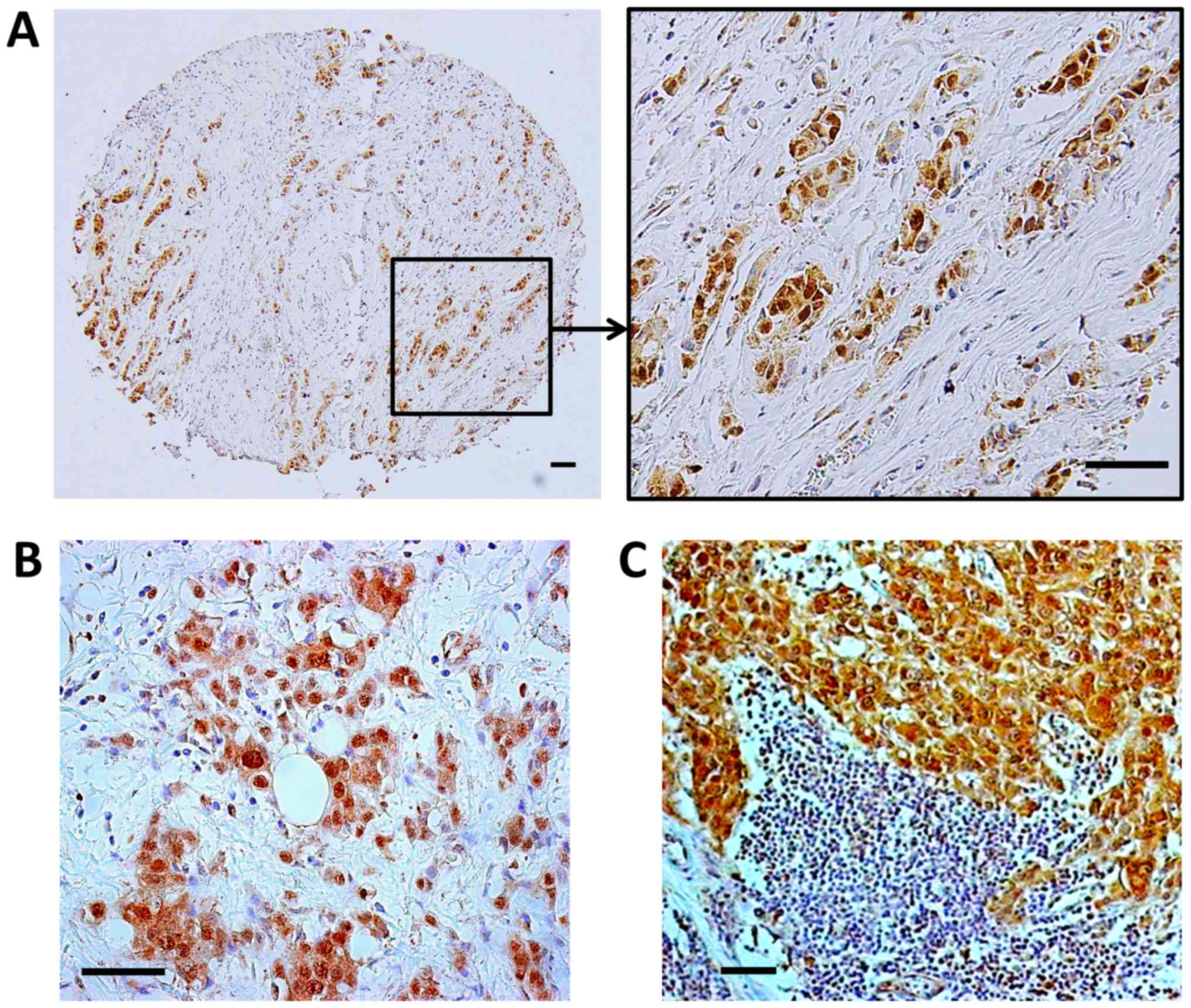
GSK-3β was observed across all breast cancer subtypes (Table I). In addition, we found nuclear
expression of GSK-3β in primary tumors and corresponding lymph node
metastases obtained from 8 breast cancer patients (4 cases were
ER+/PR+/HER2+ and 4 cases were ER-/PR-/HER2-according to the
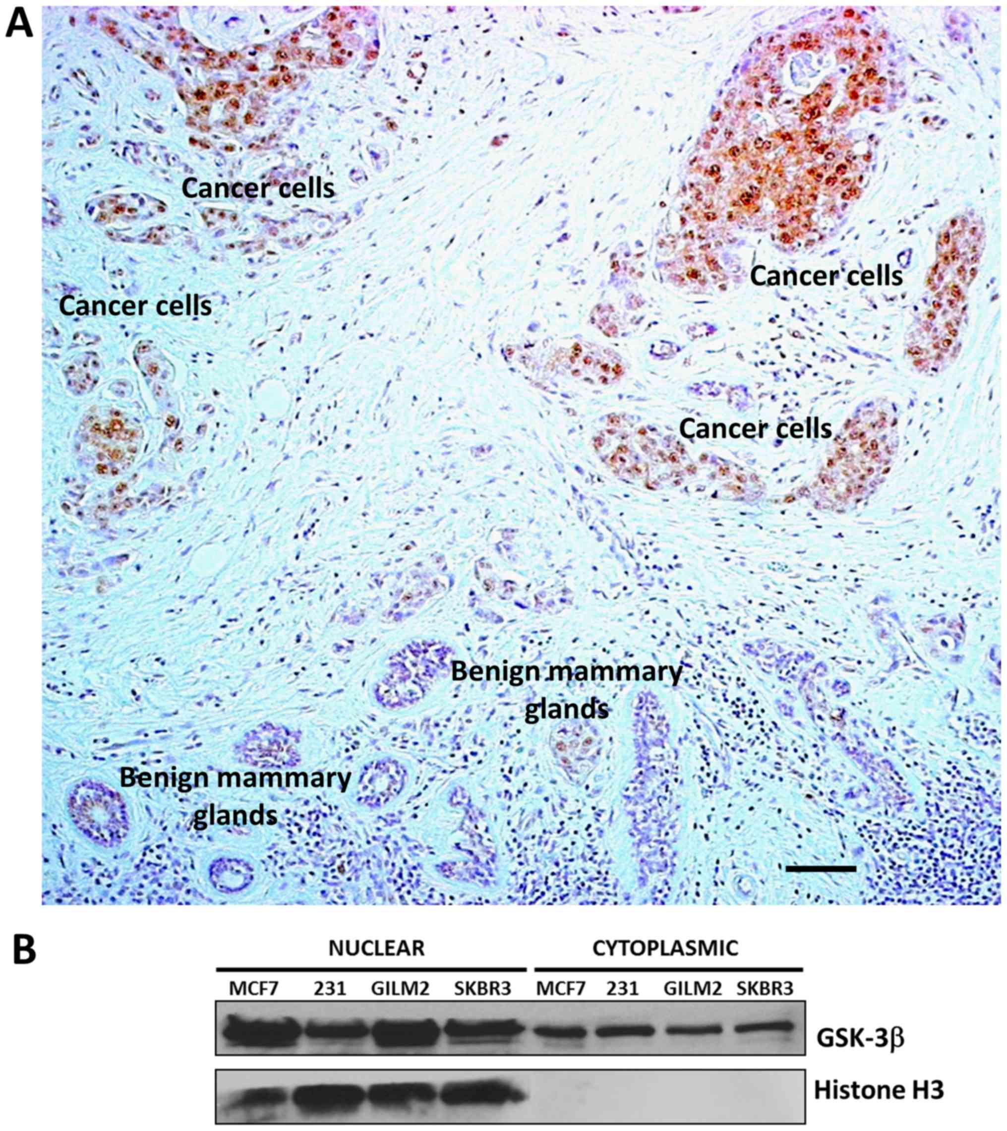
information obtained from patient's pathology report) (Fig. 2B and C). We did not observe GSK-3β
nuclear accumulation in mammary ducts, glands and other benign
cells within a breast tumor (Fig.
1A). Because it is particularly important to compare expression
of GSK-3β in benign structures adjacent to clumps of cancer cells
on the same tumor section, we selected such an area of breast
carcinoma (Fig. 1A) as a
representative picture of nuclear GSK-3β expression in breast
cancer cells and hardly detectable cytoplasmic expression of GSK-3β
in benign mammary glands. Only cytoplasmic expression of GSK-3β was
detected in breast fibroadenomas. Our results suggest that aberrant
nuclear accumulation of GSK-3β is a feature of breast cancer cells
and therefore might serve as a potential biomarker in human breast
cancer. In support of our TMA results, we also found aberrant
nuclear accumulation of GSK-3β in a panel of human breast cancer
cell lines (Fig. 1B).
 | Table I.Association between GSK-3β nuclear
expression and clinicopathological characteristics in primary
breast carcinoma. |
Table I.
Association between GSK-3β nuclear
expression and clinicopathological characteristics in primary
breast carcinoma.
|
| GSK-3β nuclear
expression |
|
|---|
|
|
|
|
|---|
|
Characteristics | Positive | Negative | P-value |
|---|
| Tumor stage |
|
| NS |
| T1 | 11 | 2 |
|
| T2 | 65 | 33 |
|
| T3 | 13 | 4 |
|
| Lymph node
metastasis |
|
| NS |
| N0 | 57 | 24 |
|
|
N1-N3 | 32 | 15 |
|
| Distant
metastasis |
|
| NS |
| M0 | 80 | 34 |
|
| M1 | 9 | 5 |
|
| Tumor
histology |
|
| NS |
| Well
differentiated | 7 | 1 |
|
|
Moderately differentiated | 64 | 25 |
|
| Poorly
differentiated | 18 | 13 |
|
| Estrogen receptor
expression |
|
| NS |
|
Positive | 19 | 11 |
|
|
Negative | 70 | 28 |
|
| HER2
expression |
|
| 0.0215 |
|
Positive | 19 | 2 |
|
|
Negative | 70 | 37 |
|
| TN carcinoma |
|
| 0.0001 |
| TN | 21 | 23 |
|
|
Non-TN | 68 | 16 |
|
Altered expression of GSK-3β in head
and neck tumors
Head and neck TMA consisted of benign tissues, 17
different benign tumors (nasal polyps, hemangioma, neurofibroma,
schwannoma and pleomorphic adenoma) and 29 different malignant
tumors including squamous cell carcinoma (15 cases), adenoid cystic
carcinoma (5 cases), sarcoma (2 cases), nasopharyngeal carcinoma (1
case), mucoepidermoid carcinoma (1 case), lymphoma (2 cases),
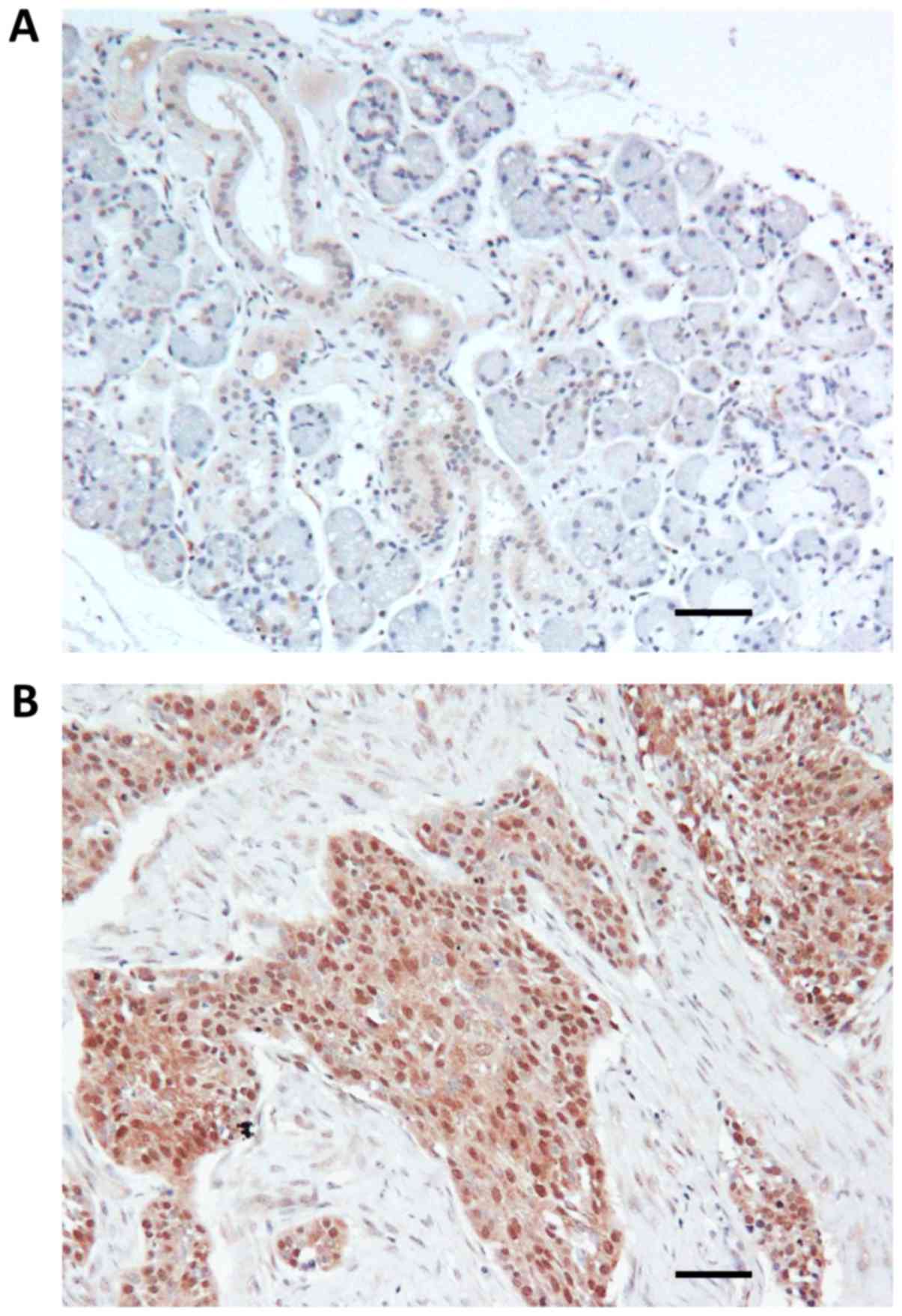
metastatic tumors (3 cases). Using immunohistochemical staining,
only weak cytoplasmic or no detectable expression of GSK-3β was
observed in benign salivary ducts, serous and mucous acini
(Fig. 3A). In great contrast to
benign tissue, GSK-3β nuclear accumulation was observed in 11 of 15
(73%) cases of squamous cell carcinoma and in 3 of 5 (60%) cases of
adenoid cystic carcinoma (Fig. 3B).
Only cytoplasmic expression of GSK-3β was observed in benign tumors
such as neurofibroma (2 cases), schwannoma (2 cases) and nasal
polyps (4 cases). However, nuclear accumulation of GSK-3β was
observed in 4 of 8 cases (50%) of pleomorphic adenoma, a common
benign tumor of salivary gland with malignant potential.
Discussion
GSK-3β has been identified as a positive regulator
of NF-κB-mediated survival and chemoresistance of cancer cells
(7,11–13).
Previous reports demonstrated that GSK-3β positively regulates
NF-κB transcriptional activity at a point downstream of the
activation of the IkB kinase complex (7), suggesting a potential role for nuclear
GSK-3β in the regulation of NF-κB transcriptional activity in the
nucleus of cancer cell. It also has been shown that only the active
form of GSK-3β accumulates in the nucleus of pancreatic cancer
cells and the inhibition of GSK-3β activity depletes its nuclear
accumulation via proteosomal degradation (10). Here, by immunohistochemical staining,
we identified aberrant nuclear accumulation of GSK-3β in five
breast cancer cell lines and in 89 of 128 (70%) human breast
carcinomas, whereas expression of GSK-3β was not detectable in
benign breast tissue. Our findings are supported by previously
published studies where aberrant GSK-3β nuclear accumulation was
found only in pancreatic, renal, and bladder cancer cells, and
malignant B-cells but not in corresponding benign cells (10–13). These
results support the hypothesis that nuclear GSK-3β may represent a
potential biomarker of breast cancer cells. Main findings of our
study are based on IHC staining of TMAs. Although we haven't
performed immunofluorescent staining of breast cancer cells in
vitro, we analyzed GSK-3β subcellular localization using
nuclear/cytosolic fractionation and Western immunoblotting with
proper control represented by Histone H3, a specific marker of
nuclear fraction (10). Our in
vitro findings of nuclear GSK-3β expression in breast cancer
cell lines are only supportive (not confirmative) for our in
vivo findings (IHC staining of TMAs) because in vitro
cancer cell culture (monoclonal) cannot truly represent
heterogenous human malignant tumor in vivo. In a previously
published immunohistochemical study of 1686 cases of breast cancer,
overexpression of GSK-3β was associated with several indicators of
poor prognosis including larger tumor size, lymph node metastasis,
high pathological grade, ER-negative disease, PR-negative disease,
increased proliferation and HER2 overexpression (27). Moreover, breast cancer patients with
GSK-3β expression in the highest quartile (246 of 1,686 cases) had
a 2.7-fold increased risk of distant relapse 5 years after tumor
resection (27). This study did not
differentiate between nuclear and cytoplasmic expression of GSK-3β
but is consistent with previously published findings showing that
GSK-3β nuclear expression was associated with high-grade tumors
(pancreatic and bladder cancer), metastasis (bladder cancer), and
poor prognosis (bladder cancer) (10,12,13). From
a technical perspective, nuclear accumulation of GSK-3β might be
overlooked in the presence of cytoplasmic GSK-3β overexpression
and/or heavy counterstaining with hematoxylin. The dark blue
staining of cancer cell nuclei by hematoxylin using standard
techniques (standard staining time is 5–8 min) might mask light
brown nuclear GSK-3β staining. In this study, we optimized the
hematoxylin counterstaining step to decrease the intensity of
hematoxylin nuclear staining by exposing the GSK-3β-stained TMA to
hematoxylin for 10 sec in order to maximize the differentiation of
the nuclear GSK-3β signal.
Because squamous cell carcinoma (common type of head
and neck cancer) is a very rare histological type of breast cancer,
we complemented our findings in breast cancer with the results of
GSK-3β staining of head and neck TMA to explore aberrant expression
of GSK-3β mainly in squamous cell carcinomas. Whereas only weak
cytoplasmic or no detectable expression of GSK-3β was observed in
benign salivary ducts, serous and mucous acini, GSK-3β nuclear
accumulation was observed in 11 of 15 (73%) cases of squamous cell
carcinoma and in 3 of 5 (60%) cases of adenoid cystic carcinoma.
Although we cannot draw a statistically significant conclusion due
to the limited number and different histotypes of head and neck
malignant tumors, we believe that it is important to present our
initial findings showing aberrant nuclear GSK-3β expression in head
and neck cancer (with focus on squamous cell carcinoma) but not in
benign tissues. This is the first exploratory IHC study to evaluate
aberrant GSK-3β expression in head and neck tumors. Further larger
scale studies of GSK-3β expression in head and neck cancer would
warrant a meaningful statistical analysis to find a correlation of
aberrant GSK-3β expression and clinicopathological parameters.
One of our translational goals was to evaluate
aberrant GSK-3β expression in breast and head and neck tumors as a
potential biomarker for selection of cancer patients for clinical
trials of GSK-3 inhibitors. To the best of our knowledge, our study
is the first to demonstrate aberrant nuclear expression of GSK-3β
as a specific marker of cancer cells in breast adenocarcinoma and
malignant head and neck tumors (squamous cell and adenoid cystic
carcinomas). To extend our initial findings to the clinic, we are
planning to evaluate GSK-3β expression in tumor tissues obtained
from cancer patients in Phase I/II clinical trials of the GSK-3
inhibitor 9-ING-41. After administration of 9-ING-41 to these
patients and subsequent evaluation of treatment response, we will
evaluate the clinical utility of nuclear GSK-3β as a potential
biomarker for selection of breast and head and neck cancer patients
for GSK-3-targeted therapy for prospective clinical trials using
paired biopsy samples.
Several reports have shown overexpression of GSK-3β
protein levels in malignant tumors of different origin. GSK-3β is
weakly expressed in the cytoplasm of benign cells and is rarely
detectable in the epithelial cells of benign prostatic tissue
whereas cytoplasmic overexpression of GSK-3β was associated with
aggressive prostate cancer (29).
Using Western blotting, a significant increase of GSK-3β protein
expression was found in ovarian adenocarcinomas compared with
normal ovaries and benign adenomas/borderline tumors (30). Another study demonstrated that the
level of GSK-3β protein expression was significantly higher in
colorectal carcinoma tissue than in their normal counterparts (18
of 20 cases), whereas inactive phosphorylated Ser9 GSK-3β was
detected in higher levels in normal tissue than in tumors in 17 of
20 cases (8). Recent studies showed
that overexpression of GSK-3β protein or its activation were
associated with adverse prognosis in patients with glioblastoma
(16), lung cancer (31,32), and
gastric cancer (33). We found only
weak cytoplasmic GSK-3β expression in salivary ducts, whereas
GSK-3β staining was hardly detectable in serous and acinar cells. A
significant finding of the present study is the presence of
aberrant nuclear accumulation of GSK-3β in cancer cells in most
cases of breast adenocarcinomas and squamous cell carcinomas. Our
results are consistent with another published study showing
aberrant nuclear GSK-3β as a specific marker of pancreatic cancer
cells whereas nuclear GSK-3β was not detectable in acinar or
epithelial pancreatic cells or in PanIN (pancreatic intraepithelial
neoplasia) lesions (10). GSK-3β
protein nuclear accumulation has also been found in colon (34), bladder (62% non-invasive and 91%
invasive) (13) and renal cancer
(12). It has been shown that only
the active form of GSK-3β accumulates in the nucleus of pancreatic
cancer cells and the inhibition of GSK-3β activity depletes its
nuclear accumulation via proteosomal degradation whereas the
expression level of cytoplasmic GSK-3β is not changed (10). It has also been demonstrated that that
nuclear GSK-3β plays an important role in regulating histone
modifications, which may contribute to NF-κB p65/p50 binding to
promoters and activating its target genes in cancer cells, leading
to increased cancer cell survival (11). Based on this published data and the
results presented herein, we hypothesize that nuclear GSK-3β might
be the actual target for GSK-3 inhibition in cancer cells, leading
to depletion of GSK-3β from the nucleus with a subsequent
inhibition of NF-κB-mediated cancer cell proliferation and
survival.
There is now extensive data credentialing GSK-3β as
a potential anticancer target in human pancreatic, colon, bladder,
breast, ovarian, lung, prostate, thyroid and renal cancer, chronic
lymphocytic leukemia, glioblastoma, and neuroblastoma (7–18,26–28,31,32,35–43).
Given the broad spectrum potential for a GSK-3 inhibitor in cancer,
the identification and validation of patient enrichment biomarkers
that select patients most likely to benefit from a GSK-3 inhibitor
will be an important component in the clinical development of novel
GSK-3 targeted drugs. Our results demonstrate aberrant nuclear
accumulation of GSK-3β in a significant number of human breast and
squamous cell carcinomas of the head and neck. As IHC staining is
an indispensable method for detection of subcellular expression and
validation of tumor markers (44,45), the
utility of nuclear GSK-3β as an enrichment biomarker will be
evaluated by IHC staining in clinical studies of the specific small
molecule inhibitor of GSK-3, 9-ING-41 (26,35,38,42,43).
Acknowledgements
Not applicable.
Funding
The present study was supported by Cancer Center
Support Grant from the Robert H. Lurie Comprehensive Cancer Center
of Northwestern University (grant no. 2 P30 CA060553-19 received by
AM and AU).
Availability of data and materials
All data generated or analyzed during the present
study are included in this published article.
Authors' contributions
AU, AM, VC and FG designed the experiments. AU
performed the experiments. AU, MM, TO and TT performed the analysis
and interpretation of data. The manuscript was prepared by AU, AM,
VC, FG and TO, and all authors contributed to writing and providing
feedback.
Ethics approval and consent to
participate
Not applicable.
Patient consent for publication
Not applicable.
Competing interests
The authors declare that there is no conflict of
interest.
References
|
1
|
Jemal A, Siegel R, Xu J and Ward E: Cancer
statistics, 2010. CA Cancer J Clin. 60:277–300. 2010. View Article : Google Scholar : PubMed/NCBI
|
|
2
|
Argiris A, Karamouzis M, Raben D and
Ferris R: Head and neck cancer. Lancet. 371:1695–1709. 2008.
View Article : Google Scholar : PubMed/NCBI
|
|
3
|
Cohen P and Frame S: The renaissance of
GSK3. Nat Rev Mol Cell Biol. 2:769–776. 2001. View Article : Google Scholar : PubMed/NCBI
|
|
4
|
Diehl JA, Cheng M, Roussel MF and Sherr
CJ: Glycogen synthase kinase-3beta regulates cyclin D1 proteolysis
and subcellular localization. Genes Dev. 12:3499–3511. 1998.
View Article : Google Scholar : PubMed/NCBI
|
|
5
|
Rubinfeld B, Albert I, Porfir E, Fiol C,
Munemitsu S and Polakis P: Binding of GSK3beta to the
APC-beta-catenin complex and regulation of complex assembly.
Science. 272:1023–1026. 1996. View Article : Google Scholar : PubMed/NCBI
|
|
6
|
Sears R, Nuckolls F, Haura E, Taya Y,
Tamai K and Nevins JR: Multiple Ras-dependent phosphorylation
pathways regulate Myc protein stability. Genes Dev. 14:2501–2514.
2000. View Article : Google Scholar : PubMed/NCBI
|
|
7
|
Ougolkov AV, Fernandez-Zapico ME, Savoy
DN, Urrutia RA and Billadeau DD: Glycogen synthase kinase-3beta
participates in nuclear factor kappaB-mediated gene transcription
and cell survival in pancreatic cancer cells. Cancer Res.
65:2076–2081. 2005. View Article : Google Scholar : PubMed/NCBI
|
|
8
|
Shakoori A, Ougolkov A, Yu ZW, Zhang B,
Modarressi M, Billadeau D, Mai M, Takahashi Y and Minamoto T:
Deregulated GSK3beta activity in colorectal cancer: Its association
with tumor cell survival and proliferation. Biochem Biophys Res
Commun. 334:1365–1373. 2005. View Article : Google Scholar : PubMed/NCBI
|
|
9
|
Gaisina IN, Gallier F, Ougolkov AV, Kim
KH, Kurome T, Guo S, Holzle D, Luchini DN, Blond SY, Billadeau DD
and Kozikowski AP: From a natural product lead to the
identification of potent and selective
benzofuran-3-yl-(indol-3-yl)maleimides as glycogen synthase kinase
3beta inhibitors that suppress proliferation and survival of
pancreatic cancer cells. J Med Chem. 52:1853–1863. 2009. View Article : Google Scholar : PubMed/NCBI
|
|
10
|
Ougolkov AV, Fernandez-Zapico ME, Bilim
VN, Smyrk TC, Chari ST and Billadeau DD: Aberrant nuclear
accumulation of glycogen synthase kinase-3beta in human pancreatic
cancer: Association with kinase activity and tumor
dedifferentiation. Clin Cancer Res. 12:5074–5081. 2006. View Article : Google Scholar : PubMed/NCBI
|
|
11
|
Ougolkov AV, Bone ND, Fernandez-Zapico ME,
Kay NE and Billadeau DD: Inhibition of glycogen synthase kinase-3
activity leads to epigenetic silencing of nuclear factor kappaB
target genes and induction of apoptosis in chronic lymphocytic
leukemia B cells. Blood. 110:735–742. 2007. View Article : Google Scholar : PubMed/NCBI
|
|
12
|
Bilim V, Ougolkov A, Yuuki K, Naito S,
Kawazoe H, Muto A, Oya M, Billadeau D, Motoyama T and Tomita Y:
Glycogen synthase kinase-3: A new therapeutic target in renal cell
carcinoma. Br J Cancer. 101:2005–2014. 2009. View Article : Google Scholar : PubMed/NCBI
|
|
13
|
Naito S, Bilim V, Yuuki K, Ugolkov A,
Motoyama T, Nagaoka A, Kato T and Tomita Y: Glycogen synthase
kinase-3beta: A prognostic marker and a potential therapeutic
target in human bladder cancer. Clin Cancer Res. 16:5124–5132.
2010. View Article : Google Scholar : PubMed/NCBI
|
|
14
|
Cao Q, Lu X and Feng Y: Glycogen synthase
kinase-3beta positively regulates the proliferation of human
ovarian cancer cells. Cell Res. 16:671–677. 2006. View Article : Google Scholar : PubMed/NCBI
|
|
15
|
Kunnimalaiyaan M, Vaccaro AM, Ndiaye MA
and Chen H: Inactivation of glycogen synthase kinase-3beta, a
downstream target of the raf-1 pathway, is associated with growth
suppression in medullary thyroid cancer cells. Mol Cancer Ther.
6:1151–1158. 2007. View Article : Google Scholar : PubMed/NCBI
|
|
16
|
Miyashita K, Kawakami K, Nakada M, Mai W,
Shakoori A, Fujisawa H, Hayashi Y, Hamada J and Minamoto T:
Potential therapeutic effect of glycogen synthase kinase 3beta
inhibition against human glioblastoma. Clin Cancer Res. 15:887–897.
2009. View Article : Google Scholar : PubMed/NCBI
|
|
17
|
Zhu Q, Yang J, Han S, Liu J, Holzbeierlein
J, Thrashe JB and Li B: Suppression of glycogen synthase kinase 3
activity reduces tumor growth of prostate cancer in vivo. Prostate.
71:835–845. 2011. View Article : Google Scholar : PubMed/NCBI
|
|
18
|
Wang Z, Smith KS, Murphy M, Piloto O,
Somervaille TC and Cleary ML: Glycogen synthase kinase 3 in MLL
leukaemia maintenance and targeted therapy. Nature. 455:1205–1209.
2008. View Article : Google Scholar : PubMed/NCBI
|
|
19
|
Hoeflich KP, Luo J, Rubie EA, Tsao MS, Jin
O and Woodgett JR: Requirement for glycogen synthase kinase-3beta
in cell survival and NF-kappaB activation. Nature. 406:86–90. 2000.
View Article : Google Scholar : PubMed/NCBI
|
|
20
|
Aggarwal BB: Nuclear factor-kappaB: The
enemy within. Cancer Cell. 6:203–208. 2004. View Article : Google Scholar : PubMed/NCBI
|
|
21
|
Tas SW, Vervoordeldonk MJ and Tak PP: Gene
therapy targeting nuclear factor-kappaB: Towards clinical
application in inflammatory diseases and cancer. Curr Gene Ther.
9:160–170. 2009. View Article : Google Scholar : PubMed/NCBI
|
|
22
|
Medina M and Castro A: Glycogen synthase
kinase-3 (GSK-3) inhibitors reach the clinic. Curr Opin Drug Discov
Devel. 11:533–543. 2008.PubMed/NCBI
|
|
23
|
Walz A, Ugolkov A, Chandra S, Kozikowski
A, Carneiro BA, O'Halloran TV, Giles FJ, Billadeau DD and Mazar AP:
Molecular pathways: Revisiting glycogen synthase kinase-3β as a
target for the treatment of cancer. Clin Cancer Res. 23:1891–1897.
2017. View Article : Google Scholar : PubMed/NCBI
|
|
24
|
Rizzieri DA, Cooley S, Odenike O, Moonan
L, Chow KH, Jackson K, Wang X, Brail L and Borthakur G: An
open-label phase 2 study of glycogen synthase kinase-3 inhibitor
LY2090314 in patients with acute leukemia. Leuk Lymphoma.
57:1800–1806. 2016. View Article : Google Scholar : PubMed/NCBI
|
|
25
|
Dignam JD, Lebovitz RM and Roeder RG:
Accurate transcription initiation by RNA polymerase II in a soluble
extract from isolated mammalian nuclei. Nucleic Acids Res.
11:1475–1489. 1983. View Article : Google Scholar : PubMed/NCBI
|
|
26
|
Ugolkov A, Gaisina I, Zhang JS, Billadeau
DD, White K, Kozikowski A, Jain S, Cristofanilli M, Giles F,
O'Halloran T, et al: GSK-3 inhibition overcomes chemoresistance in
human breast cancer. Cancer Lett. 380:384–392. 2016. View Article : Google Scholar : PubMed/NCBI
|
|
27
|
Quintayo MA, Munro AF, Thomas J, Kunkler
IH, Jack W, Kerr GR, Dixon JM, Chetty U and Bartlett JM: GSK3β and
cyclin D1 expression predicts outcome in early breast cancer
patients. Breast Cancer Res Treat. 136:161–168. 2012. View Article : Google Scholar : PubMed/NCBI
|
|
28
|
Shin S, Wolgamott L, Tcherkezian J,
Vallabhapurapu S, Yu Y, Roux PP and Yoon SO: Glycogen synthase
kinase-3β positively regulates protein synthesis and cell
proliferation through the regulation of translation initiation
factor 4E-binding protein 1. Oncogene. 33:1690–1699. 2014.
View Article : Google Scholar : PubMed/NCBI
|
|
29
|
Darrington RS, Campa VM, Walker MM,
Bengoa-Vergniory N, Gorrono-Etxebarria I, Uysal-Onganer P, Kawano
Y, Waxman J and Kypta RM: Distinct expression and activity of
GSK-3α and GSK-3β in prostate cancer. Int J Cancer. 131:E872–E883.
2012. View Article : Google Scholar : PubMed/NCBI
|
|
30
|
Rask K, Nilsson A, Brännström M, Carlsson
P, Hellberg P, Janson PO, Hedin L and Sundfeldt K: Wnt-signalling
pathway in ovarian epithelial tumours: increased expression of
beta-catenin and GSK3beta. Br J Cancer. 89:1298–1304. 2003.
View Article : Google Scholar : PubMed/NCBI
|
|
31
|
Vincent EE, Elder DJ, O'Flaherty L, Pardo
OE, Dzien P, Phillips L, Morgan C, Pawade J, May MT, Sohail M, et
al: Glycogen synthase kinase 3 protein kinase activity is
frequently elevated in human non-small cell lung carcinoma and
supports tumour cell proliferation. PLoS One. 9:e1147252014.
View Article : Google Scholar : PubMed/NCBI
|
|
32
|
Zeng J, Liu D, Qiu Z, Huang Y, Chen B,
Wang L, Xu H, Huang N, Liu L and Li W: GSK3β overexpression
indicates poor prognosis and its inhibition reduces cell
proliferation and survival of non-small cell lung cancer cells.
PLoS One. 9:e912312014. View Article : Google Scholar : PubMed/NCBI
|
|
33
|
Cho YJ, Yoon J, Ko YS, Kim SY, Cho SJ, Kim
WH, Park JW, Youn HD, Kim JH and Lee BL: Glycogen synthase
kinase-3β does not correlate with the expression and activity of
β-catenin in gastric cancer. APMIS. 118:782–790. 2010. View Article : Google Scholar : PubMed/NCBI
|
|
34
|
Salim T, Sjölander A and Sand-Dejmek J:
Nuclear expression of glycogen synthase kinase-3β and lack of
membranous β-catenin is correlated with poor survival in colon
cancer. Int J Cancer. 133:807–815. 2013. View Article : Google Scholar : PubMed/NCBI
|
|
35
|
Pal K, Cao Y, Gaisina IN, Bhattacharya S,
Dutta SK, Wang E, Gunosewoyo H, Kozikowski AP, Billadeau DD and
Mukhopadhyay D: Inhibition of GSK-3 induces differentiation and
impaired glucose metabolism in renal cancer. Mol Cancer Ther.
13:285–296. 2014. View Article : Google Scholar : PubMed/NCBI
|
|
36
|
Carter YM, Kunnimalaiyaan S, Chen H,
Gamblin TC and Kunnimalaiyaan M: Specific glycogen synthase
kinase-3 inhibition reduces neuroendocrine markers and suppresses
neuroblastoma cell growth. Cancer Biol Ther. 15:510–515. 2014.
View Article : Google Scholar : PubMed/NCBI
|
|
37
|
Duffy DJ, Krstic A, Schwarzl T, Higgins DG
and Kolch W: GSK3 inhibitors regulate MYCN mRNA levels and reduce
neuroblastoma cell viability through multiple mechanisms, including
p53 and Wnt signaling. Mol Cancer Ther. 13:454–467. 2014.
View Article : Google Scholar : PubMed/NCBI
|
|
38
|
Hilliard TS, Gaisina IN, Muehlbauer AG,
Gaisin AM, Gallier F and Burdette JE: Glycogen synthase kinase 3β
inhibitors induce apoptosis in ovarian cancer cells and inhibit
in-vivo tumor growth. Anticancer Drugs. 22:978–985. 2011.PubMed/NCBI
|
|
39
|
Kotliarova S, Pastorino S, Kovell LC,
Kotliarov Y, Song H, Zhang W, Bailey R, Maric D, Zenklusen JC, Lee
J and Fine HA: Glycogen synthase kinase-3 inhibition induces glioma
cell death through c-MYC, nuclear factor-kappaB, and glucose
regulation. Cancer Res. 68:6643–6651. 2008. View Article : Google Scholar : PubMed/NCBI
|
|
40
|
Kunnimalaiyaan M, Vaccaro AM, Ndiaye MA
and Chen H: Inactivation of glycogen synthase kinase-3beta, a
downstream target of the raf-1 pathway, is associated with growth
suppression in medullary thyroid cancer cells. Mol Cancer Ther.
6:1151–1158. 2007. View Article : Google Scholar : PubMed/NCBI
|
|
41
|
Zhu Q, Yang J, Han S, Liu J, Holzbeierlein
J, Thrasher JB and Li B: Suppression of glycogen synthase kinase 3
activity reduces tumor growth of prostate cancer in vivo. Prostate.
71:835–845. 2011. View Article : Google Scholar : PubMed/NCBI
|
|
42
|
Ugolkov A, Qiang W, Bondarenko G, Procissi
D, Gaisina I, James CD, Chandler J, Kozikowski A, Gunosewoyo H,
O'Halloran T, et al: Combination treatment with the GSK-3 inhibitor
9-ING-41 and CCNU cures orthotopic chemoresistant glioblastoma in
patient-derived xenograft models. Transl Oncol. 10:669–678. 2017.
View Article : Google Scholar : PubMed/NCBI
|
|
43
|
Ugolkov AV, Bondarenko GI, Dubrovskyi O,
Berbegall AP, Navarro S, Noguera R, O'Halloran TV, Hendrix MJ,
Giles FJ and Mazar AP: 9-ING-41, a small molecule glycogen
synthase-3 inhibitor, is active in neuroblastoma. Anticancer Drugs.
29:717–724. 2018.PubMed/NCBI
|
|
44
|
Kim SW, Roh J and Park CS:
Immunohistochemistry for pathologists: Protocols, pitfalls and
tips. J Pathol Transl Med. 50:411–418. 2016. View Article : Google Scholar : PubMed/NCBI
|
|
45
|
Cheuk W and Chan JK: Subcellular
localization of immunohistochemical signals: Knowledge of the
ultrastructural or biologic features of the antigens helps predict
the signal localization and proper interpretation of immunostains.
Int J Surg Pathol. 12:185–206. 2004. View Article : Google Scholar : PubMed/NCBI
|