Introduction
Head and neck squamous cell carcinoma (HNSCC) is one
of the most prevalent cancers and a major cause of mortality in
patients with cancer worldwide, and 330,000 mortality cases are
reported each year as a result of this disease (1). Approximately half of these cases involve
oral squamous cell carcinoma (OSCC) (2), which is a highly aggressive head and
neck tumor prone to local recurrence and metastasis (3). The development of OSCC is a long-term,
multistage and multifactorial process, and numerous regulatory
factors are involved in its carcinogenesis (4). However, the detailed molecular
pathogenesis of this type of cancer remains unclear. An
understanding of the molecular mechanisms underlying OSCC
tumorigenesis is necessary to identify tumor-specific biomarkers
and therapeutic targets for early diagnosis and treatment,
respectively.
Periostin, also known as osteoblast-specific factor
2 (fasciclin I-like), is a secreted matrix N-glycoprotein that
contains an NH2-terminal signal peptide sequence,
internal homologous repeats, a cysteine-rich domain and hydrophilic
C-terminal domain (5,6). Periostin performs an important function
in numerous biological processes, including bone development,
maturation and remodeling, cardiovascular differentiation, and
cutaneous and connective tissue remodeling, as well as in allergic
diseases, respiratory diseases and various inflammatory conditions
(7). Considering the role of
periostin in epithelial-mesenchymal transition (EMT), extracellular
matrix (ECM) restructuring and remodeling, research has been
focusing on the role of periostin in oncology (8).
Therefore, the present study examined periostin
expression and its association with the clinical characteristics of
OSCC patients, and investigated the possible underlying mechanisms.
The study findings suggested that periostin functions as an
oncogene and a potential target for OSCC therapy.
Materials and methods
Ethics statement
Experiments using human samples were approved by the
Ethics Committee of the School of Stomatology, China Medical
University (Shenyang, China), and written informed consent was
obtained from the donors.
Tissue samples
OSCC specimens were obtained from 12 patients who
underwent surgical resection in the Department of Oral and
Maxillofacial Surgery at the School of Stomatology, China Medical
University, between January 2015 and September 2016. These tumor
specimens were immediately frozen and stored at −80°C. Another set
of 90 OSCC specimens, which were fixed with formalin and embedded
in paraffin, and 20 OSCC-adjacent healthy epithelium samples were
acquired from the Department of Oral Pathology at the School of
Stomatology, China Medical University, between January 2012 and
March 2016. The specimens used in immunohistochemistry were derived
from archival wax blocks in the Department of pathology at the
School of Stomatology, China Medical University. The 12 cases used
in RT-qPCR were originally from the clinical patients. The number
of frozen tissue specimens was relatively less. So two different
sets of patients used. Patients who received radiotherapy or
chemotherapy were excluded. Clinical data, including sex, age, TNM
classification, histological grade, stage, tumor location, were
obtained from the medical records of the 90 OSCC patients. The TNM
classification was assessed according to the World Health
Organization 2010 criteria (9). The
tumor stage was based on pathological findings according to the
American Joint Committee on Cancer (AJCC) guidelines (10).
Immunohistochemical staining
Immunohistochemical staining was performed to detect
the protein localization and expression in paraffin-embedded OSCC
specimens and adjacent controls. Paraffin sections were cut at 4 mm
thickness. Briefly, the slides were stained with a rabbit
anti-periostin polyclonal antibody (ab14041; Abcam, Cambridge, MA,
USA) overnight at 4°C, which was diluted to 1:1,000, followed by
staining for 1 h at room temperature with an anti-rabbit IgG
polyclonal antibody (sc515946; Santa Cruz Biotechnology, Inc.,
Dallas, TX, USA), which was diluted to 1:10,000. Each slide was
evaluated by one of the authors under a microscope (Nikon
Corporation, Tokyo, Japan). At higher magnification (×400), five
visual fields were selected randomly, and the positive expression
signals were analyzed by means of the Image-Pro Plus 6.0 software
(Media Cybernetics, Inc., Rockville, MD, USA). Periostin protein
levels in the OSCC tissue and adjacent healthy epithelium samples
were compared in accordance with the integral optical density (IOD)
as a parameter for semi-quantitative detection.
Cell culture
Human OSCC cell lines SCC-9 and SCC-25 (ATCC,
Manassas, VA, USA) were used between passages 10 and 20. In
addition, human keratinocytes (HaCaT cells; ATCC) between passages
4 and 6 were used as the controls. The OSCC cells were maintained
in Dulbecco's modified Eagle's medium (DMEM; Gibco; Thermo Fisher
Scientific, Inc., Waltham, MA, USA) supplemented with 10% fetal
bovine serum and 1% of penicillin-streptomycin solution, whereas
HaCaT cells were cultured in DMEM and 10% fetal bovine serum
(Gibco; Thermo Fisher Scientific, Inc.). All the cell lines were
maintained at 37°C in the presence of 5% CO2.
Reverse transcription-quantitative
polymerase chain reaction (RT-qPCR)
Total RNA was extracted from the tissues and cells
with the TRIzol reagent (Sangon Biotech Co., Ltd., Shanghai,
China). Following quantification of RNA concentration using an
ultraviolet spectrophotometer, 1 µg DNase-treated RNA was used for
cDNA synthesis with an RT reagent kit (Takara Biotechnology Co.,
Ltd., Dalian, China). The RT reaction was conducted at 37°C for 15
min and 85°C for 5 min, and the RT reaction system was as follows:
1 µg RNA and 4 µl 5X PrimeScript RT Master Mix, with RNase-free
distilled H2O added up to 20 µl. Next, qPCR was
conducted by means of the SYBR Premix Ex Taq II kit (Takara
Biotechnology Co., Ltd.) with the following reaction system: 10 µl
SYBR Premix Ex Taq II, 1 µl cDNA, 0.5 µl forward primer, 0.5 µl
reverse primer and 8 µl sterile water. The cycling conditions were
as follows: 95°C for 1 min, 94°C for 30 sec, 58°C for 30 sec and
72°C for 10 sec. The sequences of the primer pairs were as follows:
Periostin, 5′-TTTACAACGGGCAAATACTGGAAAC-3′ (forward) and
5′-GATGATCTCGCGGAATATGTGAA-3′ (reverse); GAPDH,
5′-ACCACAGTCCATGCCATCAC-3′ (forward) and 5′-TCCACCACCCTGTTGCTGTA-3′
(reverse). Gene expression was normalized to GAPDH as the
internal control, and the mean relative change was determined in
triplicate or quintuplicate through relative quantification and
application of the 2−ΔΔCq method (11).
Western blotting
OSCC cells were washed three times with cold PBS,
and then with a lysis buffer containing 1 mM PMSF (Beyotime
Institute of Biotechnology Shanghai, China). The tissue samples
were collected and sonicated in a lysis buffer containing 1 mM
PMSF. Protein concentrations were determined by the bicinchoninic
acid method. Next, samples corresponding to 50 µg total protein
were subjected to SDS-PAGE in a 10% gel, and then transferred onto
polyvinylidene difluoride membranes. Subsequent to blocking with 5%
non-fat milk for 1 h at room temperature, the membranes were
incubated with anti-periostin (1:1,000, ab14041; Abcam, Cambridge,
MA, USA) at 4°C overnight. Following washing with TBST three times
at room temperature, each time for 10 min, they were incubated with
horseradish peroxidase conjugated rabbit anti-mouse secondary
antibodies (1:10,000; ab6728; Abcam, Cambridge, MA, USA) for 1 h
and then were washed again Protein bands were visualized with an
enhanced chemiluminescence reagent (Pierce; Thermo Fisher
Scientific, Inc.). GAPDH (1:10,000, ab8245; Abcam) was used as the
internal control. Image J 1.44 software (National Institutes of
Health, Bethesda, MD, USA) was used for the analysis of Periostin
expression.
Plasmid construction and siRNA
synthesis
The open reading frame of human periostin cDNA was
cloned into eukaryotic expression vector GV144 (GeneChem, Shanghai,
China). Subsequently, the amplicon of the periostin gene was
purified, digested and ligated into the respective HindIII
and EcoRI sites of the GV144 vector. The Periostin
overexpression plasmid sequences as follows:
5′-GTCCGGACTCAGATCTCGAGCTATGATTCCCTTTTTACCCATG-3′ (forward) and
5′-TATCTAGATCCGGTGGATCCTCACTGAGAACGACCTTCCCTTAATC-3′ (reverse). A
GV144 empty vector was used as control for Periostin expression.
The siPeriostin and siCtrl were synthesized by GenePharam company
(Shanghai, China). The siPeriostin sequence was as follows:
5′-GCCAUCACAUCGGACAUAUTT-3′ (forward) and
5′-AUAUGUCCGAUGUGAUGGCTT-3′ (reverse). The siCtrl sequence was as
follows: 5′-UUCUCCGAACGUGUCACGUTT-3′ (forward) and
5′-ACGUGACACGUUCGGAGAATT-3′ (reverse).
Transfection with POSTN small
interfering RNA (siRNA) and overexpression plasmid
OSCC cells were routinely cultured. OSCC cells were
transfected with the siPeriostin for Periostin expression knockdown
and with the periostin plasmid for Periostin overexpression,
using Lipofectamine 2000 (Thermo Fisher Scientific, Inc.) according
to the manufacturer's protocol. The siPeriostin sequence was as
follows: 5′-CCCAUGGAGAGCCAAUUAUTT-3′. An empty vector was
transfected into the control group. The transfection efficiency was
then assessed by western blotting and RT-qPCR analyses.
MTS cell proliferation assay
OSCC cells were transfected with siPeriostin or with
the periostin plasmid for 24 h at 37°C. Proliferation assays were
conducted using an MTS Cell Proliferation Assay kit (Promega Corp.,
Madison, WI, USA). Briefly, cells with a stable knockdown of
periostin expression or control cells were plated in 96-well plates
at a density of 2×10³ cells/well.
The culture plates were taken out at different time
periods (24, 48, and 72 h) and continuously cultured at 37°C for 2
h after the addition of 10 µl MTS in each well. The optical density
(OD) value was measured at a wavelength of 490 nm using enzyme
linked immunosorbent assay.
Cell invasion assays
OSCC cells were transfected with siPeriostin or with
the periostin plasmid for 24 h. Invasion assays were then performed
using a Cell Invasion Assay kit (BD Biosciences, Billerica, MA,
USA). Six-well plates and transwell chambers was used. The upper
chamber was pre-coated with 50 µl of 20% growth factor-reduced
Matrigel for the invasion assay. Briefly, cells were plated in the
upper well of a Boyden chamber at a concentration of
5×10⁴ cells/well in 100 µl serum-free DMEM. In the lower
chamber, 600 µl DMEM containing 10% FBS was added to serve as a
chemoattractant. After incubation at 37°C for 20 h, the invading
cells on the lower surface of the filter were fixed with 95%
ethanol and stained with the Coomassie blue dye (Leagene
Biotechnology Co., Ltd., Beijing, China). Subsequently, images of
the cells were captured, and the cell numbers were manually counted
in five random visual fields per filter (magnification, ×40).
Statistical analysis
The Mann-Whitney rank sum test was applied to
compare the periostin mRNA and protein levels between the OSCC
cells and the healthy controls. In addition, correlations between
the IOD levels of the periostin protein and clinical factors were
assessed by univariate analysis. Statistical analysis was performed
using the SPSS version 17.0 software (SPSS, Inc., Chicago, IL,
USA). P<0.05 was considered to indicate a statistically
significant difference.
Results
High expression of periostin in OSCC
tissues and cell lines
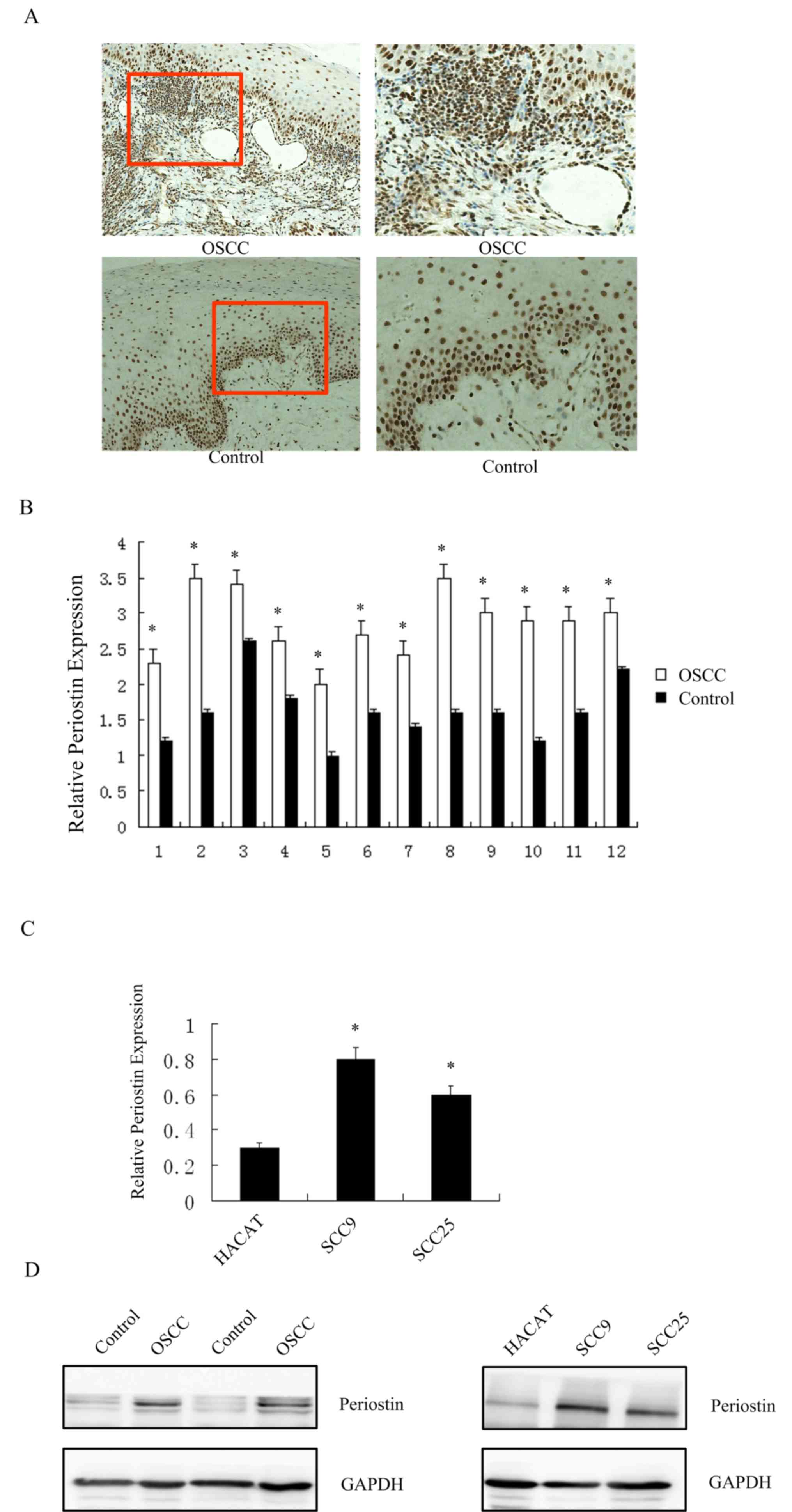
First, the expression of periostin in OSCC and
adjacent tissues, including 110 cases (90 OSCC and 20 healthy
adjacent tissues), were examined by immunohistochemistry (Fig. 1A). Periostin staining was mainly
detected in the cytoplasm and was occasionally present in the
nuclei of tumor cells. The OSCC group exhibited significantly
higher periostin expression as compared with that in the adjacent
tissue controls (Table I;
P<0.05).
 | Table I.IOD of periostin in OSCC by
immunohistochemical staining. |
Table I.
IOD of periostin in OSCC by
immunohistochemical staining.
| Tissue | IOD |
|---|
| OSCC |
156.02±7.12a |
| Adjacent | 63.23±8.15 |
To determine the expression levels of periostin, the
specimens of OSCC and healthy adjacent tissues were collected and
RT-qPCR and western blotting were then conducted. For statistical
analysis with SPSS software, the data were presented as a fold
change in the gene expression, normalized to GAPDH. As shown in
Fig. 1B and C, the mRNA (P<0.05)
and protein levels of periostin in OSCC tissue were notably higher
in comparison with those in the healthy adjacent tissue. Only 12
sets of samples were shown, as they were the fresh specimens. The
90 cases used in immunohistochemistry were derived from archival
wax blocks and are not suitable for RT-qPCR.
RT-qPCR and western blotting were also performed in
SCC9, SCC25 and HaCaT cells to measure the expression of periostin.
Similar to the tissue results, the mRNA and protein expression
levels of periostin were significantly higher in OSCC cells as
compared with those in the healthy epithelium cells (P<0.05;
Fig. 1C and D).
Association between periostin and
clinical characteristics
The association of periostin expression and the
clinical characteristics of OSCC patients was also analyzed in the
present study. Univariate analysis was conducted to evaluate any
possible associations (Table II).
The expression was considered negative if the final IOD was ≥65 and
were otherwise considered positive. There was a significant
correlation between N classification and periostin protein levels
(P=0.007). A positive periostin expression was significantly
associated with the presence of lymphovascular permeation
(P=0.007). N classification exhibited borderline significance of
correlation (P=0.053). Other factors were not found to be
associated with periostin expression.
| Table II.Association of the clinical
characteristics of oral squamous cell carcinoma patients with
positive (n=78; 86.7%) and negative (n=12; 13.3%) periostin
expression. |
Table II.
Association of the clinical
characteristics of oral squamous cell carcinoma patients with
positive (n=78; 86.7%) and negative (n=12; 13.3%) periostin
expression.
| Characteristic | N | Periostin positive, n
(%) | Periostin negative, n
(%) | P-value |
|---|
| Sex |
|
|
| 0.571 |
| Male | 55 | 50 (64.1) | 9 (75.0) |
|
|
Female | 35 | 28 (35.9) | 3 (25.0) |
|
| Age (years) |
|
|
| 0.512 |
| ≤55 | 49 | 32 (41.0) | 6 (50.0) |
|
|
>55 | 41 | 46 (59.0) | 6 (50.0) |
|
| T classification |
|
|
| 0.623 |
| T1/2 | 40 | 30 (38.5) | 3 (25.0) |
|
| T3/4 | 50 | 48 (61.5) | 9 (75.0) |
|
| N classification |
|
|
| 0.704 |
| N0 | 35 | 47 (60.3) | 5 (41.7) |
|
| N0+ | 55 | 31 (39.7) | 7 (58.3) |
|
| Histological
grade |
|
|
| 0.312 |
| Well
differentiated | 42 | 49 (62.8) | 6 (50.0) |
|
|
Poorly/moderately
differentiated | 48 | 29 (37.2) | 6 (50.0) |
|
| Stage |
|
|
| 0.235 |
| I and
II | 36 | 51 (65.4) | 5 (41.7) |
|
| III and
IV | 54 | 27 (34.6) | 7 (58.3) |
|
| Location |
|
|
| 0.412 |
| Buccal
mucosa | 20 | 16 (20.5) | 4 (33.3) |
|
|
Tongue | 45 | 40 (51.3) | 5 (41.7) |
|
| Mouth
floor | 10 | 8 (10.3) | 2 (16.7) |
|
|
Others | 15 | 14 (17.9) | 1 (8.3) |
|
| Lymphovascular
permeation |
|
|
| 0.007 |
|
Present | 82 | 72 (92.3) | 10 (83.3) |
|
|
Absent | 8 | 6 (7.7) | 2 (16.7) |
|
| Distant
metastasis |
|
|
| 0.053 |
|
Present | 55 | 45 (57.7) | 10 (83.3) |
|
|
Absent | 35 | 33 (42.3) | 2 (16.7) |
|
Periostin promotes the proliferation
and invasiveness of OSCC cells
To determine the physiological role of periostin,
the impact of periostin on the growth and motility characteristics
of OSCC cells transfected with siPeriostin or the control plasmid
was analyzed in SCC9 and SCC25 cell lines. The results revealed
that periostin mRNA and protein levels in the two cell lines
transfected with negative control were 4-fold higher in comparison
with those in cells transfected with siPeriostin according to the
RT-qPCR and western blotting data (P<0.05; Fig. 2A and B). In addition, periostin mRNA
and protein levels in cells transfected with the periostin plasmid
were 2-fold higher compared with those of cells transfected with
the empty vector, as determined by RT-qPCR and western blotting
(P<0.05; Fig. 2A and B). These
indicated that transfection with siPeriostin and plasmid
successfully resulted in the knockdown and overexpression of
periostin, respectively.
To analyze the participation of periostin in the
growth of OSCC cells, MTS cell proliferation assays were conducted
subsequent to transfection. The results demonstrated that the
knockdown of periostin by siPeriostin significantly suppressed the
proliferation of OSCC cells, whereas the upregulation of periostin
markedly increased the proliferation of OSCC cells (Fig. 2C).
In addition, to examine the potential effect of
periostin on the migration of OSCC cells, Transwell assays were
conducted in a Boyden chamber. The results revealed that the
knockdown of periostin significantly suppressed the invasiveness of
OSCC cells, whereas periostin upregulation significantly increased
the invasion by OSCC cells (Fig.
3).
Discussion
Periostin, an extracellular-matrix protein that
belongs to the fasciclin family, is encoded by the POSTN
gene and has been demonstrated to be essential for the process of
remodeling during tissue and organ development or repair, as well
as inflammation (12,13). It contains an N-terminal secretory
signal peptide, followed by a cysteine-rich domain, four internal
homologous repeats and a C-terminal hydrophilic domain. Periostin
exerts its biological activities by functioning as a matricellular
protein, affecting cell activation by binding to receptors on the
cell surface. It participates in numerous biological processes,
including the regulation of gene expression and the development of
bone, teeth and heart vessels (14,15).
Periostin upregulation has been reported in a number of solid
tumors, including colon (6,16), thyroid (17), breast (18), pancreatic (8,19) and lung
cancer (20). In addition,
accumulating evidence revealed that periostin was upregulated in
HNSCC tissue (21,22), and that it promotes invasion and
angiogenesis in OSCC (22). Certain
studies have also reported that periostin promoted tube formation
of lymphatic endothelial cells independently of vascular
endothelial growth factor C and promoted lymphangiogenesis, which
was mediated by Src and Akt activity in HNSCC (23). However, the influence of periostin on
OSCC progression and metastasis is currently poorly understood.
In the present study, periostin levels were
significantly higher in OSCC cell lines as compared with those in
HaCaT cells, which was contrary to the findings of a previous study
(21). Furthermore, the present study
reported that the knockdown of periostin expression inhibited cell
proliferation and invasiveness.
Cases of periostin overexpression were found to be
associated with lymphovascular permeation, while distant metastasis
exhibited borderline significance of correlation. Consequently,
analysis of POSTN expression may be particularly useful
during the prognostic evaluation of this cohort of patients.
Although the clinical value of periostin in OSCC was
comprehensively analyzed, there are certain limitations in the
present study. First, the sample size in the subgroup analysis was
not sufficiently large; thus, the statistical power of the results
is limited. In addition, the relevant mechanisms were identified in
the current study. In our future studies, more in-depth research
and analysis will be conducted. Furthermore, animal experiments
need to be devised to investigate the effects of periostin on tumor
growth in vitro and in vivo.
In conclusion, it was observed that the levels of
periostin were highly upregulated in the tumor tissues of patients
with OSCC, and immunohistochemical staining demonstrated increased
localization to areas of active fibrosis in the tumor tissue. The
present study provides a new line of evidence indicating that
periostin is significantly upregulated in OSCC tissues and cell
lines. The data implied that periostin, as an oncogene, serves an
important role in the pathogenesis of OSCC.
Acknowledgements
Not applicable.
Funding
This study was supported by grants from the Natural
Science Foundation of Liaoning Province (no. 20170541012) and the
Youth Research Fund of the school of Stomatology, China Medical
University (no. K101593-16-05).
Availability of data and materials
All data generated or analyzed during this study are
included in this published article.
Authors' contributions
YK and XW conceived and designed the experiments. YZ
and YS collected and analyzed the imaging and pathology data. YK
wrote the manuscript.
Ethics approval and consent to
participate
Experiments using human samples were approved by the
Ethics Committee of the School of Stomatology, China Medical
University (Shenyang, China), and written informed consent was
obtained from the donors.
Patient consent for publication
Written informed consent was obtained from the
donors for the publication of any data and associated images.
Competing interests
The authors declare that they have no competing
interests.
References
|
1
|
Karahatay S, Thomas K, Koybasi S, Senkal
CE, Elojeimy S, Liu X, Bielawski J, Day TA, Gillespie MB, Sinha D,
et al: Clinical relevance of ceramide metabolism in the
pathogenesis of human head and neck squamous cell carcinoma(HNSCC):
Attenuation of C(18)-ceramide in HNSCC tumors correlates with
lymphovascular invasion and nodal metastasis. Cancer Lett.
256:101–111. 2007. View Article : Google Scholar : PubMed/NCBI
|
|
2
|
Wang Q, Gao P, Wang X and Duan Y: The
early diagnosis and monitoring of squamous cell carcinoma via
saliva metabolomics. Sci Rep. 4:68022014. View Article : Google Scholar : PubMed/NCBI
|
|
3
|
Lo WY, Tsai MH, Tsai Y, Hua CH, Tsai FJ,
Huang SY, Tsai CH and Lai CC: Identification of over-expressed
proteins in oral squamous cell carcinoma (OSCC) patients by
clinical proteomic analysis. Clin Chim Acta. 376:101–107. 2007.
View Article : Google Scholar : PubMed/NCBI
|
|
4
|
Dong Y, Zhao Q, Ma X, Ma G, Liu C, Chen Z,
Yu L, Liu X, Zhang Y, Shao S, et al: Establishment of a new OSCC
cell line derived from OLK and identification of malignant
transformation-related proteins by differential proteomics
approach. Sci Rep. 5:126682015. View Article : Google Scholar : PubMed/NCBI
|
|
5
|
Huang Y, Liu W, Xiao H, Maitikabili A, Lin
Q, Wu T, Huang Z, Liu F, Luo Q and Ouyang G: Matricellular protein
periostin contributes to hepatic inflammation and fibrosis. Am J
Pathol. 185:786–797. 2015. View Article : Google Scholar : PubMed/NCBI
|
|
6
|
Li Z, Zhang X, Yang Y, Yang S, Dong Z, Du
L, Wang L and Wang C: Periostin expression and its prognostic value
for colorectal cancer. Int J Mol Sci. 16:12108–12118. 2015.
View Article : Google Scholar : PubMed/NCBI
|
|
7
|
Oh HJ, Bae JM, Wen XY, Cho NY, Kim JH and
Kang GH: Overexpression of POSTN in tumor stroma is a poor
prognostic indicator of colorectal cancer. J Pathol Transl Med.
51:306–313. 2017. View Article : Google Scholar : PubMed/NCBI
|
|
8
|
Baril P, Gangeswaran R, Mahon PC, Caulee
K, Kocher HM, Harade T, Zhu M, Kalthoff H, Crnogorac-Jurcevic T and
Lemoine NR: Periostin promotes invasiveness and resistance of
pancreatic cancer cells to hypoxia-induced cell death: Role of the
beta4 integrin and the PI3k pathway. Oncogene. 26:2082–2094. 2007.
View Article : Google Scholar : PubMed/NCBI
|
|
9
|
Campisi G, Giovannelli L, Calvino F,
Matranga D, Colella G, Di Liberto C, Capra G, Leao JC, Lo Muzio L,
Capogreco M and D'Angelo M: HPV infection in relation to OSCC
histological grading and TNM stage. Evaluation by traditional
statistics and fuzzy logic model. Oral Oncol. 42:638–645. 2006.
View Article : Google Scholar : PubMed/NCBI
|
|
10
|
O'Connell JB, Maggard MA and Ko CY: Colon
cancer survival rates with the new American Joint Committee on
Cancer sixth edition staging. J Natl Cancer Inst. 96:1420–1425.
2004. View Article : Google Scholar : PubMed/NCBI
|
|
11
|
Livak KJ and Schmittgen TD: Analysis of
relative gene expression data using real-time quantitative PCR and
the 2(-Delta Delta C(T)) method. Methods. 25:402–408. 2001.
View Article : Google Scholar : PubMed/NCBI
|
|
12
|
Koh SJ, Choi Y, Kim BG, Lee KL, Kim DW,
Kim JH, Kim JW and Kim JS: Matricellular protein periostin mediates
intestinal inflammation through the activation of nuclear factor κB
signaling. PLoS One. 11:e01496522016. View Article : Google Scholar : PubMed/NCBI
|
|
13
|
Rios H, Koushik SV, Wang H, Wang J, Zhou
HM, Lindsley A, Rogers R, Chen Z, Maeda M, Kruzynska-Frejtag A, et
al: Periostin null mice exhibit dwarfism, incisor enamel defects,
and an early-onset periodontal disease-like phenotype. Mol Cell
Biol. 25:11131–11144. 2005. View Article : Google Scholar : PubMed/NCBI
|
|
14
|
Snider P, Hinton RB, Moreno-Rodriguez RA,
Wang J, Rogers R, Lindsley A, Li F, Ingram DA, Menick D, Field L,
et al: Periostin is required for maturation and extracellular
matrix stabilization of noncardiomyocyte lineages of the heart.
Circ Res. 102:752–760. 2008. View Article : Google Scholar : PubMed/NCBI
|
|
15
|
Elliott CG, Wang J, Guo X, Xu SW, Eastwood
M, Guan J, Leask A, Conway SJ and Hamilton DW: Periostin modulates
myofibroblast differentiation during full-thickness cutaneous wound
repair. J Cell Sci. 125:121–132. 2012. View Article : Google Scholar : PubMed/NCBI
|
|
16
|
Xiao ZM, Wang XY and Wang AM: Periostin
induces chemoresistance in colon cancer cells through activation of
the PI3K/Akt/survivin pathway. Biotechnol Appl Biochem. 62:401–406.
2015. View
Article : Google Scholar : PubMed/NCBI
|
|
17
|
Fluge Ø, Bruland O, Akslen LA, Lillehaug
JR and Varhaug JE: Gene expression in poorly differentiated
papillary thyroid carcinomas. Thyroid. 16:161–175. 2006. View Article : Google Scholar : PubMed/NCBI
|
|
18
|
Zhang Y, Zhang G, Li J, Tao Q and Tang W:
The expression analysis of periostin in human breast cancer. J Surg
Res. 160:102–106. 2010. View Article : Google Scholar : PubMed/NCBI
|
|
19
|
Liu Y and Du L: Role of pancreatic
stellate cells and periostin in pancreatic cancer progression.
Tumour Biol. 36:3171–3177. 2015. View Article : Google Scholar : PubMed/NCBI
|
|
20
|
Xu CH, Wang W, Lin Y, Qian LH, Zhang XW,
Wang QB and Yu LK: Diagnostic and prognostic value of serum
periostin in patients with non-small cell lung cancer. Oncotarget.
8:18746–18753. 2017.PubMed/NCBI
|
|
21
|
Gonzalez HE, Guirati M, Frederick M,
Henderson Y, Arumugam J, Spring PW, Mitsudo K, Kim HW and Clayman
GL: Identification of 9 genes differentially expressed in head and
neck squamous cell carcinoma. Arch Otolaryngol Head Neck Surg.
129:754–759. 2003. View Article : Google Scholar : PubMed/NCBI
|
|
22
|
Siriwardena BS, Kudo Y, Oqawa I, Kitagawa
M, Kitajima S, Hatano H, Tilakaratne WM, Miyauchi M and Takata T:
Periostin is frequently overexpressed and enhances invasion and
angiogenesis in oral cancer. Br J Cancer. 95:1396–1403. 2006.
View Article : Google Scholar : PubMed/NCBI
|
|
23
|
Kudo Y, lizuka S, Yoshida M, Nguyen PT,
Siriwardena SB, Tsunematsu T, Ohbayashi M, Ando T, Hatakeyama D,
Shibata T, et al: Periostin directly and indirectly promotes tumor
lymphangiogenesis of head and neck cancer. PLoS One. 7:e444882012.
View Article : Google Scholar : PubMed/NCBI
|