Introduction
Liver cancer (LC) is the sixth most common type of
cancer, ranking as high as third for cancer-associated mortality
globally (1), and is particularly
prevalent in Asia (2). It has been
proposed that the incidence and mortality rates of LC has been
increasing (3). Owing to the high
prevalence of hepatitis B virus (HBV) infection in Chinese
populations, HBV-associated liver cirrhosis or LC has become a
major disease burden in China (4),
accounting for between 75 and 90% of malignant tumors in adult
livers (5). Early detection of LC
allows for curative or palliative treatment with surgical
treatments such as liver resection and liver transplantation
(6). However, owing to a lack of
detectable early symptoms, insidious onset and its high recurrence
rate following surgery, there is a relatively low reported 5-year
survival rate (7,8). It is therefore important to develop
novel methods to prevent cancer recurrence and improve the
prognosis for patients with LC. Although an increasing number of
molecular biomarkers with high sensitivity and specificity for LC
have been reported, none has so far justified its routine use in
clinical practice (9). Furthermore,
to the best of our knowledge, there has been no previous
investigation of the potential function of zonula occludens-1
(ZO-1) in LC.
ZOs are members of the membrane-associated guanylate
kinase (MAGUK) protein family, including ZO-1 (10), ZO-2 (11) and ZO-3 (12). ZO-1 is a 220-kDa scaffolding protein
which contains various domains (an Src homology 3 domain, three PDZ
domains, a proline-rich region and a guanylate kinase domain) that
allow its interaction with specialized sites of plasma membrane as
well as with other proteins (13,14). ZO-1
is associated directly with actin filaments, anchoring tight
junction transmembrane proteins to the actin cytoskeleton (15,16). ZO-1
is a characteristic factor of tight junctions, which has also been
demonstrated in epithelial (E-)cadherin junctions (17–19). In
addition, it has a scaffolding function, serving an increasingly
vital function in signal transduction by clustering critical
membrane proteins (20). Deletions or
mutations in the ZO-1 gene led to overgrowth, suggesting that ZO-1
may function as a tumor suppressor (13). For example, insulin-like growth factor
I receptor (IGF-IR) induces E-cadherin-mediated cell-cell adhesion
by upregulating ZO-1 in breast cancer cells. On the other hand, the
expression of IGF-IR and ZO-1 increased growth, and survival of the
primary tumor may decrease the occurrence of metastasis (21). Decreased ZO-1 expression has been
identified to be associated with increased invasiveness in breast
cancer (22), colorectal cancer
(23) and gastrointestinal tumors
(24). Furthermore, it is reported
that ZO-1 is involved in tumor invasion associated with
epithelial-mesenchymal transition processes (25).
In the present study, ZO-1 expression in LC tissue
samples was investigated. In addition, the effect of expression of
ZO-1 on LC cell viability, proliferation and migration were also
investigated. Furthermore, the effects of ZO-1 on the LC cell cycle
were also determined in vitro. Taken together, the results
of the present study indicated that the ZO-1 gene may act as a
tumor suppressor in LC, and serve an important function in LC
development and progression.
Materials and methods
Cell culture and transfection
HepG2 cells (an LC cell line) were purchased from
the Cell Bank of the Chinese Academy of Sciences (Shanghai, China),
and were cultured in Dulbecco's modified Eagle's medium (DMEM;
Gibco; Thermo Fisher Scientific, Inc., Waltham, MA, USA) containing
10% fetal bovine serum (FBS; Invitrogen; Thermo Fisher Scientific,
Inc.), 100 mg/ml penicillin and 100 mg/ml streptomycin. Cells were
incubated at 37°C in a humidified atmosphere containing 5%
CO2. For overexpression of ZO-1, the coding sequence of
ZO-1 was amplified and subcloned into the pcDNA3.1(+) vector
(Invitrogen; Thermo Fisher Scientific, Inc.), according to the
manufacturer's protocol. Cells were transfected with a negative
control vector or a ZO-1-expressing plasmid using Lipofectamine™
2000 (Invitrogen; Thermo Fisher Scientific, Inc.).
Patients and tissue specimens
Fresh LC and surrounding non-tumor tissue samples
were obtained from 30 randomly selected patients with LC, including
18 males and 12 females (age range, 40–60 years), all of whom had
undergone surgical resection at Liaocheng People's Hospital
(Liaocheng, China) between January 2014 and January 2015. The tumor
tissues and their adjacent normal liver tissues, which were located
>5 cm from the LC, were collected and maintained at −80°C for
reverse transcription-quantitative polymerase chain reaction
(RT-qPCR) and western blot analysis. None of the patients had
received adjuvant therapies before surgery. All of the tissues were
sampled and then verified by pathological examination. The
histopathological type and stage of LC were determined according to
the criteria of the World Health Organization classification
(26). Tumor differentiation was
assessed according to the Edmonson and Steiner grading system
(27). All LC tissues were collected
following approval by the Ethics Committee of Liaocheng People's
Hospital.
Western blot analysis
Fresh LC tissues and the surrounding non-tumor liver
tissues were treated with lysis buffer containing protease
inhibitors (Promega Corporation, Madison, WI, USA). Following
centrifugation at 20,000 × g at 4°C for 20 min, the supernatant was
collected for determination of total protein concentration using
the DC protein assay method (Bio-Rad Laboratories, Inc., Hercules,
CA, USA) to maintain equal loads (20 µg/lane). Then protein samples
were electrophoretically separated by 10% SDS-PAGE and transferred
to polyvinylidene difluoride membranes, which were then blocked at
room temperature for 1 h with 5% non-fat dried milk in
Tris-buffered saline containing Tween-20 (TBST; 50 mm Tris/HCl, 100
mm NaCl and 0.1% Tween-20, pH 7.4). Subsequently, membranes were
incubated with a polyclonal goat anti-human ZO-1 antibody (1:500;
catalog no. sc-33725; Santa Cruz Biotechnology, Inc., Dallas, TX,
USA) at 4°C overnight. Following three washes with TBST for 5 min,
the membranes were further incubated with IRDye800-conjugated
anti-goat immunoglobulin G secondary antibody (1:5,000; catalog no.
P/N 925–32210; Rockland Immunochemicals, Inc., Limerick, PA, USA)
for 2 h at room temperature. Anti-β-actin antibody (1:2,000;
catalog no. sc-70319; Santa Cruz Biotechnology, Inc.) was used as a
loading control. Finally, membranes were scanned using an Odyssey
infrared imaging system (LI-COR Biosciences, Lincoln, NE, USA) and
analyzed using PDQuest software (version 7.2.0; Bio-Rad
Laboratories, Inc.).
RT-qPCR
TRIzol® reagent (Thermo Fisher
Scientific, Inc.) was used to extract RNA from paired LC samples.
cDNA was synthesized from total RNA using an Omniscript RT kit
(Qiagen, Inc., Valencia, CA, USA), according to the manufacturer's
protocol. Subsequently, qPCR was used to determine the mRNA level
of ZO-1, which was performed using a Mastercycler Ep Realplex
instrument (Eppendorf, Hamburg, Germany). Reaction volumes of 25 µl
included 2 µl cDNA, 12 µl 2×Fast EvaGreenTM qPCR Master
mix (Biotium Inc., Freemont, CA, USA), 1 µl primers (10 mM) and 10
µl RNase/DNase-free water. Cycling parameters were as follows: Hot
start at 95°C for 10 min; 40 cycles of amplification/quantification
at 95°C for 10 sec, 60°C for 30 sec and 72°C for 30 sec during
which time fluorescence was determined. Melting curve analysis was
performed using continuous fluorescence acquisition between 65 and
97°C. These cycling parameters generated single amplicons for the
two primer sets used according to the presence of a single melt
peak. The relative expression level for each target gene was
normalized using the Cq value of GAPDH (internal reference) using
the 2−ΔΔCq relative quantification method (28). Primer sequences were as follows: GAPDH
forward, 5′-AACTTCCGTTGCTGCCAT-3′ and reverse,
5′-TTTCTTCCACAGGGCTTTG-3′; and ZO-1 forward,
5′-TATTATGGCACATCAGCACG-3′ and reverse,
5′-TGGGCAAACAGACCAAGC-3′.
Cell proliferation assay
An MTT assay was used to detect the effect of ZO-1
on cellular proliferation. In total, 5×10³ cells were
plated in each well of a 96-well plate. Following incubation for 24
h, 20 µl MTT (Sigma-Aldrich; Merck KGaA, Darmstadt, Germany) was
added, prior to incubation at 37°C for another 4 h in a 5%
CO2 incubator. Following removal of the supernatants,
the formazan crystals were dissolved in 100 µl/well
dimethylsulfoxide. A multilabel plate reader (PerkinElmer, Inc.,
Waltham, MA, USA) to determine the absorbance of each sample at 490
nm. Three independent experiments were performed.
Colony formation assay
Cells were seeded in a 6-well plate at a density of
1×10³ cells/well. Following culture for 2 weeks, cells
were fixed with 4% paraformaldehyde for 20 min and then enumerated
following staining with 1% crystal violet. Three independent
experiments were performed.
Cell cycle analysis
Cell cycle distribution was analyzed using flow
cytometry. A total of 48 h after transfection of cells with a
negative control vector or a ZO-1 overexpressing plasmid, cells
were trypsinized, rinsed with PBS, fixed with 70% ethanol at 4°C
overnight, and treated with RNase A (0.02 mg/ml) in the dark at
room temperature for 30 min. Cells were resuspended in 0.05 mg/ml
propidium iodide and analyzed using flow cytometry (BD Biosciences,
Franklin Lakes, NJ, USA). DNA histograms were analyzed using ModFit
LT (version 2.0; Verity Software House, Inc., Topsham, ME, USA).
For each sample, >104 events were recorded.
Migration assays
A Transwell chamber assay (EMD Millipore, Billerica,
MA, USA) was used to determine cell migration. Cells
(1×10⁵ cells/well) were suspended in 100 µl serum-free
DMEM. Subsequently, the upper chamber of the inserts was added, and
then DMEM containing 10% FBS was added to the lower chamber as the
chemotactic factor. Following 24 h incubation at 37°C, the cells
that migrated were fixed and stained at room temperature for 30 min
with a dye solution which contained 0.2% crystal violet and 20%
methanol. The number of migrated cells was determined under an
inverted microscope (IX71; Olympus Corporation, Tokyo, Japan) at
×200 magnification in random fields in each well.
Statistical analysis
Statistical analyses were performed using SPSS
software (version 17.0; SPSS, Inc., Chicago, IL, USA). One-way
analysis of variance followed by a post hoc Dunnett's test was used
to analyze the comparison of the means for three groups. Student's
t-test was used to evaluate the differences between two groups.
Results are presented as the mean ± standard error of the mean.
P<0.05 was considered to indicate a statistically significant
difference.
Results
ZO-1 expression is downregulated in LC
tissues
Western blotting was used to detect the protein
levels of 18 randomly selected pairs of LC and their matched
adjacent liver tissues. Fig. 1A
presents four representative cases of the western blot result. The
relative quantity of ZO-1 protein expression was normalized to the
β-actin in the same samples. Compared with their adjacent normal
liver tissues, the expression of ZO-1 protein was downregulated in
the LC tissues (13/18), and the mean ZO-1 protein level in LC
tissues was significantly decreased compared with in their adjacent
normal liver tissues (P<0.001; Fig.
1B). These results were further confirmed by determining mRNA
levels by RT-qPCR, which was used to determine the mRNA level of
ZO-1 in 30 paired LC cancerous and matched adjacent normal liver
tissues. The results indicated that the expression of the ZO-1 mRNA
level was significantly lower in 23/30 (76.7%) LC tissues compared
with the adjacent non-tumor tissues (Fig.
1C). The mean mRNA expression level of ZO-1 was significantly
decreased in LC tissues compared with that in their corresponding
normal liver tissues (P=0.006; Fig.
1D).
Overexpression of ZO-1 inhibits LC
cell viability and proliferation in vitro
Since ZO-1 was significantly decreased in LC
tissues, it was investigated whether overexpression of ZO-1
affected cell viability and proliferation of LC cells. The effects
of ZO-1 on LC cell viability and proliferation were further
evaluated using MTT and colony formation assays, respectively. The
results indicated that overexpression of ZO-1 significantly
inhibited the viability of HepG2 cells, and markedly decreased the
number of colonies compared with the control and negative control
vector cells (Fig. 2).
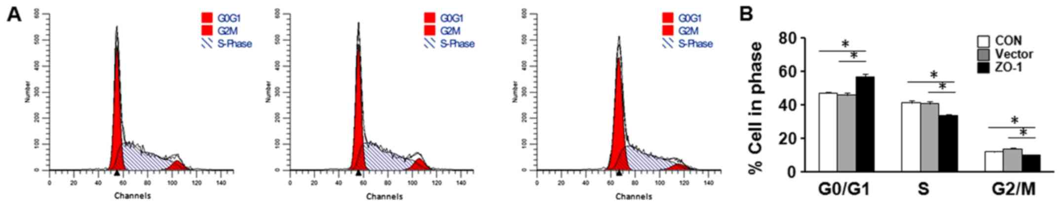
Upregulation of ZO-1 decreases LC cell
cycle and migration in vitro
To investigate the potential mechanism responsible
for the effects of ZO-1 on the proliferation of LC cells, the cell
cycle was analyzed in HepG2 cells transfected with
ZO-1-overexpressing plasmid or negative control plasmid using flow
cytometry. In cell cycle analysis, a significant increase in the
G0/G1 phase and decrease in the
S-G2 phase was identified (Fig. 3).
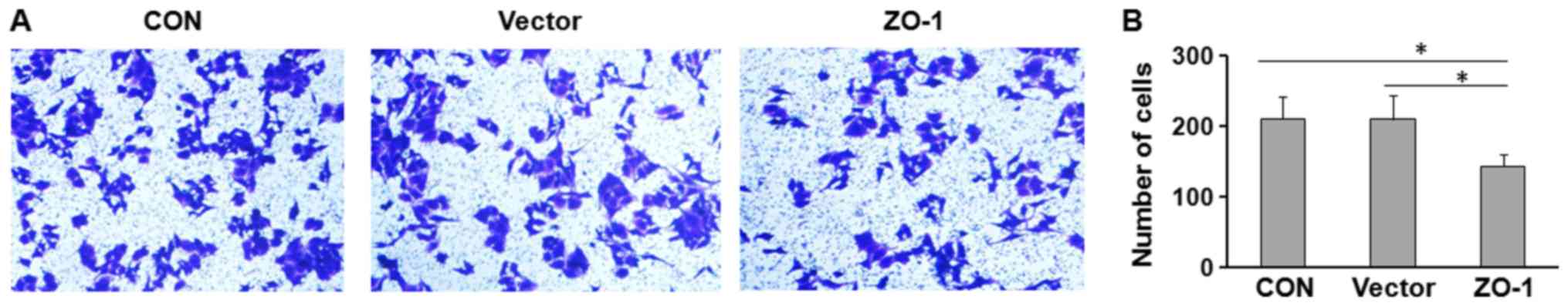
Furthermore, the potential effect of ZO-1 on cell
migration was investigated using Transwell assays. HepG2 cells were
transfected with ZO-1-overexpressing or control plasmid and seeded
in the Transwell chamber. Overexpression of ZO-1 significantly
decreased the migratory capacity of HepG2 cells (Fig. 4).
Discussion
LC is one of the most prevalent tumors globally and
the third leading cause of cancer-associated mortality (29,30).
Worldwide, ~750,000 new cases of LC are diagnosed each year.
Population-based analysis indicated that the incidence rate
continues to parallel the death rate, which indicates that the
majority of individuals who develop LC succumb to this disease
(31). Although tumor resection and
liver transplantation are effective treatments for selected
patients with LC, the prognosis of LC remains poor because the
disease is often at a fairly advanced stage at the time of
diagnosis (32). Surgical treatment
is not applicable for patients at advanced tumor stages (33). LC is involved in multiple gene
alterations including tumor suppressor inactivation, oncogene
activation and apoptosis-associated gene dysregulation (34). Therefore, there is an urgent
requirement to identify a sensitive and specific biomarker for the
detection of liver cancer at the curative stage.
ZO-1 serves as a scaffolding protein that links the
transmembrane tight junction proteins to cytoplasmic proteins and
the actin cytoskeleton (15,35). As a member of the MAGUK family of
putative signaling proteins, ZO-1 may be involved in signal
transduction, and ZO-1 has been identified to bind a target of Ras:
AF6 (36). Previous studies indicated
that epidermal growth factor and vascular endothelial growth factor
are able to increase ZO-1 tyrosine phosphorylation, modulate its
subcellular localization, and consequently lead to increased
permeability (37–39). ZO-1 serves an important function in
maintaining tight junction integrity, which is disrupted in a
number of invasive cancers and intestinal diseases (40). Consequently, studies have demonstrated
that ZO-1 downregulation is involved in tumor development and
progression (41,42).
To the best of our knowledge, the present study is
the first to investigate the expression of ZO-1 and its function in
LC progression. Using RT-qPCR and western blot analysis, it was
identified that the expression of ZO-1 was decreased at the mRNA
and protein levels in the majority of tumor tissues. Furthermore,
it was also identified that overexpression of ZO-1 significantly
inhibited cell viability and migration of LC cells in vitro.
In addition, upregulation of ZO-1 induced cell cycle arrest. These
results suggested that ZO-1 could also serve a tumor suppressor
function in LC, and that abnormal ZO-1 expression may be associated
with tumor progression and metastasis of LC. Further investigation
into the potential molecular mechanism underlying the effects of
ZO-1 are required. The mechanisms which contributed to ZO-1
downregulation in LC also require further investigation.
Acknowledgements
Not applicable.
Funding
No funding was received.
Availability of data and materials
The datasets used and/or analyzed during the current
study are available from the corresponding author on reasonable
request.
Authors' contributions
XZ, YQ and CN designed the study. XZ, YQ, LW, HZ and
FT conducted the experiments, performed the data analysis and wrote
the manuscript. CN and YQ analyzed the data and revised the
manuscript. All authors discussed the results and reviewed the
manuscript.
Ethics approval and consent to
participate
Written informed consent was obtained from each
patient and the present study was approved by the Ethics Committee
of Liaocheng People's Hospital (Shandong, China).
Patient consent for publication
Not applicable.
Competing interests
The authors declare that they have no competing
interests.
References
|
1
|
Parkin DM, Bray MF, Ferlay MJ and Pisani
P: Global cancer statistics, 2002. CA Cancer J Clin. 55:74–108.
2005. View Article : Google Scholar : PubMed/NCBI
|
|
2
|
Schütte K, Bornschein J and Malfertheiner
P: Liver cancer-epidemiological trends and risk factors. Dig Dis.
27:80–92. 2009. View Article : Google Scholar
|
|
3
|
Jemal A, Siegel R, Ward E, Murray T, Xu J
and Thun MJ: Cancer statistics, 2007. CA Cancer J Clin. 57:43–66.
2007. View Article : Google Scholar : PubMed/NCBI
|
|
4
|
Cai MY, Tong ZT, Zheng F, Liao YJ, Wang Y,
Rao HL, Chen YC, Wu QL, Liu YH, Guan XY, et al: EZH2 protein: A
promising immunomarker for the detection of liver cancers in liver
needle biopsies. Gut. 60:967–976. 2011. View Article : Google Scholar : PubMed/NCBI
|
|
5
|
Center MM and Jemal A: International
trends in liver cancer incidence rates. Cancer Epidemiol Biomarkers
Prev. 20:2362–2368. 2011. View Article : Google Scholar : PubMed/NCBI
|
|
6
|
Yuen MF, Cheng CC, Lauder IJ, Lam SK, Ooi
CG and Lai CL: Early detection of liver cancer increases the chance
of treatment: Hong Kong experience. Hepatology. 31:330–335. 2000.
View Article : Google Scholar : PubMed/NCBI
|
|
7
|
Bruix J and Sherman M: Practice Guidelines
Committee, American Association for the Study of Liver Diseases.
Hepatology. 42:1208–1236. 2005. View Article : Google Scholar : PubMed/NCBI
|
|
8
|
Chong CC, Wong GL and Lai PB: Impact of
antiviral therapy on post-hepatectomy outcome for hepatitis
B-related liver cancer. World J Gastroenterol. 20:6006–6012. 2014.
View Article : Google Scholar : PubMed/NCBI
|
|
9
|
Olsen SK, Brown RS and Siegel AB: Liver
cancer: Review of current treatment with a focus on targeted
molecular therapies. Therap Adv Gastroenterol. 3:55–66. 2010.
View Article : Google Scholar : PubMed/NCBI
|
|
10
|
Stevenson BR, Siliciano JD, Mooseker MS
and Goodenough DA: Identification of ZO-1: A high molecular weight
polypeptide associated with the tight junction (zonula occludens)
in a variety of epithelia. J Cell Biol. 103:755–766. 1986.
View Article : Google Scholar : PubMed/NCBI
|
|
11
|
Gumbiner B, Lowenkopf T and Apatira D:
Identification of a 160-kDa polypeptide that binds to the tight
junction protein ZO-1. Proc Natl Acad Sci USA. 88:3460–3464. 1991.
View Article : Google Scholar : PubMed/NCBI
|
|
12
|
Haskins J, Gu L, Wittchen ES, Hibbard J
and Stevenson BR: ZO-3, a novel member of the MAGUK protein family
found at the tight junction, interacts with ZO-1 and occludin. J
Cell Biol. 141:199–208. 1998. View Article : Google Scholar : PubMed/NCBI
|
|
13
|
Willott E, Balda MS, Fanning AS, Jameson
B, Van Itallie C and Anderson JM: The tight junction protein ZO-1
is homologous to the Drosophila discs-large tumor suppressor
protein of septate junctions. Proc Natl Acad Sci USA. 90:7834–7838.
1993. View Article : Google Scholar : PubMed/NCBI
|
|
14
|
Tsukita S, Furuse M and Itoh M: Molecular
architecture of tight junctions: Occludin and ZO-1. Soc Gen Physiol
Ser. 52:69–76. 1997.PubMed/NCBI
|
|
15
|
Fanning AS, Jameson BJ, Jesaitis LA and
Anderson JM: The tight junction protein ZO-1 establishes a link
between the transmembrane protein occludin and the actin
cytoskeleton. J Biol Chem. 273:29745–29753. 1998. View Article : Google Scholar : PubMed/NCBI
|
|
16
|
Wittchen ES, Haskins J and Stevenson BR:
Protein interactions at the tight junction. Actin has multiple
binding partners, and ZO-1 forms independent complexes with ZO-2
and ZO-3. J Biol Chem. 274:35179–35185. 1999. View Article : Google Scholar : PubMed/NCBI
|
|
17
|
Itoh M, Nagafuchi A, Moroi S and Tsukita
S: Involvement of ZO-1 in Cadherin-based cell adhesion through its
direct binding to α catenin and actin filaments. J Cell Biol.
138:181–192. 1997. View Article : Google Scholar : PubMed/NCBI
|
|
18
|
Rajasekaran AK, Hojo M, Huima T and
Rodriguez-Boulan E: Catenins and zonula occludens-1 form a complex
during early stages in the assembly of tight junctions. J Cell
Biol. 132:451–463. 1996. View Article : Google Scholar : PubMed/NCBI
|
|
19
|
Provost E and Rimm DL: Controversies at
the cytoplasmic face of the cadherin-based adhesion complex. Curr
Opin Cell Biol. 11:567–572. 1999. View Article : Google Scholar : PubMed/NCBI
|
|
20
|
Fanning AS and Anderson JM: PDZ domains:
Fundamental building blocks in the organization of protein
complexes at the plasma membrane. J Clin Invest. 103:767–772. 1999.
View Article : Google Scholar : PubMed/NCBI
|
|
21
|
Mauro L, Bartucci M, Morelli C, Andò S and
Surmacz E: IGF-I receptor-induced cell-cell adhesion of MCF-7
breast cancer cells requires the expression of junction protein
ZO-1. J Biol Chem. 276:39892–39897. 2001. View Article : Google Scholar : PubMed/NCBI
|
|
22
|
Hoover KB, Liao SY and Bryant PJ: Loss of
the Tight Junction MAGUK ZO-1 in breast cancer: Relationship to
glandular differentiation and Loss of heterozygosity. Am J Pathol.
153:1767–1773. 1998. View Article : Google Scholar : PubMed/NCBI
|
|
23
|
Kaihara T, Kusaka T, Nishi M, Kawamata H,
Imura J, Kitajima K, Itoh-Minami R, Aoyama N, Kasuga M, Oda Y, et
al: Dedifferentiation and decreased expression of adhesion
molecules, E-cadherin and ZO-1, in colorectal cancer are closely
related to liver metastasis. J Exp Clin Cancer Res. 22:117–123.
2003.PubMed/NCBI
|
|
24
|
Kimura Y, Shiozaki H, Hirao M, Maeno Y,
Doki Y, Inoue M, Monden T, Ando-Akatsuka Y, Furuse M, Tsukita S, et
al: Expression of occludin, tight-junction-associated protein, in
human digestive tract. Am J Pathol. 151:45–54. 1997.PubMed/NCBI
|
|
25
|
Polette M, Mestdagt M, Bindels S,
Nawrocki-Raby B, Hunziker W, Foidart JM, Birembaut P and Gilles C:
Beta-catenin and ZO-1: Shuttle molecules Involved in tumor
Invasion-associated epithelial-mesenchymal transition processes.
Cells Tissues Organs. 185:61–65. 2007. View Article : Google Scholar : PubMed/NCBI
|
|
26
|
Akiba J, Nakashima O, Hattori S, Tanikawa
K, Takenaka M, Nakayama M, Kondo R, Nomura Y, Koura K, Ueda K, et
al: Clinicopathologic analysis of combined
hepatocellular-cholangiocarcinoma according to the latest WHO
classification. Am J Surg Pathol. 37:496–505. 2013. View Article : Google Scholar : PubMed/NCBI
|
|
27
|
Pirisi M, Leutner M, Pinato DJ, Avellini
C, Carsana L, Toniutto P, Fabris C and Boldorini R: Reliability and
reproducibility of the edmondson grading of hepatocellular
carcinoma using paired core biopsy and surgical resection
specimens. Arch Pathol Lab Med. 134:1818–1822. 2010.PubMed/NCBI
|
|
28
|
Yu S, Liu H and Luo L: Analysis of
relative gene expression using different real-time quantitative
PCR. Acta Agronomica Sinica. 33:1214–1218. 2007.(In Chinese).
|
|
29
|
Yang Y, Liu YM, Wei MY, Wu YF, Gao JH, Liu
L, Zhou WP, Wang HY and Wu MC: The liver tissue bank and clinical
database in China. Front Med China. 4:443–447. 2010. View Article : Google Scholar : PubMed/NCBI
|
|
30
|
Fares N and Péron JM: Epidemiology,
natural history, and risk factors of liver cancer. Rev Prat.
63(216-217): 220–222. 2013.(In French).
|
|
31
|
Jemal A, Bray F, Center MM, Ferlay J, Ward
E and Forman D: Global cancer statistics. CA Cancer J Clin.
61:69–90. 2011. View Article : Google Scholar : PubMed/NCBI
|
|
32
|
Ng KK, Lo CM, Chan SC, Chok KS, Cheung TT
and Fan ST: Liver transplantation for liver cancer: The Hong Kong
experience. J Hepatobiliary Pancreat Sci. 17:548–554. 2010.
View Article : Google Scholar : PubMed/NCBI
|
|
33
|
Amram ML, Benamran DA and Roth AD:
Targeted therapies in digestive oncology. Rev Med Suisse.
7:1131-1132–1134-1136. 2011.(In French).
|
|
34
|
Jain S, Singhal S, Lee P and Xu R:
Molecular genetics of hepatocellular neoplasia. Am J Transl Res.
2:105–118. 2010.PubMed/NCBI
|
|
35
|
Furuse M, Itoh M, Hirase T, Nagafuchi A,
Yonemura S and Tsukita S and Tsukita S: Direct association of
occludin with ZO-1 and its possible involvement in the localization
of occludin at tight junctions. J Cell Biol. 127:1617–1626. 1994.
View Article : Google Scholar : PubMed/NCBI
|
|
36
|
Yamamoto T, Harada N, Kano K, Taya S,
Canaani E, Matsuura Y, Mizoguchi A, Ide C and Kaibuchi K: The Ras
target AF-6 interacts with ZO-1 and serves as a peripheral
component of tight junctions in epithelial cells. J Cell Biol.
139:785–795. 1997. View Article : Google Scholar : PubMed/NCBI
|
|
37
|
Van Itallie CM, Balda MS and Anderson JM:
Epidermal growth factor induces tyrosine phosphorylation and
reorganization of the tight junction protein ZO-1 in A431 cells. J
Cell Sci. 108:1735–1742. 1995.PubMed/NCBI
|
|
38
|
Antonetti DA, Barber AJ, Hollinger LA,
Wolpert EB and Gardner TW: Vascular endothelial growth factor
induces rapid phosphorylation of tight junction proteins occludin
and zonula occluden 1. A potential mechanism for vascular
permeability in diabetic retinopathy and tumors. J Biol Chem.
274:23463–23467. 1999. View Article : Google Scholar : PubMed/NCBI
|
|
39
|
Merwin JR, Anderson JM, Kocher O, Van
Itallie CM and Madri JA: Transforming growth factor beta 1
modulates extracellular matrix organization and cell-cell
junctional complex formation during in vitro angiogenesis. J Cell
Physiol. 142:117–128. 1990. View Article : Google Scholar : PubMed/NCBI
|
|
40
|
Giri S, Poindexter KM, Sundar SN and
Firestone GL: Arecoline induced disruption of expression and
localization of the tight junctional protein ZO-1 is dependent on
the HER 2 expression in human endometrial Ishikawa cells. BMC Cell
Biol. 11:532010. View Article : Google Scholar : PubMed/NCBI
|
|
41
|
Németh Z, Szász AM, Somorácz Á, Tátrai P,
Németh J, Gyorffy H, Szíjártó A, Kupcsulik P, Kiss A and Schaff Z:
Zonula occludens-1, occludin, and E-cadherin protein expression in
biliary tract cancers. Pathol Oncol Res. 15:533–539. 2009.
View Article : Google Scholar : PubMed/NCBI
|
|
42
|
Martin TA, Watkins G, Mansel RE and Jiang
WG: Loss of tight junction plaque molecules in breast cancer
tissues is associated with a poor prognosis in patients with breast
cancer. Eur J Cancer. 40:2717–2725. 2004. View Article : Google Scholar : PubMed/NCBI
|