Introduction
Cytotoxic T-lymphocyte antigen-4 (CTLA-4) is a
characteristic inhibitory regulator expressed on the membrane of
activated T cells, which serves as an immunoregulatory role and is
responsible for T-cell expansion by suppressing the action of
cluster of differentiation (CD)28 (1). CTLA-4 may terminate activation of the
T-cell response by downregulating ligand expression levels and
emitting inhibitory signals (2,3). A
previous study confirmed that the CTLA-4 signaling pathway is a
strong inducer in regulating the function of regulatory T cells
(Tregs), which serve a key role in immunity and tolerance. In
humans, the major cell type expressing CTLA-4 are Tregs (4), which are a potent controller of the
immune response. CTLA-4 is constitutively expressed in Tregs and
upregulated in activated conventional T cells, which are classified
as suppressor T-lymphocyte subgroups (5). Previous studies have suggested that
CTLA-4+ T-cell subgroups are disrupted when the human
immune system is weakened, the occurrence of CTLA-4+ T
cells is imbalanced at tumor sites and in the peripheral blood of
patients with cancer, and this may be associated with clinical
diagnosis and prognosis (6–8). Consequently, the detection and analysis
of this specific CTLA-4+ lymphocyte subgroup serves a
significant role.
Multiple methods have been employed to detect
CTLA-4+ T cells, and fluorescent-conjugated monoclonal
antibodies (mAbs) are the most commonly utilized. Due to the low
proportion of these CTLA-4+ T-cell subgroups in the
peripheral blood and tumor tissues, and interference from the tumor
microenvironment, their detection is difficult and costly.
Therefore, a simpler and more sensitive assay is urgently required
to monitor CTLA-4+ T-cell subgroups. Nanobodies (Nbs)
are a special type of single-domain antibodies consisting of
different regions of heavy chain antibodies, which are present in
the blood of camels. Compared with conventional antibodies, Nbs
possess several inherent characteristics, including high
specificity, high physiochemical stability, lack of immunogenicity,
high yield and low cost, which make them suitable for
immune-targeted diagnosis and treatment of cancer (9). In our previous study, a CTLA-4-specific
Nb (Nb36) was screened (10). Nb36
may recognize unique CTLA-4 epitopes and effectively bind with
CTLA-4+ T cells. Due to their relatively complex
structures and large molecular masses, traditional antibodies
cannot effectively penetrate the barrier of blood vessels and tumor
tissues to successfully bind the tumor antigen. Nbs possess a
molecular mass of 15 kDa, no chemical hydrophobicity, potent heat
resistance and high resistance to acids and alkalis (11). Therefore, it has been hypothesized
that Nbs may be used in the detection of certain targets with
reduced expression. Recently, nanometer-sized optic-functional
semiconductors, particularly fluorescent carbon quantum dots (QDs),
have attracted widespread attention, and have been utilized as
photographic developers in diagnostic instruments (12,13).
QDs, 2.5–6 nm in size, have a wider excitation spectrum and
narrower emission spectrum than traditional organic fluorescent
dyes (14); therefore, QDs have been
widely used as optical imaging probes. Furthermore, QDs exhibit
excellent biocompatibility, non-toxicity and high light stability,
and are convenient to modify (15),
which are useful in detecting biomarkers or cell surface markers,
when combined with a targeting probe (16). Due to their specific optical
characteristics, the use of QDs leads to high sensitivity in
multiple techniques, including immunohistochemical staining, flow
cytometry, fluorescence in situ hybridization and biochips,
widening their application range in clinical practice.
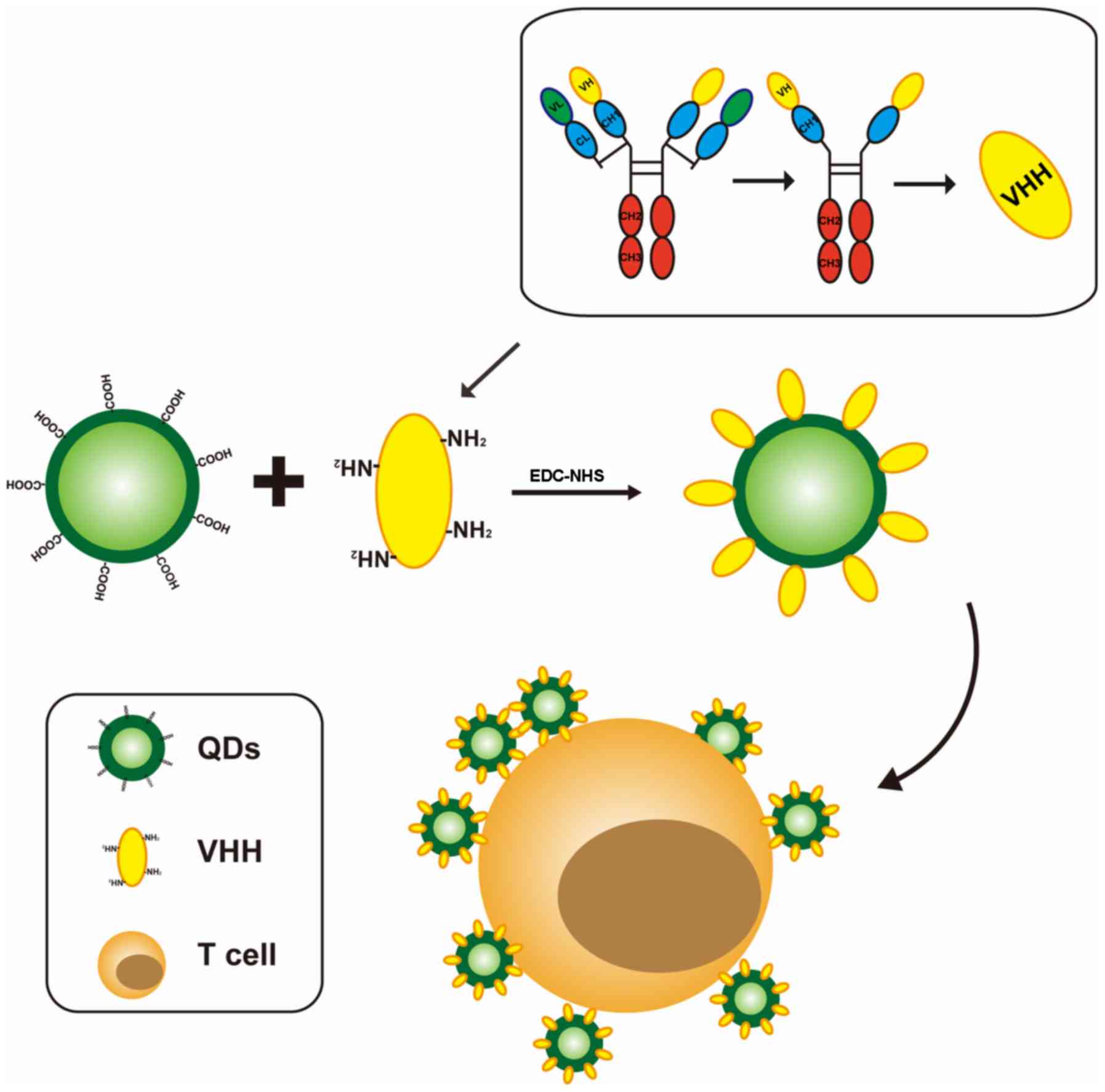
To the best of our knowledge, the construction of
detection tools based on QDs combined with Nbs has yet to be
reported. In the present study, a novel complex based on QDs in
combination with Nbs was synthesized to detect CTLA-4+ T
cells (QDs-Nb36; Fig. 1). The
efficacy of the novel complex and conventional mAbs in target
detection was statistically compared.
Materials and methods
Cells and animals
Peripheral blood T cells were isolated from the
whole blood samples of healthy donors (2 males and 2 females, aged
between 23 and 33, with normal blood routine, normal liver and
kidney function, and no serious diseases such as malignant tumors)
using Ficoll-Hypaque density-gradient centrifugation (20 min, 24°C,
800 × g). Written informed consent was obtained from all donors.
The study procedures were approved by The Local Ethics Committee of
Guangxi Medical University (Nanning, China). The 293-cell line was
purchased from American Type Culture Collection (Manassas, VA,
USA).
A total of 60 female C57BL/6 mice, aged 4–6 weeks,
between 13 and 18 g, were purchased from Guangxi Laboratory Animal
Center (Guangxi, China) and raised in laminar flow cabinets in a
specific pathogen-free environment (temperature, 23±1°C; humidity,
50±10%; 12-h light/dark cycle starting at 7:00 a.m.; with free
access to food and water). All protocols were approved by the
Animal Ethics Committee of Guangxi Medical University (Guangxi,
China).
Patients
A total of five patients (aged between 35 and 63
years old; 3 males and 2 females) admitted to The First Affiliated
Hospital Guangxi Medical University (Guangxi, China) between
October 2016 and January 2018, and eligible for surgical resection
of hepatocellular carcinoma were enrolled in the present study.
Written informed consent was obtained from all patients. Paired
tumor tissue and adjacent mucosa (≥3 cm from the tumor) samples
were obtained after hepatectomy and stored in sterile PBS solution
at room temperature. The study procedures were approved by The
Local Ethics Committee of Guangxi Medical University.
Reagents
QDs were purchased from Beijing Zhongke Wu Yuan
Biotechnology Co., Ltd. (Beijing, China). Ficoll-Paque™
Plus was purchased from GE Healthcare Life Sciences (Shanghai,
China). 1-ethyl-3-(3-dimethylaminopropyl) carbodiimide
hydrochloride (EDC) and N-hydroxysulfosuccinimide sodium salt
(sulfo-NHS) were purchased from Sigma-Aldrich (Merck KGaA,
Darmstadt, Germany). The nuclear dye DAPI was purchased from Thermo
Fisher Scientific, Inc. (Waltham, MA, USA). Anti-CTLA-4 mAb (cat.
no. Ab7222, 100 µg/100 µl) and Anti-CD3 mAb (cat. no. AA3600, 100
µg/100 µl) were purchased from Abcam (Cambridge, MA, USA; both used
at a 1:1000 dilution). Sterile PBS (10 mM, pH 7.4) was used as a
buffer.
Preparation of the QDs-Nb36
complex
The quantum dot suspension droplets were dropped on
a copper mesh, and left at room temperature until dry enough for
transmission electron microscopy (TEM; model: H-7650; Hitachi,
Ltd., Tokyo, Japan) analysis to confirm that the sample was
qualified. Previously, Nb36 protein was obtained from a high
quality dromedary camel immune library by phage display technology
and expression in E. coli WK6 electrocompetent cells. After further
purification, the Nb36 protein solution was obtained (10). First, Nb36 protein solution was
dissolved in the PBS (to a final concentration of 1 mg/ml).
Subsequently, 50 µl QDs solution and 100 µl EDC solution (0.8 mM)
were mixed using a vortex mixer for 10 min at room temperature,
then supplemented with 100 µl NHS (0.8 mM) and 300 µl Nbs (1
mg/ml). The mixture was co-cultured for 1 h with gentle agitation
at room temperature. Finally, unconjugated Nb36s were discarded.
The conjugated QDs-Nb36 complexes were collected by centrifugation
(1,000 × g, 20 min) at room temperature. Subsequently, the
complexes were resuspended in PBS containing 1% (m/v) bovine serum
albumin (BSA; cat. no. A7030; Sigma-Aldrich; Merck KGaA).
In vitro specificity of QDs-Nb36 for
polyhydroxyalkanoates (PHA)-stimulated human T cells
The peripheral blood mononuclear cells were isolated
and cultured in RPMI-1640 medium (cat. no. 31800-021; Gibco; Thermo
Fisher Scientific, Inc.) containing 10% fetal bovine serum (FBS;
cat. no. 10099-141; Gibco; Thermo Fisher Scientific, Inc.) at 37°C
for 2 h. Subsequently, the adherent cells were removed and T cells
were isolated using nylon-wool. PHA (10 µg/ml; cat. no. L8754;
Sigma-Aldrich; Merck KGaA)-stimulated T cells (1×10⁶)
were incubated with 2% BSA solution for 30 min at 24°C with
agitation to avoid non-specific binding. PHA is a
phytohemagglutinin-specific polyclonal stimulator that stimulates
human T cell proliferation through CD3-TCR on the cell membrane. A
total of 4×10⁵ T cells were resuspended in 300 µl PBS
for each sample. These samples were incubated with QDs-Nb36 in
RPMI-1640 medium at 4°C for 30 min, followed by washing, suspension
in PBS (300 µl) and analysed using a Beckman Coulter flow cytometer
with ~10,000 cells (Beckman Coulter, Inc., Brea, CA, USA). The
fluorescence signal was detected on the fluorescein isothiocyanate
channel. In the control groups, an equivalent dose of anti-CTLA-4
mAb and QDs were administered. Flowjo v10.07 software (FlowJo LLC,
Ashland, OR, USA) was utilized for statistical analysis.
The PHA-stimulated T cells were washed with PBS
three times and then fixed with 4% polyoxymethylene for 15 min at
room temperature. After washing with PBS, the cell samples were
incubated with QDs-Nb36 (100 nM), anti CTLA-4 mAb (5 µl) and QDs
(100 nM) in binding buffer at 4°C for 50 min and washed with PBS
three times. Subsequently, the samples were stained with DAPI (1
µg/ml) for 5 min at 4°C. Images were captured using confocal
microscopy (magnification, ×400).
In vitro specificity of QDs-Nb36 for
tumor-infiltrated CTLA-4+ T cells
The tumor tissues and adjacent mucosa were immersed
in optimal cutting temperature compound and processed into frozen
sections (−20°C). QDs-Nb36, anti CTLA-4 mAb and QDs were used for
immunofluorescent staining. DAPI was used for nuclear staining. The
frozen sections were observed using a confocal microscope (FV1000;
Olympus Corporation, Japan; magnification, 400×). After the sample
tissues were cut up, collagenase II (Sigma-Aldrich; Merck KGaA) was
added for digestion. The tumor-infiltrated T cells isolated from
tumor tissues and adjacent mucosa were processed using the
anti-human CD3 monoclonal antibody immunomagnetic bead kit (cat.
no. BMCD3; BioMag Corp., Wu Xi, China), as described previously
(17,18), then analyzed by flow cytometry. The
specific antibodies used to detect the expression of CD3 and CTLA-4
were as follows: Anti-human CD3 mAb and QDs-Nb36 (100 nM),
anti-CTLA-4 mAb or QDs (100 nM), incubated at 4°C for 30 min. Data
were analyzed using FlowJo v10.0.7 software.
In vitro toxicity of QDs-Nb36
Toxicity of QDs-Nb36 against 293 cells was
determined by MTT assay. The 293 cells (2×10⁵) were
cultured in RPMI-1640 medium containing 10% FBS, in a 96-well plate
overnight in a cell incubator (5% CO2). Subsequently,
the samples were treated with QDs-Nb36 (10, 20, 30, 40 and 50 nM)
for 24 or 48 h. In the control group, an equivalent volume of PBS
was administered. A total of 10 µl MTT (5 mg/ml) was added to each
well, and the plates were incubated at room temperature for 4 h in
the dark. Subsequently, the medium was discarded, 150-µl
ethanol/dimethyl sulfoxide was added to each well and the cells
were further incubated for 10 min. The optical density of the
samples was analyzed using an ELISA microplate reader (Thermo
Fisher Scientific, Inc.) at a wavelength of 570 nm.
In vivo toxicity of QDs-Nb36
The mice received an intravenous injection of
QDs-Nb36 (15 mg/kg) or PBS through the tail vein, once a week for 4
weeks. Alterations in the biological behavior and weight of the
mice were monitored daily. A total of 7 days after treatment, serum
was collected from the mice for biochemical examination with an
AU5800 Clinical Chemistry Analyzer (Beckman Coulter, Inc.) The
alanine aminotransferase, aspartate transaminase, serum creatinine
and blood urea nitrogen levels were measured to assess liver and
kidney function in mice. Subsequently, the animals were sacrificed.
The primary organs, including spleen, kidney, heart, lung, liver
and brain were immersed and preserved in 10% formaldehyde solution
at 4°C for 4 h, dehydrated and embedded in paraffin. The
paraffin-embedded sections (4 µm) were stained with hematoxylin and
eosin (HE) (1 min each) at room temperature. Images were captured
using a confocal microscope (magnification, 400×).
Statistical analysis
All experiments were repeated 3 times. Statistical
analysis was performed using GraphPad Prism 6.02 software (GraphPad
Software, Inc., La Jolla, CA, USA). Data were analyzed by Student's
t-test or a Levene's test followed by a one-way analysis of
variance and Newman-Keuls post-hoc test. P<0.05 was considered
to indicate a statistically significant difference. All data are
expressed as the mean ± standard deviation.
Results
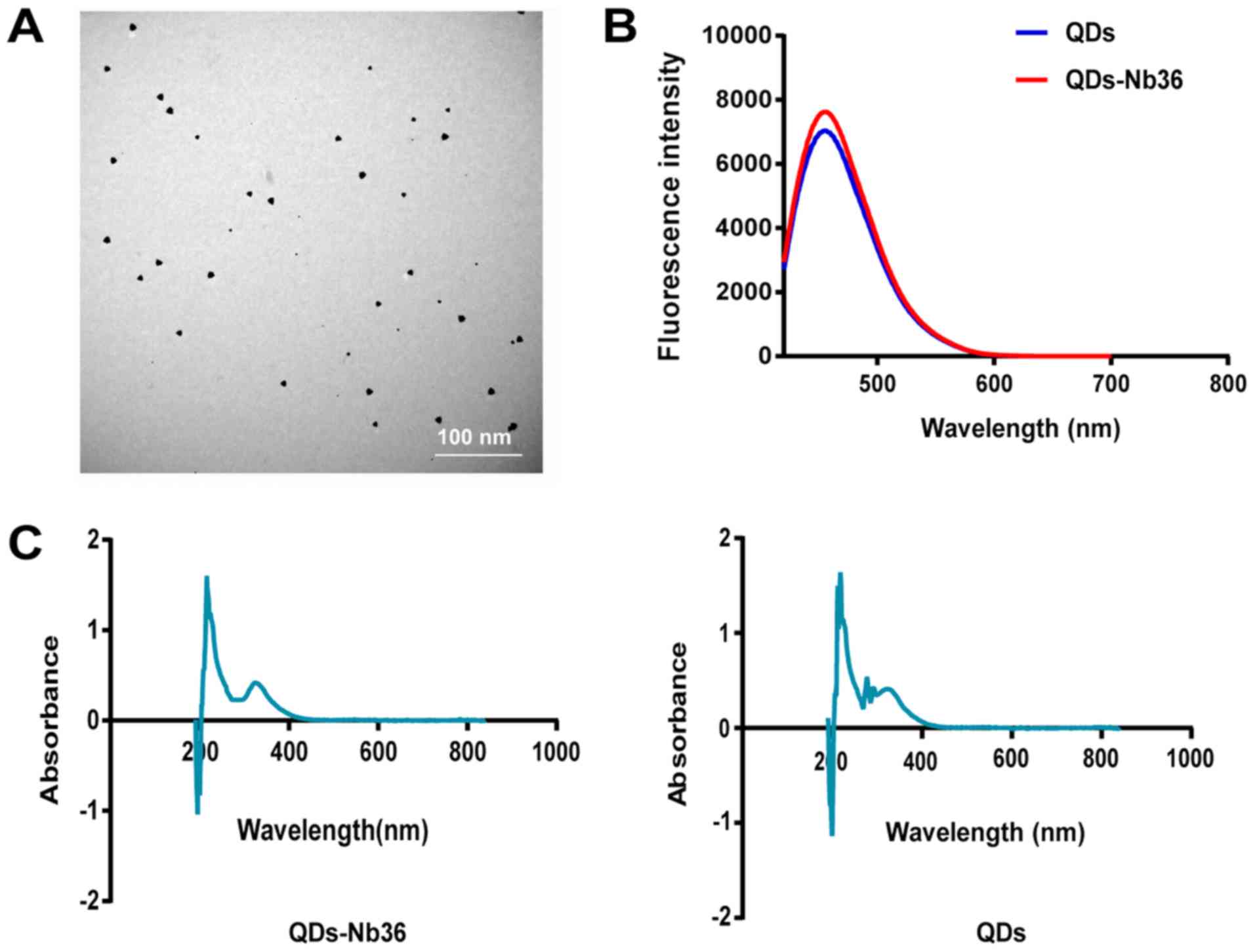
Characterization of QDs-Nb36
The characteristics of QDs-Nb36 were analyzed. TEM
data demonstrated that the dissemination of QDs-Nb36 was almost
monodisperse (Fig. 2A). Their
photoluminescence was measured using a fluorescence
spectrophotometer (FL-7000; Perkin Elmer, Waltham, MA, USA). The
fluorescence emission spectra peak of the QDs-Nb36 and QDs was 450
nm (Fig. 2B), and the absorbance
peak was 350 nm (Fig. 2C).
Highly sensitive detection of
CTLA-4+ T cells from human peripheral blood using
QDs-Nb36
To characterize QDs-Nb36 binding to
CTLA-4+ T cells, binding was analyzed using
PHA-stimulated and normal T cells. The binding efficiency of
QDs-Nb36 to target samples was evaluated by flow cytometry.
QDs-Nb36 and anti-CTLA-4 mAb specifically bound to
CTLA-4+ T cells. A higher binding rate was identified
for QDs-Nb36 compared with CTLA-4 mAb in PHA-stimulated cells and
minimal binding of QDs was observed for all target cell lines
(Fig. 3A and B). To obtain a more
direct visualization of CTLA-4+ T cells using the
QDs-Nb36 complex, confocal microscopy was performed to examine
PHA-stimulated human T cells incubated with QDs-Nb36, anti-CTLA-4
mAb or QDs. The fluorescent images revealed that PHA-stimulated T
cells exerted a significantly stronger fluorescence at the cell
membrane in the presence of QDs-Nb36 compared with anti-CTLA-4 mAb,
and no fluorescent signal was observed for QDs alone. When normal T
cells were used as the sample, a small quantity of positive cells
could be observed (Fig. 3C),
suggesting that QDs-Nb36 complexes could be used in the detection
of CTLA-4+ T cells with high sensitivity due to the
specificity of Nb36 to CTLA-4.
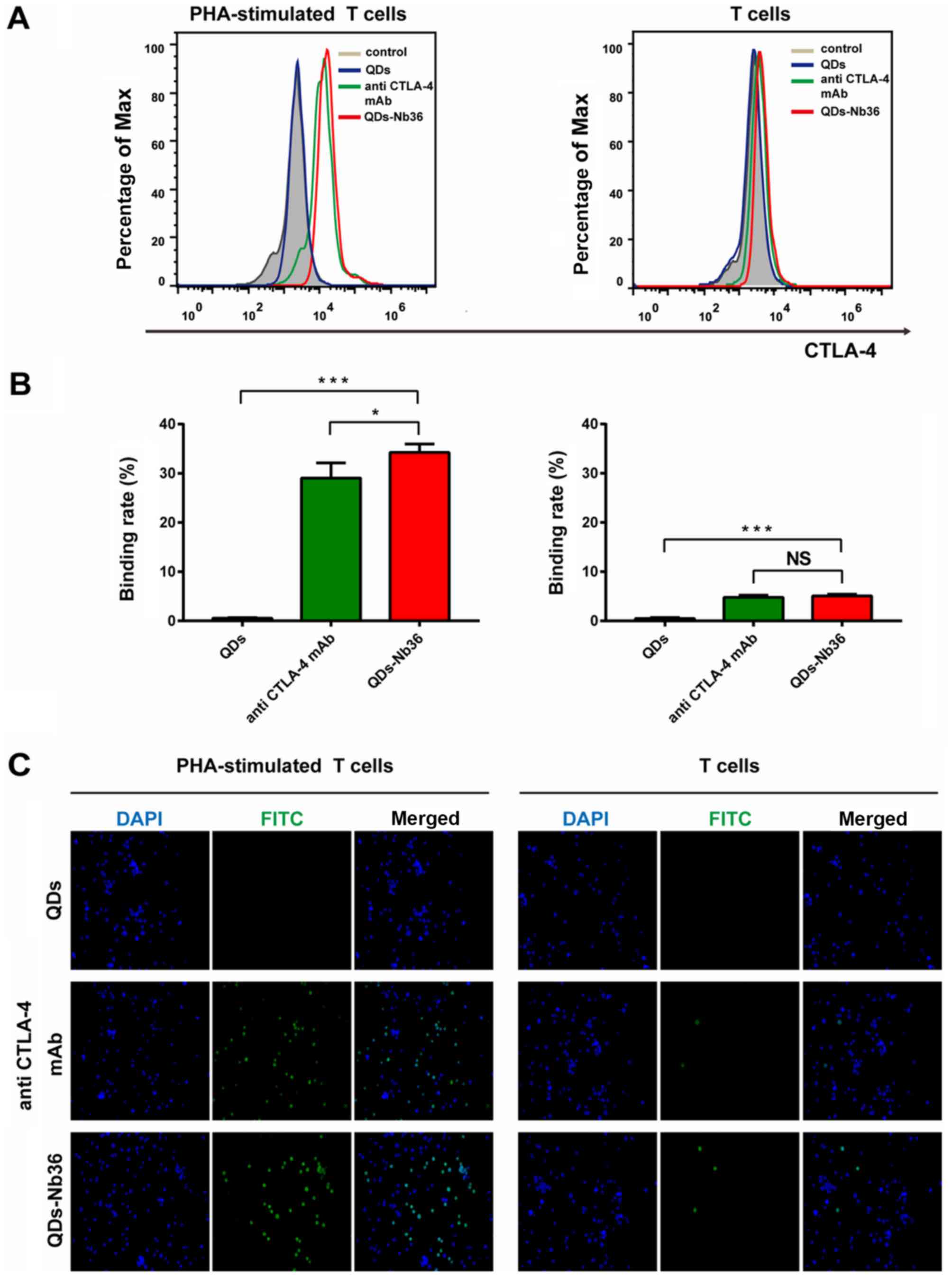 | Figure 3.Specific detection of
CTLA-4+ T cells with the QDs-Nb36 complex. (A) Flow
cytometry of PHA-stimulated T cells and normal T cells following
incubation with QDs, anti-CTLA-4 mAb or QDs-Nb36. (B) Quantitative
analysis of PHA-stimulated and normal T cells. (C) Fluorescent
micrographs of PHA-stimulated T cells and normal T cells following
incubation with QDs, anti-CTLA-4 mAb or QDs-Nb36. Fluorescence was
detected on the FITC channel, whereas DAPI-stained nuclei were
detected on the blue channel. Magnification, ×400. *P<0.05,
***P<0.001. CTLA-4, cytotoxic T-lymphocyte antigen-4; FITC,
fluorescein isothiocyanate; mAb, monoclonal antibody; NS, not
significant; PHA, polyhydroxyalkanoates; QDs, quantum dots;
QDs-Nb36, QDs-CTLA-4-specific nanobody. |
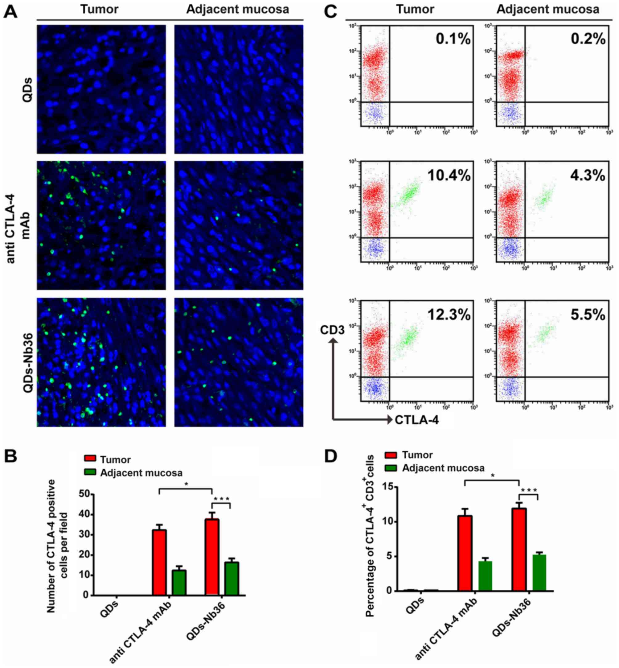
Highly sensitive detection of
tumor-infiltrated CTLA-4+ T cells using QDs-Nb36
Next, immunofluorescent staining of the tumor
tissues and adjacent mucosa was performed to investigate whether
the fluorescence signal of infiltrated CTLA-4+ T cells
in the tumor tissues was significantly higher than that in adjacent
mucosa for QDs-Nb36 or anti-CTLA-4 mAb. The number of positive
cells detected using QDs-Nb36 was higher compared with anti-CTLA-4
mAb (Fig. 4A and B). Following
isolation of tumor-infiltrated mononuclear leukocytes from the
cancer tissue and adjacent mucosa, the expression levels of CTLA-4
were measured by flow cytometry and the results were consistent
with the findings of immunofluorescent staining (Fig. 4C and D).
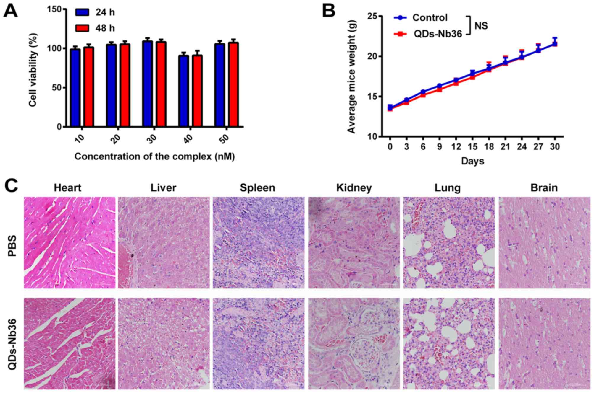
QDs-Nb36 exhibits non-toxicity in
vitro and in vivo
An MTT assay confirmed that different concentrations
of QDs-Nb36 exhibited no toxic effect on the normal cell line 293
after 24 and 48 h (Fig. 5A). The
cytotoxicity of QDs-Nb36 in mice was subsequently analyzed. HE
staining and serum biochemical examination were performed 1week
after the last injection. As illustrated in Fig. 5C, no evident inflammation or necrosis
was observed in the tissue sections. In addition, no significant
differences in body weight gain and serum biochemistry of the
treatment and control groups were identified (Fig. 5B and Table
I). These results confirmed that QDs-Nb36 injection caused
almost no cytotoxicity in mice.
 | Table I.Serum biochemical examination of mice
in the QDs-Nb36 group compared with the PBS group. |
Table I.
Serum biochemical examination of mice
in the QDs-Nb36 group compared with the PBS group.
| Group | ALT, U/ml | AST, U/ml | SCr, µmol/l | BUN, µmol/l |
|---|
| PBS | 20.66±1.73 | 41.27±2.94 | 21.73±3.75 | 11.51±1.36 |
| QDs-Nb36 | 18.34±1.68 | 45.21±4.31 | 20.96±3.41 | 12.97±1.55 |
Discussion
Nanoparticle probes based on graphene, liposome,
silicon and the rare earth ions have been widely used in
biomolecular imaging and diagnosis; however, the majority of these
probes rely on complex synthesis methods and cannot provide direct
fluorescence signals (19–22). Due to their specific optical and
electronic features, semiconductor QDs serve as novel nanoparticle
probes in bioimaging and bio-diagnostics (23). Nevertheless, the QD complexes retain
certain limitations. It is of great significance to reduce the size
of the complexes to lower steric hindrance and prevent the
absorption of non-specific proteins (24). Heavy-chain antibodies (HCAbs) are
unique in that they lack alight chain and are present in the serum
of alpacas and llamas (25). In
HCAbs, the antigen-binding fragments consist of one variable
domain. A type of antibody, termed Nbs, may be obtained by cloning
the genes encoding this variable domain (26). It is the smallest antigen-binding
antibody fragment compared with conventional antibodies, with a
molecular weight of ~15 kDa, 4.8 nm length and 2.2 nm diameter. Due
to their small molecular mass, Nbs may diffuse more efficiently
throughout solid tumors. Additionally, Nbs possess several inherent
characteristics, including high physiochemical stability, high
binding specificity, high affinity (KD<10 nM) and
rapid clearance in the bloodstream, which are applicable in
quantitative bioassays (27).
The synthesis of the CTLA-4-specificNbs and QDs
complex is a simple and efficient strategy for detecting
CTLA-4+ T cells. Anti-CTLA-4 Nbs specifically bind to
CTLA-4+ T cells, whereas QDs provide a sensitive
fluorescent signal for accurate detection. Anamide bond between a
carboxylic acid group on QDs and an amine group on NBs maybe used
to construct the QDs-Nb36 complex. In the present study, in
vitro imaging demonstrated that the fluorescent intensity in
PHA-stimulated human T cells and tumor sections was significantly
higher for the QDs-Nb36 complex compared with that of the
anti-CTLA-4 mAb. In addition, significant differences in the
binding rates between the two groups were also identified in
tumor-infiltrated T cells, which demonstrated the specific binding
of the QDs-Nb36 complex to CTLA-4+ cells in
vitro. These findings suggested that the efficiency of QDs-Nb36
detection was higher than that of anti-CTLA-4 mAb. The method of
QDs-Nb36 synthesis may increase the binding of Nbs and QDs to
enhance specificity and fluorescence intensity.
The toxicity of QDs-Nb36 in vitro and in
vivo was assessed by MTT assay, HE staining and serum
biochemical examination. QDs-Nb36 yielded no toxicity to normal
human cells and the primary organs of mice 4 weeks post-injection.
In addition, it exerted no effect on the body weight of mice,
indicating that the QDs-Nb36 complex is not only a safe in
vitro detection tool, but has the potential for in vivo
assessment or treatment of tumor suppressor T cells.
In conclusion, a novel complex was developed to
detect CTLA-4+ T cells based on a CTLA-4-specific Nb and
QDs. The specificity of the Nb for CTLA-4+ human T cells
coupled with QDs, which provided a strong fluorescent signal, led
to high specificity and sensitivity in the detection of the
CTLA-4+ T-cell ratio. The fluorescent intensity of QDs
was significantly higher compared with a mAb. The complex was
superior to mAbs in monitoring CTLA-4+ cell number.
Furthermore, this method may be used for the detection of other
biological targets using Nbs specific to other targets.
Acknowledgements
Not applicable.
Funding
The present study was supported, in part, by grants
from The National Natural Scientific Foundation of China (grant no.
81773254); Programs for Changjiang Scholars and Innovative Research
Team in University (grant no. IRT_15R13); International Cooperation
Project of the Ministry of Science and Technology of China (grant
no. 2015DFA31320); Project for Innovative Research Team in Guangxi
Natural Science Foundation (grant no. 2015GXNSFFA139001); and The
Project for International Nanobody Research Center of Guangxi
(grant no. GuiKe-AD17195001).
Availability of data and materials
The datasets used and/or analyzed during the present
study are available from the corresponding author on reasonable
request.
Authors' contributions
WW, XH, XY, AL, ZT, FM and SY performed the
experiments. XL performed the statistical analysis and wrote the
manuscript.
Ethics approval and consent to
participate
The present animal study was approved by Guangxi
Medical University. The study procedures were approved by The
Animal Ethics Committee of Guangxi Medical University (Guangxi,
China).
Patient consent for publication
Not applicable.
Competing interests
The authors declare that they have no competing
interests.
References
|
1
|
Lenschow DJ, Walunas TL and Bluestone JA:
CD28/B7 system of T cell costimulation. Annu Rev Immunol.
14:233–258. 1996. View Article : Google Scholar : PubMed/NCBI
|
|
2
|
Teft WA, Kirchhof MG and Madrenas J: A
molecular perspective of ctla-4 function. Annu Rev Immunol.
24:65–97. 2006. View Article : Google Scholar : PubMed/NCBI
|
|
3
|
Walker LS and Sansom DM: The emerging role
of ctla4 as a cell-extrinsic regulator of T cell responses. Nat Rev
Immunol. 11:852–863. 2011. View
Article : Google Scholar : PubMed/NCBI
|
|
4
|
Wakamatsu E, Mathis D and Benoist C:
Convergent and divergent effects of costimulatory molecules in
conventional and regulatory CD4+ T cells. Proc Natl Acad
Sci USA. 110:1023–1028. 2013. View Article : Google Scholar : PubMed/NCBI
|
|
5
|
Krummel MF and Allison JP: CD28 and ctla-4
have opposing effects on the response of T cells to stimulation. J
Exp Med. 182:459–465. 1995. View Article : Google Scholar : PubMed/NCBI
|
|
6
|
Ichihara F, Kono K, Takahashi A, Kawaida
H, Sugai H and Fujii H: Increased populations of regulatory T cells
in peripheral blood and tumor-infiltrating lymphocytes in patients
with gastric and esophageal cancers. Clin Cancer Res. 9:4404–4408.
2003.PubMed/NCBI
|
|
7
|
Aggarwal S, Sharma SC and N Das S:
Dynamics of regulatory T cells (Tregs) in patients with oral
squamous cell carcinoma. J SurgOncol. 116:1103–1113. 2017.
|
|
8
|
Benevides L, Cardoso CR, Tiezzi DG, Marana
HR, Andrade JM and Silva JS: Enrichment of regulatory T cells in
invasive breast tumor correlates with the upregulation of IL-17A
expression and invasiveness of the tumor. Eur J Immunol.
43:1518–1528. 2013. View Article : Google Scholar : PubMed/NCBI
|
|
9
|
Steeland S, Vandenbroucke RE and Libert C:
Nanobodies as therapeutics: Big opportunities for small antibodies.
Drug Discov Today. 21:1076–1113. 2016. View Article : Google Scholar : PubMed/NCBI
|
|
10
|
Wan R, Liu A1, Hou X, Lai Z, Li J, Yang N,
Tan J, Mo F, Hu Z, Yang X, et al: Screening and antitumor effect of
an anti-CTLA-4 nanobody. Oncol Rep. 39:511–518. 2018.PubMed/NCBI
|
|
11
|
Muyldermans S: Nanobodies: Natural
single-domain antibodies. Annual Review of Biochemistry. 775–797.
2013. View Article : Google Scholar : PubMed/NCBI
|
|
12
|
Zhang M, Liu H, Chen L, Yan M, Ge L, Ge S
and Yu J: A disposable electrochemiluminescence device for
ultrasensitive monitoring of K562 leukemia cells based on aptamers
and simpleZnO@carbon quantum
dots. Biosens Bioelectron. 49:79–85. 2013. View Article : Google Scholar : PubMed/NCBI
|
|
13
|
Yu Y, Duan S, He J, Liang W, Su J, Zhu J,
Hu N, Zhao Y and Lu X: Highly sensitive detection of leukemia cells
based on aptamer and quantum dots. Oncol Rep. 36:886–892. 2016.
View Article : Google Scholar : PubMed/NCBI
|
|
14
|
Qu K, Wang J, Ren J and Qu X: Carbon dots
prepared by hydrothermal treatment of dopamine as an effective
fluorescent sensing platform for the label-free detection of iron
(III) ions and dopamine. Chemistry. 19:7243–7249. 2013. View Article : Google Scholar : PubMed/NCBI
|
|
15
|
Yang ST, Wang X, Wang H, Lu F, Luo PG, Cao
L, Meziani MJ, Liu JH, Liu Y, Chen M, et al: Carbon dots as
nontoxic and high-performance fluorescence imaging agents. J Phys
Chem C Nanomater Interfaces. 113:18110–18114. 2009. View Article : Google Scholar : PubMed/NCBI
|
|
16
|
Xing Y and Rao J: Quantum dot
bioconjugates for in vitro diagnostics & in vivo
imaging. Cancer Biomark. 4:307–319. 2008. View Article : Google Scholar : PubMed/NCBI
|
|
17
|
Peter PA, Durflinger KH, Wunderlich JR,
Rosenberg SA and Dudley ME: Enrichment of CD8+ cells from melanoma
tumor-infiltrating lymphocyte cultures reveals tumor reactivity for
use in adoptive cell therapy. J Immunother. 33:547–556. 2010.
View Article : Google Scholar : PubMed/NCBI
|
|
18
|
Tran KQ, Zhou J, Durflinger KH, Langhan
MM, Shelton TE, Wunderlich JR, Robbins PF, Rosenberg SA and Dudley
ME: Minimally cultured tumor-infiltrating lymphocytes display
optimal characteristics for adoptive cell therapy. J Immunother.
31:742–751. 2008. View Article : Google Scholar : PubMed/NCBI
|
|
19
|
Zhang L, Wang Z, Lu Z, Xia K, Deng Y, Li
S, Zhang C, Huang Y and He N: Synthesis of LiYF4:Yb, Erupconversion
nanoparticles and its fluorescence properties. J
NanosciNanotechnol. 14:4710–4713. 2014. View Article : Google Scholar
|
|
20
|
Heo NS, Lee SU, Rethinasabapathy M, Lee
EZ, Cho HJ, Oh SY, Choe SR, Kim Y, Hong WG, Krishnan G, et al:
Visible-light-driven dynamic cancer therapy and imaging using
graphitic carbon nitride nanoparticles. Mater Sci Eng C Mater Biol
Appl. 90:531–538. 2018. View Article : Google Scholar : PubMed/NCBI
|
|
21
|
Ji X, Wang H, Song B, Chu B and He Y:
Silicon nanomaterials for biosensing and bioimaging analysis. Front
Chem. 6:382018. View Article : Google Scholar : PubMed/NCBI
|
|
22
|
Lamichhane N, Udayakumar TS, D'Souza WD,
Simone CB II, Raghavan SR, Polf J and Mahmood J: Liposomes:
Clinical applications and potential for image-guided drug delivery.
Molecules. 23:2882018. View Article : Google Scholar
|
|
23
|
Alivisatos P: The use of nanocrystals in
biological detection. Nat Biotechnol. 22:47–52. 2004. View Article : Google Scholar : PubMed/NCBI
|
|
24
|
Baker M: Nanotechnology imaging probes:
Smaller and more stable. Nat Methods. 7:957–962. 2010. View Article : Google Scholar
|
|
25
|
Hamers-Casterman C, Atarhouch T,
Muyldermans S, Robinson G, Hamers C, Songa EB, Bendahman N and
Hamers R: Naturally occurring antibodies devoid of light chains.
Nature. 363:446–448. 1993. View
Article : Google Scholar : PubMed/NCBI
|
|
26
|
Deffar K, Shi H, Li L, Wang X and Zhu X:
Nanobodies-the new concept in antibody engineering. Afr J
Biotechnol. 8:2645–2652. 2009.
|
|
27
|
Hisada H, Tsutsumi H, Ishida H and Hata Y:
High production of llama variable heavy-chain antibody fragment
(VHH) fused to various reader proteins by Aspergillusoryzae.
Appl Microbiol Biotechnol. 97:761–766. 2013. View Article : Google Scholar : PubMed/NCBI
|