Introduction
Epidemiological statistics have revealed that the
morbidity and mortality of patients with colon cancer are both
ranked third in the world of all cancer types (1). Numerous Chinese patients are diagnosed
with advanced colon carcinoma due to not receiving colonoscopy and
a lack of obvious early symptoms (2). A large number of patients initially
visit their doctor due to the occurrence of blood in their stool,
along with symptoms of stomach pain and a change in bowel movements
(2). Although surgical therapy is
widely performed, the 5-year relative survival rate of patients
with colon cancer was 63.2%, and the overall survival rate was 52%
in the Swiss population between 1996 and 2008 (3). It is well-known that aberrant cell
proliferation induces tumor initiation, with subsequent metastasis
aggravating the deterioration of patients with cancer. Metastasis
has been recognized as the most fatal feature of advanced
malignancy, leading to 66.7% of deaths caused by solid tumors
according to population-based data in Norway between 2005 and 2015
(4).
Over the past few decades, chemotherapy has been
considered one of the most effective therapies for colon cancer
except for surgery. Conventionally in China, 5-fluorouracil (5-Fu)
alone or in combination with other anticancer agents (such as
irinotecan or oxaliplatin) has been frequently administered and has
been shown to improve the survival rate of patients with partial
colon cancer (5,6). In addition, the anticarcinogenic effect
of several traditional Chinese medicines has also been emphasized
in colon cancer (7,8). The authors' previous study demonstrated
that modified Si-Jun-Zi Decoction (SJZ), a Chinese medicinal
formula, could inhibit colon cancer liver metastasis by increasing
the content of macrophage cells (9).
A second Chinese medicine, acorus calamus, or its bioactive
phytochemical β-asarone, has been widely reported to possess
antitumor and chemopreventive activities in multiple carcinomas,
including lung cancer (10), gastric
cancer (11) and glioma (12). The suppressive role of β-asarone in
gastric cancer cell proliferation has also been indicated (13). A previous study demonstrated that
β-asarone could induce LoVo colon cancer cell apoptosis in
vitro and in vivo, suggesting its anticancer properties
in colon cancer (14). Furthermore,
the study determined that LoVo cell proliferation was reliant on
β-asarone in a time and dose-dependent manner. Moreover, apoptosis
could be induced through the mitochondrial/caspase pathway in
vitro. Nude mice xenografts with LoVo cells have previously
been established to demonstrate growth-suppressing and
apoptosis-promoting β-asarone activity in vivo (14). However, the functional role of
β-asarone on HCT116 colon cancer cells in vitro and in
vivo has not yet been elucidated.
The current study aimed to identify the
antineoplastic effect of β-asarone in HCT116 colon cancer cells
using in vitro and in vivo experiments. Nude mice are
a group of mutant mice with a T lymphocyte deficiency, but with a
healthy and undamaged system of innate immunocyte, including
macrophage cells, neutrophile granulocytes and natural killer cells
(15,16). Therefore, the aim of the study was to
focus on the effect of β-asarone on the innate immune system and
perform xenograft tumor and intrasplenic transplantation assays in
nude mice. Taken together, the results of the current study further
revealed the anticancer effects of β-asarone in HCT116 colon cancer
cells, supporting the possibility of its multi-system regulation,
as well as providing supporting information for future
β-asarone-associated research using LoVo colon cancer cells.
Materials and methods
Chemicals and preparation
β-asarone was purchased from Sigma-Aldrich (Merck
KGaA; cat. no. 221074; batch no. STBF1732V; purity, 96.7%) and
dissolved in DMSO (Beyotime Institute of Biotechnology) for in
vitro assays and edible oil solvent for in vivo assays.
In particular, β-asarone dissolved in edible oil was made into
concentrations of 50 or 100 mg/kg body weight, and used for
intragastric administration in nude mice. Control mice received the
same volume of normal saline by intragastric administration.
α-asarone and β-asarone are isomerides (17). α-asarone was also used in the
xenograft tumor assay and the intrasplenic implantation model to
distinguish if different structures could affect the inhibitory
effect of the drug.
Cell culture and cell proliferation
assay
The human colon cancer cell line, HCT116, was
purchased from The Type Culture Collection of the Chinese Academy
of Sciences and cultured in RPMI-1640 medium supplemented with 10%
bovine serum (both Biological Industries), penicillin (100 U/ml)
and streptomycin (100 µg/ml) in a water-saturated atmosphere at
37°C with 5% CO2. The Real-Time Cell Analyzer (RTCA;
ACEA Bioscience, Inc.; Agilent) was used to determine the effect of
β-asarone on HCT116 cell proliferation according to the instruction
manual. HCT-116 cells (~5,000) were seeded in each well of an
e-plate (ACEA Bioscience, Inc.; Agilent) and incubated at 37°C with
5% CO2. After 24 h, two-fold serial dilutions of
β-asarone ranging from 500, 250, 125, 62.5 and 31 µmol/l were added
to the wells accompanied with blank and DMSO controls. RTCA
Software 2.0 (ACEA Bioscience, Inc.; Agilent) was used to evaluate
the Cell Index (CI) value, which reflected the cell proliferation
profile based on impedance measurement. The CI values were
normalized at the time of treatment and continuously monitored for
>72 h.
Gene set enrichment analysis
We obtained the RNA transcriptome sequencing results
from LC Sciences LLC (https://www.lcsciences.com/), which offers a
sequencing service. The differentially expressed genes (DEGs)
identified following β-asarone treatment for 24 and 48 h were
analyzed via pathway analysis [including Gene Ontology (GO) and
Kyoto Encyclopedia of Genes and Genomes (KEGG)] using the edge R
package. (http://www.bioconductor.org/packages/release/bioc/html/edgeR.html)
implemented in R version 4.0.1 (18). Analysis was performed based on the
Wallenius non-central hyper-geometric distribution.
Animals and ethics statement
In total, 88 male Balb/c nu/nu mice aged 4–6 weeks
with an average weight of ~22 g were purchased from the Comparative
Medicine Centre of Yangzhou University (animal certificate no.
0038475). Mice were maintained under specific pathogen-free
conditions at 25°C with a 12 h light-dark cycle. Food and water
were available ad libitum. All experimental procedures were
approved by The Animal Ethics Committee of the Affiliated Hospital
of Nanjing University of Chinese Medicine (Nanjing, China; approval
no. 2018 DW-01-03). Experiments were conducted after a 1-week
acclimatization period. For surgical anesthesia, 120 mg/kg ketamine
and 10 mg/kg xylazine were applied intraperitoneally. For
euthanasia, an intraperitoneal injection of 200 mg/kg sodium
pentobarbital (overdose) was administered. After confirming the
cessation of murine heartbeats, the tumors were collected for
further analysis. The humane endpoints will come when either the
maximum diameter of tumor is larger than 2.0 cm or the loss is more
than 20% of the beginning body weight.
Animal groups and treatment in the
xenograft tumor assay
HCT116 cancer cells (2 million) were suspended with
PBS and subcutaneously transplanted into the right posterior flank
of every mouse of 60 nude mice, which were then randomly divided
into the following four groups (n=10 mice/group): i) Negative
control group; ii) β-asarone 50 mg/kg group; iii) β-asarone 100
mg/kg group; and iv) α-asarone 50 mg/kg group; v) α-asarone 100
mg/kg group and vi) 5-Fu 25 mg/kg group. All treatments were
administered from the 3rd day after injection of HCT116 cells.
Tumor growth was examined every 4 days, and tumor volumes were
calculated using the following equation: Volume=0.5 × longitudinal
diameter × latitudinal diameter2. At 4 weeks after
treatment, mice were euthanized as aforementioned, subcutaneous
tumors were imaged and tumor weights were measured.
Plasma collection for cytokine
analysis
Following euthanasia, the peripheral blood of mice
was collected. Blood samples in each mouse were aliquoted for
cytokine and flow cytometry analysis. Half the plasma obtained from
a single mouse within the same group was used to analyze plasma
cytokines using the Mouse Cytokine Array Q5 kit (cat. no.
QAM-CYT-5-1; Raybiotech, Inc.) as previously described (19).
Flow cytometry analysis of macrophages
and neutrophil granulocytes
In addition to samples used in cytokines analysis,
the remaining blood samples obtained from each mouse were used for
the flow cytometry analysis of macrophages and neutrophil
granulocytes. Additionally, spleen and liver cells were obtained
from each mouse after sacrifice and used for flow cytometric
analysis. Accuri™ C6 flow cytometry and BD Accuri™ C6 software
(version 1.0.264.21) from Becton, Dickinson and Company were used
for above flow cytometry analysis. For cell surface marker
staining, splenocytes and liver cells from mice were cut into cell
suspensions. The cell suspensions were sequentially filtered
through 70-µm (cat no. 340635) and 50-µm (cat no. 340592) cell
strainers (both BD Biosciences), and the single cells were
collected by centrifugation at 300 × g for 5 min at 4°C. After
treatment with 5 µl FcR blocking buffer in 100 µl reaction system
for 15 min at 4°C (cat no. 130-092-575; Miltenyi Biotec, Inc.), the
cells were immediately prepared for staining. All the cells were
incubated 15 min at 4°C with PE-Vio770-conjugated anti-mouse CD45
mAb (cat no. 130-117-529; Miltenyi Biotec, Inc.) for chosen the
leukocytes, followed by stained other antibodies for 30 min at 4°C
in the dark. Double positive CD11b-APC (cat no. 17-0112-83) and
F4/80-PE mAb (cat no. 12-4801-82) (both eBioscience; Thermo Fisher
Scientific, Inc.) staining was for macrophages and double positive
CD11b-APC and Ly-6G-FITC mAb (Biolegend, cat no. 127606) staining
was for neutrophiles (9).
Intrasplenic transplantation and
animal treatment
The fluorescent signals of GFP-HCT116 cells (Nanjing
Tran-Medical Inc., http://www.tranmedical.com/xbx.html) were confirmed
using flow cytometry. Before intrasplenic implantation, the flow
cytometry results showed that 95.9% cells were GFP positive.
GFP-HCT116 cells were diluted with the complete RPMI-1640 medium to
a final concentration of 1×10⁸/ml and intrasplenically
transplanted according to the method proposed by Giavazzi et
al (20). An intraperitoneal
injection of ketamine and xylazine were administered as anesthetic,
after which 20 µl cell suspension was injected into the spleen of
each nude mouse. Animals were subsequently left to recover on a
heating pad after surgery and returned to housing cages.
Intrasplenically transplanted nude mice (n=48) were then randomly
divided into the following four groups: i) Negative control (n=12);
ii) 5-Fu (n=12); iii) α-asarone (n=12); and iv) β-asarone (n=12)
groups. The 5-Fu group received a 15 mg/kg body weight
intraperitoneal injection twice a week (on the first day and fourth
day). The α-asarone and β-asarone group were administered a 100
mg/kg body weight intragastric injection once per day. All
treatments were applied for a total of 12 consecutive days,
followed by an 8-day break of treatment for two cycles. Following
sacrifice as aforementioned, animals that did not exhibit a tumor
of the spleen or liver were excluded. Of the included mice, the
orthotopic splenic tumors were dissected and weighed.
Tumor fluorescence imaging
A fluorescence optical imaging system was used to
examine colon cancer liver metastasis in vivo at the day of
sacrifice. Mice were euthanized as aforementioned and dissected to
observe the liver metastasis of GFP-HCT116 cells. Fluorescent
images were acquired using a fluorescence stereo-microscope (model,
NSZ-608T; Nanjing Jiangnan Novel Optics Co., Ltd.) equipped with a
D510 long-pass emission filter (Chroma Technology Corporation) and
a cooled color charge-coupled device camera (Teledyne Technologies
Inc.). Image Pro plus 6.0 software (Media Cybernetics, Inc.) was
used to process and analyze fluorescent images. HCT116 cells were
labeled with GFP, which indicated that green fluorescence on the
liver represented metastatic HCT116 cells.
Statistical analysis
Data are presented as the mean ± SD. One-way ANOVA,
unpaired t-tests or Fisher's exact tests were used to analyze the
differences between groups. Dunnett's post hoc tests were used
following one-way ANOVA. P<0.05 was considered to indicate a
statistically significant difference.
Results
β-asarone significantly inhibits the
proliferation of HCT116 colon cancer cells
To determine the biological effect of β-asarone in
HCT116 cell malignant behavior, HCT116 cell proliferation was
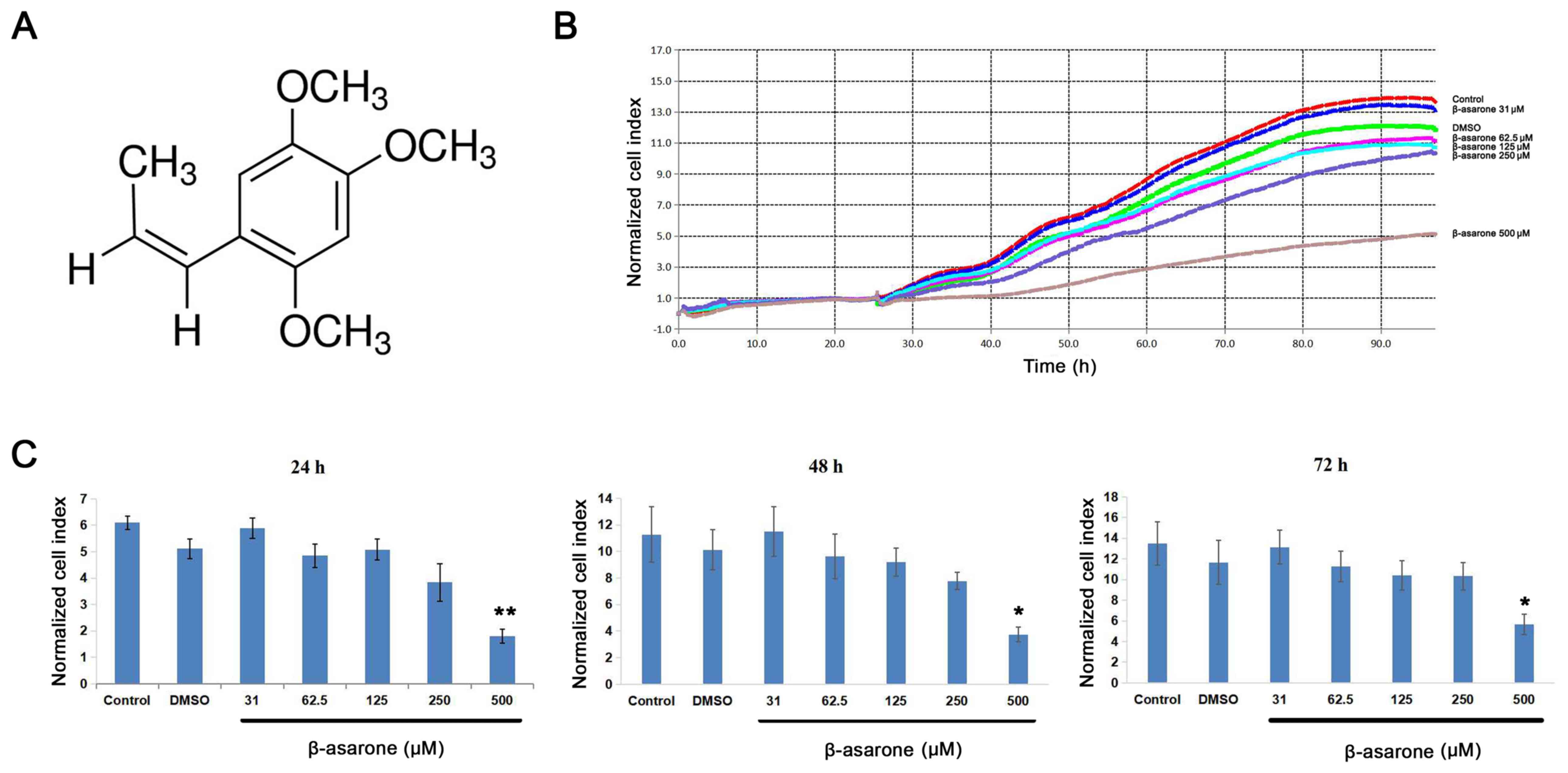
assessed after β-asarone treatment. As presented in Fig. 1B, human HCT116 colon cancer cells
were treated with 31, 62.5, 125, 250 or 500 µM β-asarone. The
results indicated that β-asarone effectively decreased HCT116 cell
proliferation in a dose-dependent manner. Moreover, cells
administered 500 µM β-asarone exhibited a 50% reduction in cell
proliferation compared with the control group (Fig. 1B). The grouped means of the
normalized CI following 24, 48 and 72 h β-asarone treatment are
presented in Fig. 1C. The results
indicated that only 500 µM β-asarone was statistically significant
compared with the control.
GO analysis and DEG pathway enrichment
following β-asarone inhibition in HCT116 cells
To investigate the potential target genes and
associated pathways involved in HCT116 cell β-asarone inhibition on
an unbiased basis, RNA transcriptome sequencing following β-asarone
treatment for 24 and 48 h was performed. The results identified 100
DEGs at 24 and 48 h treatment (fold-change >2; P<0.05). Among
the results obtained at 24 h, 66 genes were significantly
upregulated, while 34 genes were downregulated (Fig. S1A). Analogous data were obtained
following 48 h treatment; 61 genes were upregulated and 39 genes
were downregulated (Fig. S1C).
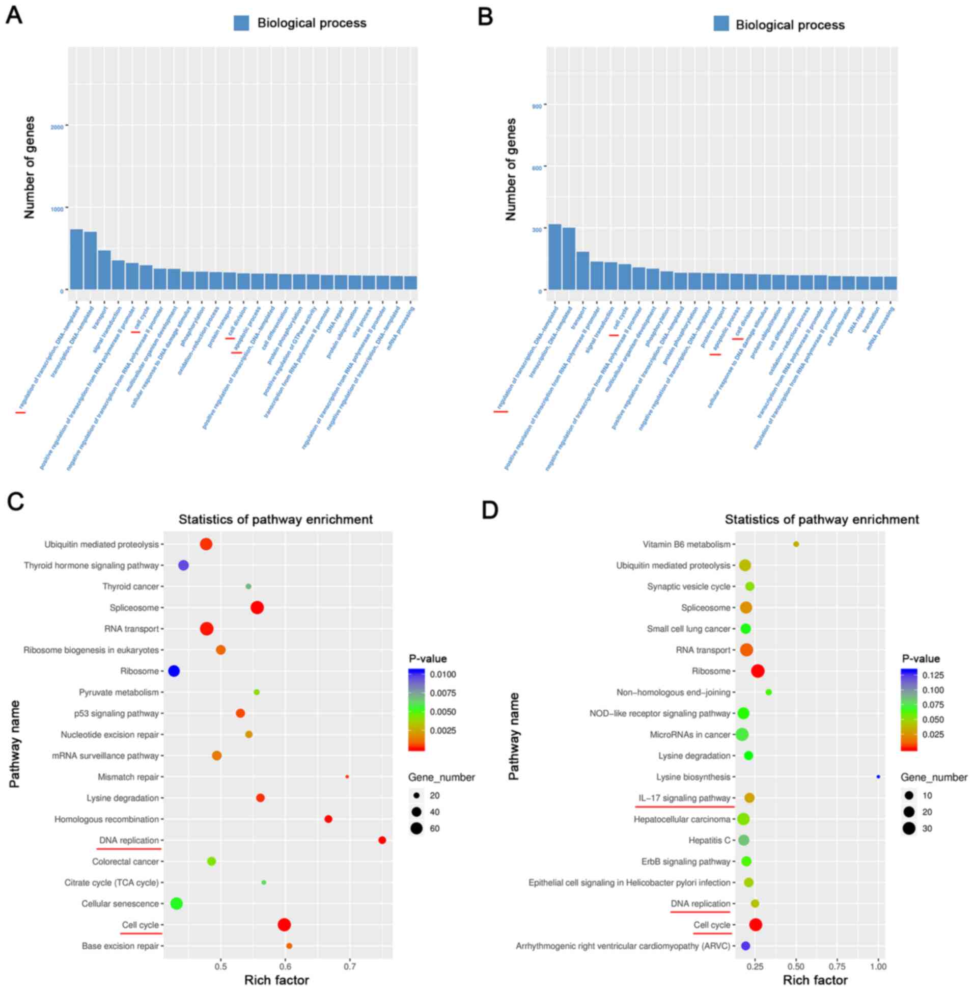
Pathway analysis (Fig. 2) revealed
that the ‘regulation of transcription’, ‘cell cycle’, ‘cell
division’, ‘apoptosis’ and ‘DNA replication’ were prominent
pathways involved in the modulatory effects of β-asarone after
treatment for 24 and 48 h, which may be involved in β-asarone
inhibition-mediated HCT116 cell proliferation. The ‘IL-17 signaling
pathway’ was identified as a regulatory pathway involved in
β-asarone treatment for 48 h, but not in treatment for 24 h,
indicating that β-asarone may have only activated the innate immune
system in HCT116 cancer cells at a later point in time (Fig. 2D).
β-asarone significantly inhibits
HCT116 cell tumorigenesis in vivo
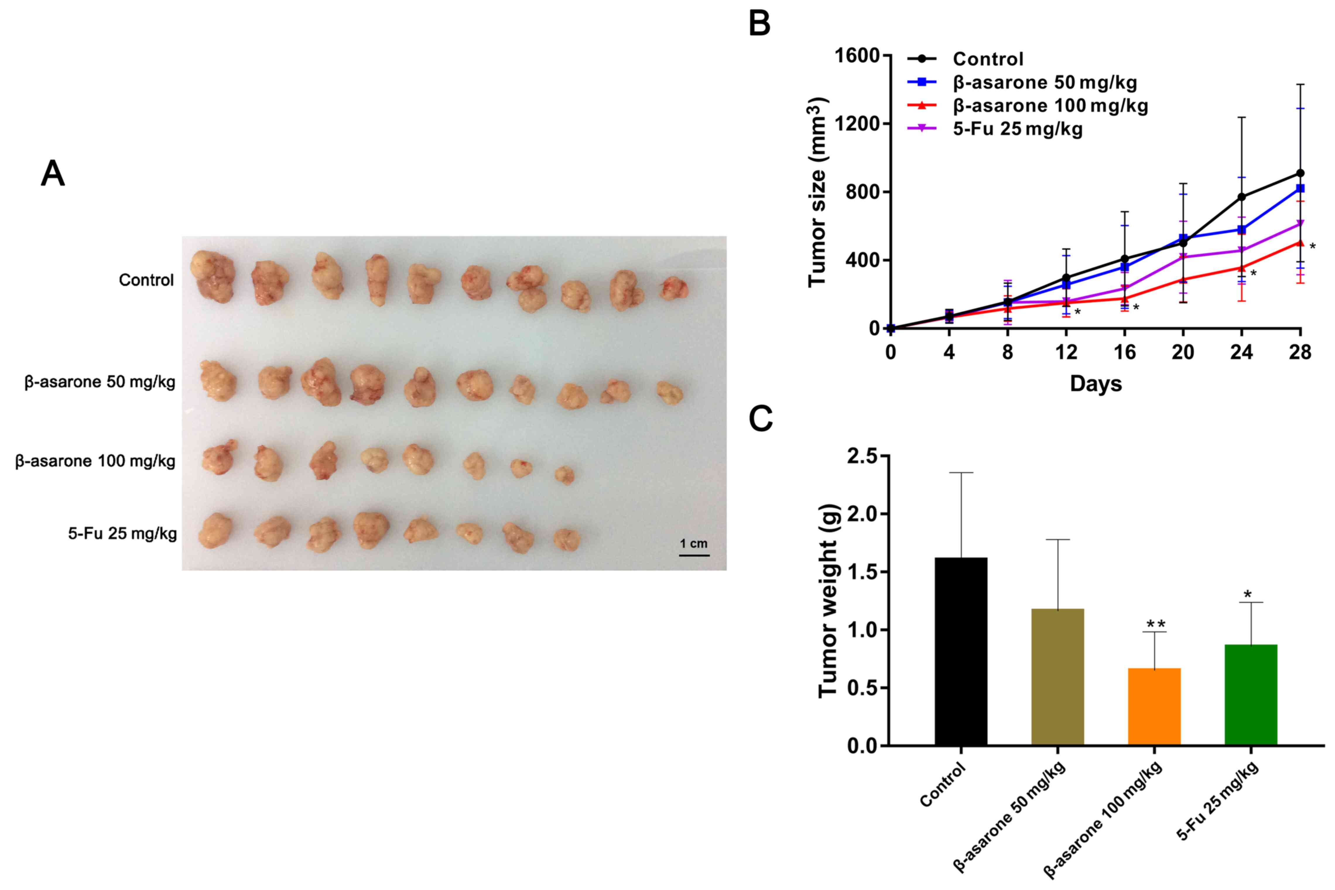
To further investigate whether β-asarone inhibited
HCT116 cell tumorigenesis in vivo, HCT116 cells were
subcutaneously injected into nude mice. As presented in Fig. 3B, tumor growth following 100 mg/kg
β-asarone and 25 mg/kg 5-Fu treatment was markedly reduced compared
with the control group. The maximum diameter and volume of a single
tumor were 19.30 mm and 1,758.71 mm3, respectively.
Furthermore, images revealed that the tumors of the treatment
groups were generally smaller than those of the control group,
particularly in mice treated with 100 mg/kg β-asarone (Fig. 3A). In addition, the mean tumor
weights of the treatment groups were reduced compared with matched
control. Moreover, the 100 mg/kg β-asarone lavage group exhibited a
50% reduction in weight compared with the control (Fig. 3C). α-asarone was also used in
xenograft model experiment (Fig.
S2A). However, the statistical P-value of tumor weight between
control and α-asarone groups was >0.05, which revealed no
statistical significance (Fig.
S2B).
β-asarone may activate the innate
immune system of nude mice
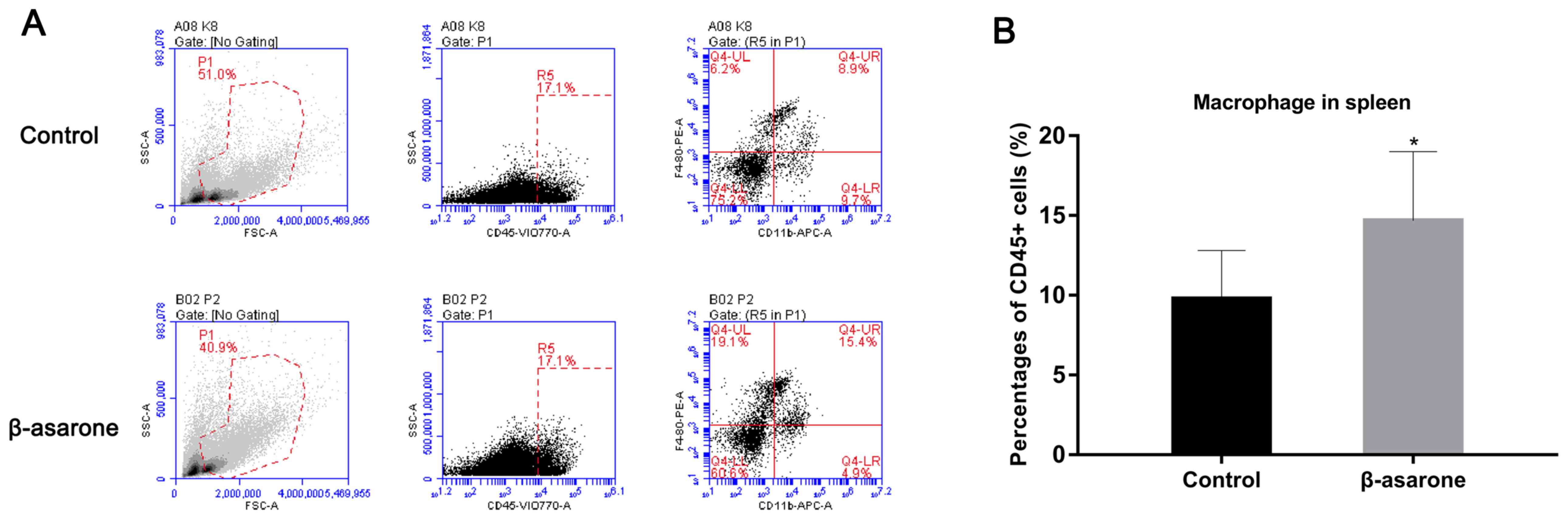
The results of cytokine analysis revealed that
granulocyte colony-stimulating factor (G-CSF) levels in the
peripheral blood of nude mice were significantly increased after
β-asarone treatment (fold-change, 1.78; P=0.02). Additionally, the
results of flow cytometry demonstrated that the number of splenic
macrophage cells increased by 50% (from 10 to 15%) after β-asarone
treatment (Fig. 4A and B;
P<0.05). However, no significant difference in neutrophil
granulocyte levels was observed among the treatment groups in
murine livers, spleens and peripheral blood (Fig. S3), indicating that the functional
role of β-asarone in tumor suppression may also depend on splenic
immune regulation.
Effect of β-asarone on colon cancer
liver metastasis and orthotopic splenic tumors in vivo
Green fluorescence indicated that colon cancer
tissues were present. As presented in Fig. 5, the fluorescence of tissue following
5-Fu and β-asarone treatment was markedly reduced in murine livers
compared with the original splenic injection site. Tumor
fluorescence images revealed no metastatic tumor tissue in the
liver following β-asarone treatment, indicating the strong
inhibitory effect of β-asarone on colon cancer liver metastasis
in vivo. Furthermore, metastatic rate dropped from 90.9%
(10/11 in the control group) to 41.7% (5/12 in the
β-asarone-treated group; P=0.027), suggesting that β-asarone
exerted a strong inhibitory effect on colon cancer liver metastasis
in vivo (Table I). Moreover,
orthotopic splenic tumor weights were measured, the results of
which revealed that the mean tumor weights of the β-asarone
treatment group were significantly decreased compared with the
negative control group (0.19±0.12 g vs. 0.78±0.66 g; P=0.022). The
results further supported the inhibitory effect of β-asarone on
orthotopic splenic tumors (Table
II).
 | Table I.Effect of different treatments on
colon cancer liver metastasis. |
Table I.
Effect of different treatments on
colon cancer liver metastasis.
| Group | Na |
Nnon-metastasis |
Nmetastasis | Rate, % | OR | P-value |
|---|
| Negative
control | 11 | 1 | 10 | 90.9 | – | – |
| Positive
controlb | 7 | 2 | 5 | 71.4 | 3.68 | 0.528 |
| α-asarone | 10 | 2 | 8 | 80.0 | 2.39 | 0.586 |
| β-asarone | 12 | 7 | 5 | 41.7 | 12.34 | 0.027 |
| Table II.Effects of different treatments on
orthotopic splenic tumor weight in liver metastatic nude mice
models. |
Table II.
Effects of different treatments on
orthotopic splenic tumor weight in liver metastatic nude mice
models.
| Treatment
groups | Tumor
weighta, g | P-value |
|---|
| Negative
control | 0.78±0.66 | – |
| Positive
controlc | 0.51±0.48 | 0.573 |
| α-asarone | 0.66±0.59 | 0.918 |
| β-asarone | 0.19±0.12 |
0.022b |
Discussion
Several conventional Chinese medicines have been
commonly used in clinical practice to treat cancer, including
shenqi fuzheng, kanglaite, huachansu and cantharidin sodium
injections. These treatments have been demonstrated to reduce tumor
metastasis and recurrence, and improve the quality of life and
survival of patients with hepatocellular carcinoma, non-small cell
lung cancer and colorectal cancer (21). Both α- and β-asarone, the most
studied bioactive phytochemicals of acorus calamus, have been
reported to have multiple pharmacological activities such as
antidepressant, antianxiety, anti-Alzheimer's, anti-Parkinson's,
antiepileptic, anticancer, antihyperlipidemic, antithrombotic,
anticholestatic and radioprotective activities through its
interaction with multiple molecular targets (22). α-asarone and β-asarone are isomerides
(23). A large number of clinical
studies in China have indicated the effectiveness of α-asarone
against respiratory disorders and epilepsy (24,25);
while β-asarone has been reported to exert biological effects on
different human body systems, serving inhibitory functions in
numerous human carcinomas (10,26). For
instance, β-asarone exerts antifungal and anthelmintic activity,
regulates the nervous system and blocks cholesterol synthesis
(27–29). In addition, previous studies have
also highlighted the inhibitory function of β-asarone on tumor
metastasis in glioma, lung cancer and gastric cancer cells
(10–12). Furthermore, the antiproliferative
property of β-asarone has been reported in gastric cancer and LoVo
colon cancer cells in a previous study (13,14). Liu
et al (30) revealed that
β-asarone could induce senescence in colorectal cancer cells by
increasing the expression of Lamin B1. However, its suppressive
function on the proliferation of HCT116 colon cancer cells and
subsequent liver metastasis is yet to be fully elucidated.
The current study aimed to determine the
antineoplastic role of β-asarone in HCT116 cells, the results of
which revealed that the inhibitory effect of β-asarone was greater
compared with α-asarone. The results of the present study
demonstrated that β-asarone effectively repressed HCT116 cell
proliferation in a dose-dependent manner. Subsequent GO and pathway
analysis revealed that the DEGs identified following β-asarone
inhibition were involved in the ‘cell cycle’, ‘cell division’,
‘cell proliferation’ and ‘apoptosis’. Furthermore, xenograft tumor
assays indicated the inhibitory role of β-asarone on HCT116 cell
tumorigenesis in vivo. In view of that α-asarone and
β-asarone are both isomerides, α-asarone was also used in xenograft
model experiment to distinguish if different structures could
affect the inhibitory effectiveness of the drug. However, there was
no statistically significant difference. A nude mouse model of
HCT116 cell splenic-transplantation was established to assess liver
metastasis, which mimicked the pathogenesis of colon cancer.
Based on ancient Chinese medicinal theory, TCM
regulates the human immune system to maintain homeostasis (31,32).
Trichosanthin, which is extracted from the Chinese medicinal herb
Trichosanthes kirilowi, improves antitumor immunity through the
interaction between tumor suppressor in lung cancer 1 and cytotoxic
and regulatory T cells (33). Chang
and Shen (34) determined that
linalool stimulates IFN-γ, IL-13, IL-2, IL-21, IL-21R, IL-4, IL-6sR
and TNF-α secretion, indicating that it exerts cytotoxic effects in
the antitumor immunity process. Additionally, our previous study
demonstrated that modified SJZ inhibited colon cancer liver
metastasis by activating the innate immune system (9). It was similarly demonstrated that
plasma GM-CSF and macrophage levels are significantly increased
following modified SJZ treatment (9). Nevertheless, the functional role of
β-asarone in colon cancer immunity has not yet been fully
elucidated, to the best of our knowledge. The current study
assessed the influence of β-asarone on the innate immune response
of HCT116 cells. Cytokine analysis revealed that G-CSF levels in
the peripheral blood of nude mice were significantly increased
after β-asarone treatment. G-CSF stimulates the proliferation of
neutrophil granulocytes and macrophages (35,36).
Neutrophil granulocytes are therefore the major effectors of acute
inflammation, as they are one of the first responders during the
immune response, migrating to inflammation sites to target bacteria
or infiltrating cancer tissue (37,38). An
increase in macrophage numbers could also be used to estimate
immune system changes. It is well-known that macrophage cells
engulf and digest cellular debris, foreign substances and cancer
cells, indicating its vital role in non-specific defense (39). Therefore, the current study focused
on the innate immune system following β-asarone treatment and
verified β-asarone's anticancer immunoregulation. However, the
downstream target genes of β-asarone and the potential associated
mechanisms remain unclear in HCT116 colon cancer cells, which
requires further study.
Previous studies have not provided the evidence for
the adverse effect of β-asarone on normal cells, which could also
be a limitation of the present study. Certainly, the possible
cytotoxic activity of β-asarone in normal cells needs to be further
explored to improve our understanding of β-asarone's inhibitory
effect on these tumorigenic cells. Additionally, to develop our
understanding of tumor growth and metastasis-related biomarkers in
tumor tissues/metastases, it would be important to elucidate the
mechanisms by which β-asarone exerts its anti-growth and
anti-metastasis activities in vivo. The lack of such
investigation is another limitation of the present study, but could
be a further area of future research.
In conclusion, the present study demonstrated that
β-asarone exerted an inhibitory effect on the proliferation and
metastasis of HCT116 colon cancer cells in vitro and in
vivo. In addition, β-asarone may be involved in the antitumor
immune response by stimulating G-CSF and increasing the number of
macrophages in the spleen. Collectively, the current data verified
the anticancer effects of β-asarone, both functionally and
immunologically, supporting the possibility of its multi-system
regulation and providing a basis for future research into colon
cancer.
Supplementary Material
Supporting Data
Acknowledgements
Not applicable.
Funding
This work was funded by The Priority Academic
Program Development of Jiangsu Higher Education Institutions, the
National Natural Science Foundation of China (grant no. 81202954),
The Natural Science Fund of Jiangsu Province, China (grant no.
BK20201499), The Peak Academic Talents Plan in Jiangsu Province
Hospital of Chinese Medicine (grant no. k2018yrc25), The Six One
Project in Jiangsu Province (grant no. LGY2018062), The State
Administration of Traditional Chinese Medicine of the People's
Republic of China (National Famous Chinese Medicine Doctor Studio
grant no. 2018-119) and The National Traditional Chinese Medicine
Inheritance and Innovation Platform Construction Project by
National Administration of Traditional Chinese Medicine (grant no.
20085-9-5).
Availability of data and materials
The datasets used and/or analyzed during the current
study are available from the corresponding author on reasonable
request.
Authors' contributions
MC and YWZ designed experiments. MC, YWZ and JYZ
performed the experiments. MC, CEW, HYP and JQ analyzed the
results. CEW and JYZ wrote the manuscript. JQ and JYZ conceived and
supervised the project. JYZ and JQ confirm the authenticity of all
the raw data. All authors read and approved the final
manuscript.
Ethics approval and consent to
participate
All experimental procedures were approved by The
Animal Ethics Committee of Affiliated Hospital of Nanjing
University of Chinese Medicine (approval no. 2018 DW-01-03)
according to the Regulation of Experimental Animal Management
(State Scientific and Technological Commission of the People's
Republic of China; approval no. 2, 1988) and the Jiangsu Province
Experimental Animal Management (Jiangsu province government, China;
approval no. 45, 2008).
Patient consent for publication
Not applicable.
Competing interests
The authors declare that they have no competing
interests.
References
|
1
|
Siegel RL, Miller KD, Fuchs HE and Jemal
A: Cancer statistics, 2021. CA Cancer J Clin. 71:7–33. 2021.
View Article : Google Scholar : PubMed/NCBI
|
|
2
|
Wu XD, Zeng YY, Wu XJ and Qin HY: The
prevalence and correlates of prehospital delay and health belief in
chinese patients with colorectal cancer. Gastroenterol Nurs.
43:186–195. 2020.PubMed/NCBI
|
|
3
|
Ukegjini K, Zadnikar M, Warschkow R,
Muller S, Schmied BM and Marti L: Baseline mortality-adjusted
survival in colon cancer patients. Langenbecks Arch Surg.
401:633–641. 2016. View Article : Google Scholar : PubMed/NCBI
|
|
4
|
Dillekås H, Rogers MS and Straume O: Are
90% of deaths from cancer caused by metastases? Cancer Med.
8:5574–5576. 2019. View Article : Google Scholar : PubMed/NCBI
|
|
5
|
Wu C: Systemic therapy for colon cancer.
Surg Oncol Clin N Am. 27:235–242. 2018. View Article : Google Scholar : PubMed/NCBI
|
|
6
|
Park JH, Zhao M, Han Q, Sun Y, Higuchi T,
Sugisawa N, Yamamoto J, Singh SR, Clary B, Bouvet M and Hoffman RM:
Efficacy of oral recombinant methioninase combined with
oxaliplatinum and 5-fluorouracil on primary colon cancer in a
patient-derived orthotopic xenograft mouse model. Biochem Biophys
Res Commun. 518:306–310. 2019. View Article : Google Scholar : PubMed/NCBI
|
|
7
|
Yan S, Yue Y, Wang J, Li W, Sun M, Zeng L
and Wang X: Banxia Xiexin decoction, a traditional Chinese
medicine, alleviates colon cancer in nude mice. Ann Transl Med.
7:3752019. View Article : Google Scholar : PubMed/NCBI
|
|
8
|
Sun X, Ng TTH, Sham KWY, Zhang L, Chan
MTV, Wu WKK and Cheng CHK: Bufalin, a traditional Chinese medicine
compound, prevents tumor formation in two murine models of
colorectal cancer. Cancer Prev Res (Phila). 12:653–666. 2019.
View Article : Google Scholar : PubMed/NCBI
|
|
9
|
Zhou JY, Chen M, Wu CE, Zhuang YW, Chen YG
and Liu SL: The modified Si-Jun-Zi Decoction attenuates colon
cancer liver metastasis by increasing macrophage cells. BMC
Complement Altern Med. 19:862019. View Article : Google Scholar : PubMed/NCBI
|
|
10
|
Wang TL, Ouyang CS and Lin LZ: β-Asarone
suppresses Wnt/β-catenin signaling to reduce viability, inhibit
migration/invasion/adhesion and induce mitochondria-related
apoptosis in lung cancer cells. Biomed Pharmacother. 106:821–830.
2018. View Article : Google Scholar : PubMed/NCBI
|
|
11
|
Tao H, Ding X, Wu J, Liu S, Sun W, Nie M,
Pan X and Zou X: β-Asarone increases chemosensitivity by inhibiting
tumor glycolysis in gastric cancer. Evid Based Complement Alternat
Med. 2020:69815202020. View Article : Google Scholar : PubMed/NCBI
|
|
12
|
Wang N, Han Y, Luo L, Zhang Q, Ning B and
Fang Y: β-Asarone induces cell apoptosis, inhibits cell
proliferation and decreases migration and invasion of glioma cells.
Biomed Pharmacother. 106:655–664. 2018. View Article : Google Scholar : PubMed/NCBI
|
|
13
|
Wu J, Zhang XX, Sun QM, Chen M, Liu SL,
Zhang X, Zhou JY and Zou X: β-Asarone inhibits gastric cancer cell
proliferation. Oncol Rep. 34:3043–3050. 2015. View Article : Google Scholar : PubMed/NCBI
|
|
14
|
Zou X, Liu SL, Zhou JY, Wu J, Ling BF and
Wang RP: Beta-asarone induces LoVo colon cancer cell apoptosis by
up-regulation of caspases through a mitochondrial pathway in vitro
and in vivo. Asian Pac J Cancer Prev. 13:5291–5298. 2012.
View Article : Google Scholar : PubMed/NCBI
|
|
15
|
Prehn LM and Outzen HC: Primary tumor
immunity in nude mice. Int J Cancer. 19:688–691. 1977. View Article : Google Scholar : PubMed/NCBI
|
|
16
|
Kenyon RH and Pedersen CE Jr: Immune
responses to Rickettsia akari infection in congenitally athymic
nude mice. Infect Immun. 28:310–313. 1980.PubMed/NCBI
|
|
17
|
Uebel T, Hermes L, Haupenthal S, Müller L
and Esselen M: α-Asarone, β-asarone, and γ-asarone: Current status
of toxicological evaluation. J Appl Toxicol. Nov 25–2020.(Epub
ahead of print). View
Article : Google Scholar : PubMed/NCBI
|
|
18
|
R Core Team, . R: A language and
environment for statistical computing. R Foundation for Statistical
Computing; Vienna: 2012, http://www.R-project.org/
|
|
19
|
Correnti JM, Cook D, Aksamitiene E, Swarup
A, Ogunnaike B, Vadigepalli R and Hoek JB: Adiponectin fine-tuning
of liver regeneration dynamics revealed through cellular network
modelling. J Physiol. 593:365–383. 2015. View Article : Google Scholar : PubMed/NCBI
|
|
20
|
Giavazzi R, Garofalo A, Damia G, Garattini
S and D'Incalci M: Response to flavone acetic acid (NSC 347512) of
primary and metastatic human colorectal carcinoma xenografts. Br J
Cancer. 57:277–280. 1988. View Article : Google Scholar : PubMed/NCBI
|
|
21
|
Qi F, Zhao L, Zhou A, Zhang B, Li A, Wang
Z and Han J: The advantages of using traditional Chinese medicine
as an adjunctive therapy in the whole course of cancer treatment
instead of only terminal stage of cancer. Biosci Trends. 9:16–34.
2015. View Article : Google Scholar : PubMed/NCBI
|
|
22
|
Chellian R, Pandy V and Mohamed Z:
Pharmacology and toxicology of α- and β-Asarone: A review of
preclinical evidence. Phytomedicine. 32:41–58. 2017. View Article : Google Scholar : PubMed/NCBI
|
|
23
|
Oprean R, Tamas M and Roman L: Comparison
of GC-MS and TLC techniques for asarone isomers determination. J
Pharm Biomed Anal. 18:227–234. 1998. View Article : Google Scholar : PubMed/NCBI
|
|
24
|
Huang C, Li WG, Zhang XB, Wang L, Xu TL,
Wu D and Li Y: α-Asarone from acorus gramineus alleviates epilepsy
by modulating A-type GABA receptors. Neuropharmacology. 65:1–11.
2013. View Article : Google Scholar : PubMed/NCBI
|
|
25
|
Yu X, Zhe Z, Tang B, Li S, Tang L, Wu Y,
Chen X and Fang H: α-Asarone suppresses the proliferation and
migration of ASMCs through targeting the lncRNA-PVT1/miR-203a/E2F3
signal pathway in RSV-infected rats. Acta Biochim Biophys Sin
(Shanghai). 49:598–608. 2017. View Article : Google Scholar : PubMed/NCBI
|
|
26
|
Wang N, Wang H, Li L, Li Y and Zhang R:
β-Asarone inhibits amyloid-β by promoting autophagy in a cell model
of Alzheimer's disease. Front Pharmacol. 10:15292020. View Article : Google Scholar : PubMed/NCBI
|
|
27
|
Lee JY, Lee JY, Yun BS and Hwang BK:
Antifungal activity of beta-asarone from rhizomes of acorus
gramineus. J Agric Food Chem. 52:776–780. 2004. View Article : Google Scholar : PubMed/NCBI
|
|
28
|
Liu XC, Zhou LG, Liu ZL and Du SS:
Identification of insecticidal constituents of the essential oil of
acorus calamus rhizomes against Liposcelis bostrychophila badonnel.
Molecules. 18:5684–5696. 2013. View Article : Google Scholar : PubMed/NCBI
|
|
29
|
Lee SH, Kim KY, Ryu SY, Yoon Y, Hahm DH,
Kang SA, Cho SH, Lim JS, Moon EY, Yoon SR, et al: Asarone inhibits
adipogenesis and stimulates lipolysis in 3T3-L1 adipocytes. Cell
Mol Biol (Noisy-le-grand). 56 (Suppl):OL1215–OL1222.
2010.PubMed/NCBI
|
|
30
|
Liu L, Wang J, Shi L, Zhang W, Du X, Wang
Z and Zhang Y: β-Asarone induces senescence in colorectal cancer
cells by inducing lamin B1 expression. Phytomedicine. 20:512–520.
2013. View Article : Google Scholar : PubMed/NCBI
|
|
31
|
Wang JH: Traditional Chinese medicine and
the positive correlation with homeostatic evolution of human being:
Based on medical perspective. Chin J Integr Med. 18:629–634. 2012.
View Article : Google Scholar : PubMed/NCBI
|
|
32
|
Meng MB, Wen QL, Cui YL, She B and Zhang
RM: Meta-analysis: Traditional Chinese medicine for improving
immune response in patients with unresectable hepatocellular
carcinoma after transcatheter arterial chemoembolization. Explore
(NY). 7:37–43. 2011. View Article : Google Scholar : PubMed/NCBI
|
|
33
|
Cai Y, Xiong S, Zheng Y, Luo F, Jiang P
and Chu Y: Trichosanthin enhances anti-tumor immune response in a
murine Lewis lung cancer model by boosting the interaction between
TSLC1 and CRTAM. Cell Mol Immunol. 8:359–367. 2011. View Article : Google Scholar : PubMed/NCBI
|
|
34
|
Chang MY and Shen YL: Linalool exhibits
cytotoxic effects by activating antitumor immunity. Molecules.
19:6694–6706. 2014. View Article : Google Scholar : PubMed/NCBI
|
|
35
|
Hara M, Yuasa S, Shimoji K, Onizuka T,
Hayashiji N, Ohno Y, Arai T, Hattori F, Kaneda R, Kimura K, et al:
G-CSF influences mouse skeletal muscle development and regeneration
by stimulating myoblast proliferation. J Exp Med. 208:715–727.
2011. View Article : Google Scholar : PubMed/NCBI
|
|
36
|
Rapoport AP, Abboud CN and DiPersio JF:
Granulocyte-macrophage colony-stimulating factor (GM-CSF) and
granulocyte colony-stimulating factor (G-CSF): Receptor biology,
signal transduction, and neutrophil activation. Blood Rev. 6:43–57.
1992. View Article : Google Scholar : PubMed/NCBI
|
|
37
|
Waugh DJ and Wilson C: The interleukin-8
pathway in cancer. Clin Cancer Res. 14:6735–6741. 2008. View Article : Google Scholar : PubMed/NCBI
|
|
38
|
De Larco JE, Wuertz BR and Furcht LT: The
potential role of neutrophils in promoting the metastatic phenotype
of tumors releasing interleukin-8. Clin Cancer Res. 10:4895–4900.
2004. View Article : Google Scholar : PubMed/NCBI
|
|
39
|
Gordon S: The macrophage: Past, present
and future. Eur J Immunol. 37 (Suppl 1):S9–S17. 2007. View Article : Google Scholar : PubMed/NCBI
|