Introduction
Parkinson's disease (PD) is a chronic and
progressive movement disorder that is mediated by the degeneration
of nigrostriatal dopaminergic (DA) neurons, which causes motor
symptoms, including tremor at rest, rigidity, bradykinesia and
postural instability (1–4). Although the etiology of PD is poorly
understood and therefore cannot guide the development of
knowledge-based targeted therapeutics, investigators have attempted
to develop targeted therapies using viral vector technologies to
transfer specific genes to neurons, with the aim of improving the
function of the degenerating DA system (5, 6).
Striatal delivery of adeno-associated virus (AAV) 2-mediated
cerebral dopamine neurotrophic factor produced neuroprotective and
functional restorative effects in the 6-hydroxydopamine
(6-OHDA)-induced animal model of PD (7). In addition, our previous studies
(8, 9) demonstrated that AAV1 transduction with
a gene encoding the constitutively active form of ras homolog
enriched in the brain, with a mutation of the serine to histidine
at the 16 position [Rheb(S16H)], induced trophic effects. These
effects resulted in the protection and restoration of DA neurons in
the 6-OHDA-induced model of PD via activation of the mammalian
target of rapamycin complex 1 (mTORC1), indicating that the
activation of mTORC1 by a specific gene delivery, such as
Rheb(S16H) to DA neurons, may be a useful strategy in protecting
the DA systems in the adult brain.
Accumulating evidence suggests that the use of
various growth factors, such as glial cell line-derived
neurotrophic factor (GDNF) (10–13)
and brain-derived neurotrophic factor (BDNF) (10, 12,
14, 15), may have a therapeutic potential
against PD. GDNF is a trophic factor involved in the survival of DA
neurons (10–13) and has been identified in numerous
types of neurons, including DA neurons (16). BDNF is a neurotrophin that can be
synthesized by DA neurons in the substantia nigra (SN) of the adult
brain (17) and is also involved in
the survival of DA neurons (18).
The study by Chauhan et al (19) reported that DA neurons in the SN of
PD brains express decreased levels of GDNF and BDNF. In animal
models of PD, experimental results using GDNF (10–13)
and BDNF (10, 18) have consistently shown
neuroprotective effects on DA neurons. Thus, replacement strategies
to supplement neurotrophic factors are considered potential
therapeutic approaches for PD. Our recent study found that the
activation of mTORC1 by Rheb(S16H) transduction of DA neurons
induced the production of trophic factors, such as GDNF and BDNF,
and that the endogenous production of those factors contributed to
the neuroprotection by Rheb(S16H) transduction in a neurotoxin
model of PD (20). The present
study describes the importance of Rheb(S16H) transduction of DA
neurons in the adult brain, indicating a potential therapeutic
method for PD.
mTORC1 activation for survival of DA neurons
in the adult brain
Neurotrophic factors, such as GDNF and BDNF,
regulate the development, maintenance, function and plasticity of
mature neurons and they have emerged as promising therapeutic
agents for PD (21). The cellular
effects of GDNF and BDNF, which show the most evident reduction in
DA neurons in PD brains (19), are
initiated by their binding to GNDF family receptor α1 (22) and tropomyosin receptor kinase B
(12), respectively. The
stimulation of these receptors by treatment with GDNF and BDNF
activates the Akt/mTOR signaling pathway, which is involved in the
downstream activation of the pro-survival pathway in neurons
(23). In addition, consistent with
the decreased levels of GDNF and BDNF in PD brains, the decreased
levels of Akt phosphorylation, resulting in a loss of mTORC1
activation, are observed in the SN of patients with PD and in a
1-methyl-4-phenylpyridinium (MPP+) model of PD, which
mimics the phenotype of patients with PD (24). Numerous studies have also reported
that the activation of the Akt/mTOR signaling pathway enhances the
activity of intracellular cell survival pathways under a variety of
conditions, such as ischemic shock, oxidative stress and the
withdrawal of trophic factors (25–27).
These results indicate that the activation of mTORC1 signaling
pathway may be necessary for the survival of DA neurons and the
functional maintenance of the DA system in the adult brain.
Rheb(S16H) transduction of DA neurons for
mTORC1 activation
Rheb, a key upstream regulator of mTORC1 activation,
is regulated via GTPase activity of tuberous sclerosis complex
(TSC)1/TSC2 and it is involved in cellular processes, such as
protein synthesis, cell growth, proliferation, survival and
synaptic plasticity (8, 28). Furthermore, the serine at position
16 of Rheb has sensitivity to TSC activation and Rheb(S16H), a
mutation of the serine to histidine, exhibits resistance to GTPase
activation by TSC (8, 29), indicating that Rheb(S16H) is a
strong activator of mTORC1. Our recent studies demonstrated that
Rheb(S16H) expression in DA neurons induced an increase in
phosphorylation of the mTORC1 substrates, 4E-BP1 and p70S6K,
indicating activation of mTORC1 and showed apparent neurotrophic
effects in the nigrostriatal DA system in the adult brains
(8, 20). Additionally, the activation of
mTORC1 by Rheb(S16H) transduction of DA neurons resulted in
neuroprotective effects on the nigrostriatal DA projection in rat
and mouse models of PD, indicating that Rheb(S16H) transduction of
SN pars compacta neurons may have a common effect against different
neurotoxins and models associated with the degeneration of the
nigrostriatal DA projection in the adult brain.
GDNF and BDNF against PD
GDNF and BDNF are important trophic factors involved
in the survival of neurons (10–15).
The neurotrophic properties of GDNF were first reported by Lin
et al (11), in a study that
demonstrated that GDNF promotes cell survival and increases
dopamine uptake in the DA neuron cultures derived from the
embryonic rodent midbrain. Subsequent studies have demonstrated
that treatment with GDNF has neurotrophic and protective effects in
animal models of PD (10, 12, 13)
and conditional ablation of GDNF in adult mice results in a delayed
and progressive loss of DA neurons (30). Similar to GDNF, the experimental
results have shown that BDNF has neuroprotective effects in animal
models of PD (10, 18) and the infusion of an antisense
oligonucleotide specific to BDNF results in anatomical,
neurochemical and behavioral deficits that are characteristic of
neurotoxic models of PD (31).
Although these results indicate that neurotrophic factors are
indispensable for the survival and protection of DA neurons and the
support of diverse neurotrophic factors may mediate the survival of
DA neurons, the use as a treatment for PD is impeded due to one
particular factor. GDNF and BDNF do not cross the blood-brain
barrier; therefore, direct application of these factors to the
brain is essential. However, clinical trials of
intracerebroventricular injection and intraputaminal infusion of
GDNF failed in patients with PD (32, 33),
possibly due to the limited penetration and distribution to the
target brain areas. Thus, despite the potential clinical importance
of these factors, the utilization in clinical pharmacology and
other therapeutics for PD is dependent upon sustained delivery of
the appropriate amounts of the factors to the target areas in a
safe and efficacious manner, without producing adverse effects.
Mechanisms of Rheb(S16H)-induced
neuroprotection
As reported in our previous studies (8, 9), the
activation of mTORC1 by Rheb(S16H) transduction of DA neurons
resulted in the SN-induced neurotrophic effects in the DA neurons,
as well as in the protection and restoration of the nigrostriatal
DA projection in a neurotoxin model of PD. These results indicate
that an alternative to delivering neurotrophic molecules to the
brain extracellular spaces, such as viral vector approaches to
transduction of neurons, is to directly activate the intracellular
signaling pathways responsible for their effects. However, it is
largely unknown whether the activated mTORC1 induced the production
of neurotrophic factors, such as GDNF and BDNF, thereby
contributing to the protection of the nigrostriatal DA projection
by intracellular signaling pathways in neurons of the adult brain.
Our recent study found that Rheb(S16H) expression induced GDNF and
BDNF, which contributed to the protection of the nigrostriatal DA
projection in adult DA neurons in vivo and the induction of
those factors was significantly attenuated by treatment with
rapamycin, a specific mTORC1 inhibitor (20). Additionally, Rheb(S16H) expression
induced an increase in the levels of the phospho-cAMP response
element-binding protein (p-CREB) in DA neurons, which may be
involved in the production of GDNF and BDNF (20). In addition to the induction of
neurotrophic factors, our results using neutralizing antibodies
against GDNF and BDNF showed that the Rheb(S16H)-induced trophic
factors contributed to the protection of the nigrostriatal DA
projection through synergistic effects in the
MPP+-treated rat model of PD (20). These observations indicate that the
observed upregulation of neurotrophic factors may be mediated by
the Rheb(S16H)/mTORC1/CREB signaling pathway in DA neurons, and
Rheb(S16H) expression in DA neurons protects the nigrostriatal DA
projections through synergistic trophic effects mediated by GDNF
and BDNF (Fig. 1).
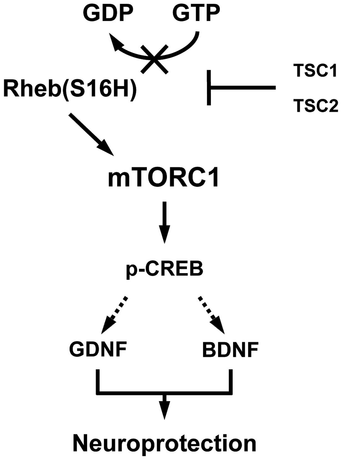 | Figure 1.Schematic representation of the
Rheb(S16H)/mTORC1/p-CREB, GDNF and BDNF signaling pathways. The
serine at position 16 of Rheb has sensitivity to TSC GTPase
activation (20, 29) and Rheb(S16H) results in the
persistence of the GTP-bound Rheb as an activated from. The
accumulation of GTP-bound Rheb by hRheb(S16H) stimulates mTORC1 to
produce p-CREB, GDNF and BDNF, resulting in protection of the
nigrostriatal DA projection. The dotted arrows indicate that p-CREB
may mediate the production of GDNF and BDNF. Rheb(S16H), ras
homolog enriched in brain, which has a S16H mutation; mTORC1,
mammalian target of rapamycin complex 1; p-CREB, phospho-cAMP
response element-binding protein; GDNF, glial cell line-derived
neurotrophic factor; BDNF, brain-derived neurotrophic factor; TSC,
tuberous sclerosis complex. |
Conclusion
Our recent studies showed that Rheb(S16H)
transduction of DA neurons induced the sustained production of GDNF
and BDNF, which contributed to the neuroprotective effect of
Rheb(S16H) in a neurotoxin model of PD. Additionally, the decreased
levels of GDNF, BDNF and Akt phosphorylation, which resulted in a
loss of mTORC1 activation, are observed in the SN of PD patients.
Therefore, even though the optimum gene delivery system without any
side-effects for the clinical trial remains to be found, our
results indicate that Rheb(S16H) transduction may be a useful
strategy to protect the nigrostriatal DA pathway in the adult
brain, particularly considering that the activation of mTORC1 by a
specific gene delivery to DA neurons in the SN results in the
sustained production of diverse neurotrophic factors, such as GDNF
and BDNF, involved in the maintenance and protection of the
nigrostriatal DA system in the adult brain.
Acknowledgements
The present study was supported by the National
Research Foundation of Korea grant funded by the Korean government
(nos. 2008-0061888 and 2012R1A1A1039140).
References
|
1
|
Burke RE and O'Malley K: Axon degeneration
in Parkinson's disease. Exp Neurol. 246:72–83. 2013. View Article : Google Scholar : PubMed/NCBI
|
|
2
|
Jung UJ, Leem E and Kim SR: Naringin: a
protector of the nigrostriatal dopaminergic projection. Exp
Neurobiol. 23:124–129. 2014. View Article : Google Scholar : PubMed/NCBI
|
|
3
|
Leem E, Nam JH, Jeon MT, et al: Naringin
protects the nigrostriatal dopaminergic projection through
induction of GDNF in a neurotoxin model of Parkinson's disease. J
Nutr Biochem. 25:801–806. 2014. View Article : Google Scholar : PubMed/NCBI
|
|
4
|
Savitt JM, Dawson VL and Dawson TM:
Diagnosis and treatment of Parkinson disease: molecules to
medicine. J Clin Invest. 116:1744–1754. 2006. View Article : Google Scholar : PubMed/NCBI
|
|
5
|
Bartus RT, Weinberg MS and Samulski RJ:
Parkinson's disease gene therapy: success by design meets failure
by efficacy. Mol Ther. 22:487–497. 2014. View Article : Google Scholar : PubMed/NCBI
|
|
6
|
Coune PG, Schneider BL and Aebischer P:
Parkinson's disease: gene therapies. Cold Spring Harb Perspect Med.
2:a0094312012. View Article : Google Scholar : PubMed/NCBI
|
|
7
|
Ren X, Zhang T, Gong X, Hu G, Ding W and
Wang X: AAV2-mediated striatum delivery of human CDNF prevents the
deterioration of midbrain dopamine neurons in a 6-hydroxydopamine
induced parkinsonian rat model. Exp Neurol. 248:148–156. 2013.
View Article : Google Scholar : PubMed/NCBI
|
|
8
|
Kim SR, Kareva T, Yarygina O, Kholodilov N
and Burke RE: AAV transduction of dopamine neurons with
constitutively active Rheb protects from neurodegeneration and
mediates axon regrowth. Mol Ther. 20:275–286. 2012. View Article : Google Scholar : PubMed/NCBI
|
|
9
|
Kim SR, Chen X, Oo TF, Kareva T, Yarygina
O, Wang C, During M, Kholodilov N and Burke RE: Dopaminergic
pathway reconstruction by Akt/Rheb-induced axon regeneration. Ann
Neurol. 70:110–120. 2011. View Article : Google Scholar : PubMed/NCBI
|
|
10
|
Allen SJ, Watson JJ, Shoemark DK, Barua NU
and Patel NK: GDNF, NGF and BDNF as therapeutic options for
neurodegeneration. Pharmacol Ther. 138:155–175. 2013. View Article : Google Scholar : PubMed/NCBI
|
|
11
|
Lin LF, Doherty DH, Lile JD, Bektesh S and
Collins F: GDNF: a glial cell line-derived neurotrophic factor for
midbrain dopaminergic neurons. Science. 260:1130–1132. 1993.
View Article : Google Scholar : PubMed/NCBI
|
|
12
|
Manfredsson FP, Okun MS and Mandel RJ:
Gene therapy for neurological disorders: challenges and future
prospects for the use of growth factors for the treatment of
Parkinson's disease. Curr Gene Ther. 9:375–388. 2009. View Article : Google Scholar : PubMed/NCBI
|
|
13
|
Siegel GJ and Chauhan NB: Neurotrophic
factors in Alzheimer's and Parkinson's disease brain. Brain Res
Brain Res Rev. 33:199–227. 2000. View Article : Google Scholar : PubMed/NCBI
|
|
14
|
Howells DW, Porritt MJ, Wong JY, Batchelor
PE, Kalnins R, Hughes AJ and Donnan GA: Reduced BDNF mRNA
expression in the Parkinson's disease substantia nigra. Exp Neurol.
166:127–135. 2000. View Article : Google Scholar : PubMed/NCBI
|
|
15
|
Studer L, Spenger C, Seiler RW, Othberg A,
Lindvall O and Odin P: Effects of brain-derived neurotrophic factor
on neuronal structure of dopaminergic neurons in dissociated
cultures of human fetal mesencephalon. Exp Brain Res. 108:328–336.
1996. View Article : Google Scholar : PubMed/NCBI
|
|
16
|
Pochon NA, Menoud A, Tseng JL, Zurn AD and
Aebischer P: Neuronal GDNF expression in the adult rat nervous
system identified by in situ hybridization. Eur J Neurosci.
9:463–471. 1997. View Article : Google Scholar : PubMed/NCBI
|
|
17
|
Seroogy KB, Lundgren KH, Tran TM, Guthrie
KM, Isackson PJ and Gall CM: Dopaminergic neurons in rat ventral
midbrain express brain-derived neurotrophic factor and
neurotrophin-3 mRNAs. J Comp Neurol. 342:321–334. 1994. View Article : Google Scholar : PubMed/NCBI
|
|
18
|
Hyman C, Hofer M, Barde YA, Juhasz M,
Yancopoulos GD, Squinto SP and Lindsay RM: BDNF is a neurotrophic
factor for dopaminergic neurons of the substantia nigra. Nature.
350:230–232. 1991. View
Article : Google Scholar : PubMed/NCBI
|
|
19
|
Chauhan NB, Siegel GJ and Lee JM:
Depletion of glial cell line-derived neurotrophic factor in
substantia nigra neurons of Parkinson's disease brain. J Chem
Neuroanat. 21:277–288. 2001. View Article : Google Scholar : PubMed/NCBI
|
|
20
|
Nam JH, Leem E, Jeon MT, et al: Induction
of GDNF and BDNF by hRheb(S16H) transduction of SNpc neurons:
neuroprotective mechanisms of hRheb(S16H) in a model of Parkinson's
disease. Mol Neurobiol. May 25–2014.(Epub ahead of print).
View Article : Google Scholar : PubMed/NCBI
|
|
21
|
Aron L and Klein R: Repairing the
parkinsonian brain with neurotrophic factors. Trends Neurosci.
34:88–100. 2011. View Article : Google Scholar : PubMed/NCBI
|
|
22
|
Kholodilov N, Kim SR, Yarygina O, Kareva
T, Cho JW, Baohan A and Burke RE: Glial cell line-derived
neurotrophic factor receptor-α1 expressed in striatum in trans
regulates development and injury response of dopamine neurons of
the substantia nigra. J Neurochem. 116:486–498. 2011. View Article : Google Scholar : PubMed/NCBI
|
|
23
|
Creedon DJ, Tansey MG, Baloh RH, Osborne
PA, Lampe PA, Fahrner TJ, Heuckeroth RO, Milbrandt J and Johnson EM
Jr: Neurturin shares receptors and signal transduction pathways
with glial cell line-derived neurotrophic factor in sympathetic
neurons. Proc Natl Acad Sci USA. 94:7018–7023. 1997. View Article : Google Scholar : PubMed/NCBI
|
|
24
|
Selvaraj S, Sun Y, Watt JA, Wang S, Lei S,
Birnbaumer L and Singh BB: Neurotoxin-induced ER stress in mouse
dopaminergic neurons involves downregulation of TRPC1 and
inhibition of AKT/mTOR signaling. J Clin Invest. 122:1354–1367.
2012. View
Article : Google Scholar : PubMed/NCBI
|
|
25
|
Datta SR, Brunet A and Greenberg ME:
Cellular survival: a play in three Akts. Genes Dev. 13:2905–2927.
1999. View Article : Google Scholar : PubMed/NCBI
|
|
26
|
Brunet A, Datta SR and Greenberg ME:
Transcription-dependent and -independent control of neuronal
survival by the PI3K-Akt signaling pathway. Curr Opin Neurobiol.
11:297–305. 2001. View Article : Google Scholar : PubMed/NCBI
|
|
27
|
Chang N, El-Hayek YH, Gomez E and Wan Q:
Phosphatase PTEN in neuronal injury and brain disorders. Trends
Neurosci. 30:581–586. 2007. View Article : Google Scholar : PubMed/NCBI
|
|
28
|
Karassek S, Berghaus C, Schwarten M, et
al: Ras homolog enriched in brain (Rheb) enhances apoptotic
signaling. J Biol Chem. 285:33979–33991. 2010. View Article : Google Scholar : PubMed/NCBI
|
|
29
|
Yan L, Findlay GM, Jones R, Procter J, Cao
Y and Lamb RF: Hyperactivation of mammalian target of rapamycin
(mTOR) signaling by a gain-of-function mutant of the Rheb GTPase. J
Biol Chem. 281:19793–19797. 2006. View Article : Google Scholar : PubMed/NCBI
|
|
30
|
Pascual A, Hidalgo-Figueroa M, Piruat JI,
Pintado CO, Gomez-Diaz R and Lopez-Barneo J: Absolute requirement
of GDNF for adult catecholaminergic neuron survival. Nat Neurosci.
11:755–761. 2008. View
Article : Google Scholar : PubMed/NCBI
|
|
31
|
Porritt MJ, Batchelor PE and Howells DW:
Inhibiting BDNF expression by antisense oligonucleotide infusion
causes loss of nigral dopaminergic neurons. Exp Neurol.
192:226–234. 2005. View Article : Google Scholar : PubMed/NCBI
|
|
32
|
Nutt JG, Burchiel KJ, Comella CL, et al:
Randomized, double-blind trial of glial cell line-derived
neurotrophic factor (GDNF) in PD. Neurology. 60:69–73. 2003.
View Article : Google Scholar : PubMed/NCBI
|
|
33
|
Peterson AL and Nutt JG: Treatment of
Parkinson's disease with trophic factors. Neurotherapeutics.
5:270–280. 2008. View Article : Google Scholar : PubMed/NCBI
|


