Introduction
N-methyl-N'-nitro-N-nitrosoguanidine (MNNG) is
a well-known alkylating agent that can induce gastric carcinoma
(1, 2). As a carcinogenic chemical, the
mechanism of carcinogenesis induced by MNNG is not well understood.
Previous studies have focused on the methylation aspect (1, 3,
4) and presently, MNNG has been
recognized as a methylating agent universally (5, 6).
Thus far, MNNG-induced DNA epigenetic changes have
been mostly reported in rats (1,
2, 4–6), no
relevant studies in humans found. Therefore, to improve the insight
into the mechanism of human gastric carcinogenesis induced by MNNG,
the epigenetic status of the human telomerase reverse transcriptase
(hTERT) promoter, the rate-limiting subunit of telomerase whose
activity is considered to be an early step in gastric
carcinogenesis (7–9), was investigated in normal human
gastric mucosal epithelial cells (nhGMECs) following MNNG
exposure.
Materials and methods
Chemical
MNNG was purchased from Tokyo Chemical Industry Co.,
Ltd., (Tokyo, Japan) dissolved in redistilled water at 25˚C and
formulated with a concentration of 0.1%. Subsequently, the
MNNG-containing water was confected into 6.8 and 68 µM in
Dulbecco's modified Eagle's medium (DMEM) - F12 medium (Gibco-BRL,
Life Technologies, Carlsbad, CA, USA). The MNNG was freshly
prepared for the experiment.
Cell culture
Following the approval by the The Third Military
Medical University and the affiliated Southwest Hospital Ethical
Committees and informed consent from the patient, nhGMECs were
isolated from the specimen obtained by routine surgery with our
previously developed method (10).
The patient that provided the gastric sample was aged 42 years and
underwent surgery for a gastric ulcer complicating perforation. The
possibility of gastric cancer was excluded by postoperative
pathology. BGC-823, SGC-7901 and MKN-28 cell lines were maintained
in the laboratory and routinely cultured. All the cells were grown
in DMEM - F12 medium supplemented with 10% fetal bovine serum (FBS)
and without any antibiotics.
MNNG-treated cells
nhGMECs were cultured primarily for 48 h. The
supernatant was discarded and the cells were washed three times in
warmed phosphate-buffered saline (PBS). Exposure for 4 h to MNNG at
different concentrations of 6.8 and 68 µM was carried out. The
cells were cultured in DMEM-F12 medium without MNNG and FBS as a
negative control. Following treatment, the cells were washed five
times with warmed PBS to remove any residual MNNG and reincubated
in fresh DMEM-F12 medium supplemented with FBS. At intervals of 48
h (≤ 100 h post-isolation), the cells were harvested and studies
were performed as described below.
hTERT promoter methylation assay
DNA were isolated from nhGMECs that were treated
with and without MNNG, BGC-823, SGC-7901 and MKN-28 cells using
E.Z.N.A.® SQ DNA kit (Omega Bio-Tek Inc., Norcross, GA,
USA). The hTERT promoter methylation status was assessed with
bisulfite sequencing polymerase chain reaction (PCR) using the EZ
DNA methylation kit (Zymo Research, Irvine, CA, USA) as follows:
Genomic DNA was modified by sodium bisulfite. A segment of 290
basepairs (bp) of the hTERT promoter (GeneBank accession no.
AF097365) was amplified by PCR with primers: Forward,
5′-TTTGAGAATTTGTAAAGAGAAATG-3′ and reverse,
5′-AATATAAAAACCCTAAAAACAAATAC-3′; under 32 cycles of 94˚C for 50
sec, 53˚C for 50 sec and 72˚C for 1 min, followed by 8 min at 72˚C.
Sequencing of all the PCR products following cloning was performed
by Sangon, Shanghai, China using the ABI Prism 3730 sequenator. All
the sequence comparisons were carried out with the DNA Star
software (DNASTAR Inc., Madison, WI, USA).
Results
MNNG treatment and the methylation
status
The nhGMECs were dissociated and cultured. DNA were
isolated from nhGMECs that were treated with and without MNNG,
BGC-823, SGC-7901 and MKN-28 cells. Bisulfite treatment of genomic
DNA can cause unmethylated cytosines to be completely converted
into uracil, which are detected as thymine following PCR
amplification, whereas methylated cytosines remain unchanged. The
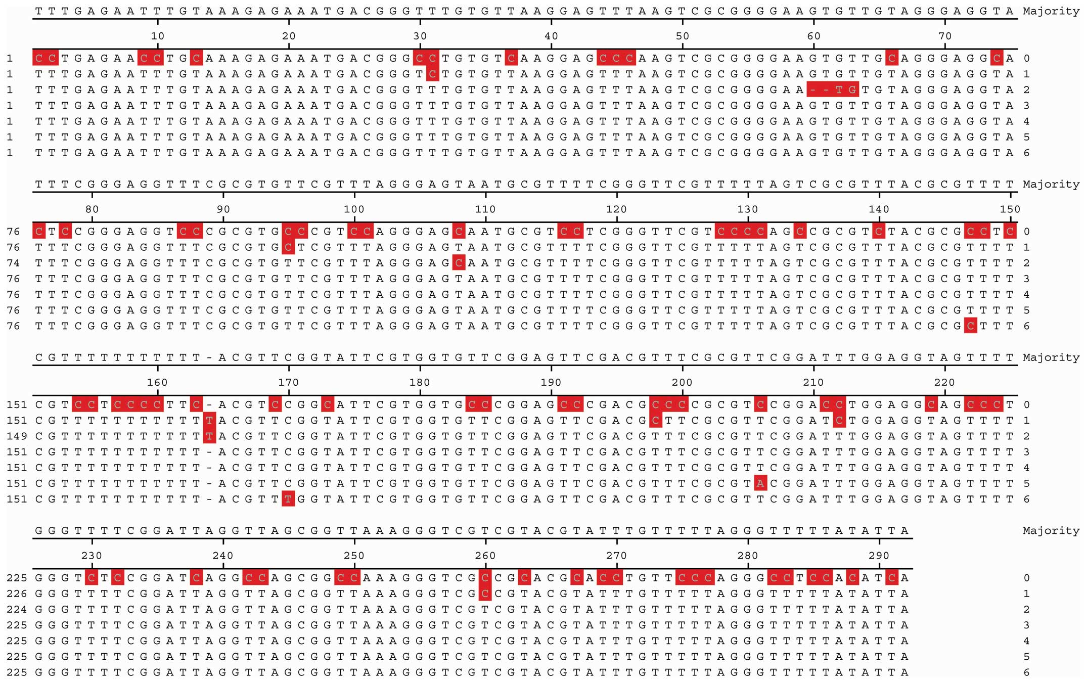
six sequences of the bisulfite-treated DNA were compared to those
published in GenBank respectively. All 5′-CpG-3′ dinucleotides were
found to be methylated, with only five cytosines at positions 31,
95, 198, 212 and 260 outside the CpG dinucleotides methylated in
the 290-bp fragment from the hTERT promoter region of the nhGMECs.
Following 6.8 and 68 µM MNNG exposure, all the methylated 5′-CpG-3′
dinucleotides remained intact, the five cytosines were all
demethylated and the methylation status of the target region was
extremely similar to those of the BGC-823, SGC-7901 and MKN-28
lines (Fig. 1). The demethylated
cytosines were at position 31, 95, 198, 212 and 260, which
corresponded respectively to the −716, −652, −550, −536 and −488
positions, relative to the ATG translation initiation site
(GeneBank accession no. AF097365).
Discussion
Primary cultured nhGMECs have a clear similarity
with their corresponding cells in vivo and are taken as the
ideal tool for gastric pathological studies. The methylation status
of the hTERT promoter plays a role in hTERT expression and
subsequent telomerase activation. The process may be an early step
in gastric carcinogenesis (9). The
aim of the present study, using nhGMECs, was to elucidate the
mechanism of human gastric carcinogenesis induced by MNNG from the
point of the epigenetic state of the hTERT promoter. As a
carcinogenic agent, MNNG has been reported to produce DNA
methylation and is usually recognized as a methylating agent
(1, 4–6). The
present study observed that the epigenetic change of MNNG-treated
nhGMECs was in agreement with those in previous studies. By
contrast, it showed a selective demethylation in the 290-bp
fragment from the hTERT promoter region. Furthermore, the
demethylation of cytosine occured at position cytosine outside the
CpG dinucleotides, and the methylated cytosine in the CpG
dinucleotides remained intact. Notably, such epigenetic status of
the target region following MNNG exposure was extremely similar to
those of BGC-823, SGC-7901 and MKN-28 lines; the three cell lines
from human gastric adenocarcinoma. This experiment could not be
repeated due to the limited gastric specimens from similar
patients. Therefore, the mechanism of human gastric carcinogenesis
induced by MNNG may be correlated with demethylation in the hTERT
promoter region or another undetected region. Previous studies on
the mechanism of carcinogenesis have focused on more methylation
and less demethylation. In neoplasia, demethylation of the genome
as a whole occurs in vivo (11). Additionally, increasing the
incidence of cancer during aging is accompanied by decreasing DNA
methylation (12, 13), although there is controversy
regarding this (8). From another
perspective, the promoter of the hTERT gene becomes
methylated during the development of some but not all tumors
(14). All these indicate a
possible role for demethylation in carcinogenesis. The epigenetic
state of cytosine outside the CpG dinucleotides may be involved in
the carcinogenesis. By computational prediction it has been
estimated that 29,000 CpG-rich regions are distributed in the human
genome (15); therefore, the
majority of studies on gene methylation focused on cytosine in the
CpG dinucleotides and revealed that the CpG islands within gene
promoters generally become methylated during human carcinogenesis.
As cytosine in the CpG dinucleotides, cytosine outside the CpG
dinucleotides are also distributed in the promoter and first exon
of genes and contain putative binding motifs, such as
myeloid-specific zinc finger protein 2 (16, 17).
Their epigenetic state may also affect the binding of transcription
factors (16) and possibly result
in carcinogenesis. Therefore, the epigenetic status of cytosine
outside the CpG dinucleotides requires further study.
In the present study, to prevent interference MNNG
was dissolved in redistilled water and not dimethylsulphoxide and
was freshly prepared for the experiment. nhGMECs were all primary,
not passage cells (8, 12) and were cultured in DMEM-F12 medium
without any antibiotics. In general, alkylating agents, such as
MNNG, produce increased G → A, not C → T, transition mutations. All
five C → T outside the CpG dinucleotides are not considered to
result from the mutagenic effect of MNNG (5).
In conclusion, a selective demethylation in the
hTERT promoter in nhGMECs was observed following exposure to
different MNNG doses in vitro. Demethylation in cytosine
outside the CpG dinucleotides may be an early molecular lesion with
the potential for impacting malignant transformation and a possible
underlying carcinogenic mechanism of MNNG. Thus, it may provide
another insight into the mechanisms of MNNG carcinogenesis.
Acknowledgements
The present study was supported by The National
Natural Science Foundation of China (grant no. 30270609).
References
|
1
|
Gombar CT and Magee PN: DNA-methylation by
nitrosocimetidine and N-methyl-N′-nitro-N-nitrosoguanidine in the
intact rat. Chem Biol Interact. 40:149–157. 1982. View Article : Google Scholar : PubMed/NCBI
|
|
2
|
Manikandan P, Murugan RS, Priyadarsini RV,
Vinothini G and Nagini S: Eugenol induces apoptosis and inhibits
invasion and angiogenesis in a rat model of gastric carcinogenesis
induced by MNNG. Life Sci. 86:936–941. 2010. View Article : Google Scholar : PubMed/NCBI
|
|
3
|
Haerlin R, Sussmuth R and Lingens F:
Mechanism of mutagenesis by N-methyl-N′-nitro-N-nitrosoguanidine
(MNNG) v. methylation of DNA by
N-trideuteriomethyl-N′-nitro-N-nitrosoguanidine (D3-MNNG). FEBS
Lett. 9:175–176. 1970. View Article : Google Scholar : PubMed/NCBI
|
|
4
|
Bhalla A, Sachdeva G and Bamezai R: T-cell
receptor-gamma rearrangement and c-myb methylation in MNNG exposed
Bloom syndrome B-lymphoblastoid cells. Cancer Lett. 126:1–6. 1998.
View Article : Google Scholar : PubMed/NCBI
|
|
5
|
Schroering AG and Williams KJ: Rapid
induction of chromatin-associated DNA mismatch repair proteins
after MNNG treatment. DNA Repair (Amst). 7:951–969. 2008.
View Article : Google Scholar : PubMed/NCBI
|
|
6
|
Tomicic MT, Christmann M, Fabian K and
Kaina B: Apaf-1 deficient mouse fibroblasts are resistant to MNNG
and MMS-induced apoptotic death without attenuation of Bcl-2
decline. Toxicol Appl Pharmacol. 207 (Suppl 2):117–122. 2005.
View Article : Google Scholar : PubMed/NCBI
|
|
7
|
Sabah M, Cummins R, Leader M and Kay E:
Expression of human telomerase reverse transcriptase in
gastrointestinal stromal tumors occurs preferentially in malignant
neoplasms. Hum Pathol. 35:1231–1235. 2004. View Article : Google Scholar : PubMed/NCBI
|
|
8
|
Neumeister P, Albanese C, Balent B,
Greally J and Pestell RG: Senescence and epigenetic dysregulation
in cancer. Int J Biochem Cell Biol. 34:1475–1490. 2002. View Article : Google Scholar : PubMed/NCBI
|
|
9
|
Gulmann C, Lantuejoul S, Grace A, Leader
M, Patchett S and Kay E: Telomerase activity in proximal and distal
gastric neoplastic and preneoplastic lesions using
immunohistochemical detection of hTERT. Dig Liver Dis. 37:439–445.
2005. View Article : Google Scholar : PubMed/NCBI
|
|
10
|
Cheng YB, Fang DC, Guo LP, Wang ZQ, Luo YH
and Zhou JC: A convenient primary culture of human gastric
epithelial cells. Chin J Gastroenterol. 12:31–35. 2007.
|
|
11
|
Issa JP: Aging, DNA methylation and
cancer. Crit Rev Oncol Hematol. 32:31–43. 1999. View Article : Google Scholar : PubMed/NCBI
|
|
12
|
Suzuki T, Fujii M and Ayusawa D:
Demethylation of classical satellite 2 and 3 DNA with chromosomal
instability in senescent human fibroblasts. Exp Gerontol.
37:1005–1014. 2002. View Article : Google Scholar : PubMed/NCBI
|
|
13
|
Liu L, Lai S, Andrews LG and Tollefsbol
TO: Genetic and epigenetic modulation of telomerase activity in
development and disease. Gene. 340:1–10. 2004. View Article : Google Scholar : PubMed/NCBI
|
|
14
|
Devereux TR, Horikawa I, Anna CH, Annab
LA, Afshari CA and Barrett JC: DNA methylation analysis of the
promoter region of the human telomerase reverse transcriptase
(hTERT) gene. Cancer Res. 59:6087–6090. 1999.PubMed/NCBI
|
|
15
|
Santourlidis S, Wernet P, Ghanjati F,
Graffmann N, Springer J, Kriegs C, Zhao X, Brands J, Arauzo-Bravo
MJ, Neves R, et al: Unrestricted somatic stem cells (USSC) from
human umbilical cord blood display uncommitted epigenetic
signatures of the major stem cell pluripotency genes. Stem Cell
Res. 6:60–69. 2011. View Article : Google Scholar : PubMed/NCBI
|
|
16
|
Poole JC, Andrews LG and Tollefsbol TO:
Activity, function, and gene regulation of the catalytic subunit of
telomerase (hTERT). Gene. 269:1–12. 2001. View Article : Google Scholar : PubMed/NCBI
|
|
17
|
Fujimoto K, Kyo S, Takakura M, Kanaya T,
Kitagawa Y, Itoh H, Takahashi M and Inoue M: Identification and
characterization of negative regulatory elements of the human
telomerase catalytic subunit (hTERT) gene promoter: possible role
of MZF-2 in transcriptional repression of hTERT. Nucleic Acids Res.
28:2557–2562. 2000. View Article : Google Scholar : PubMed/NCBI
|