Introduction
Antioxidant defenses help to prevent free
radical-mediated injury (1,
2). These substances, including
enzymatic (catalase, superoxide dismutases, glutathione peroxidase
and glutathione reductase) and non-enzymatic [vitamin E, vitamin C,
glutathione (GSH) and uric acid] categories, protect an organism
from oxygen free radicals (3). Red
blood cells (RBCs) are the main source of antioxidants; in
vitro and in vivo tests have been performed to
demonstrate the importance of RBC antioxidant enzymes in protecting
target cells from reactive oxygen species (ROS) (4, 5).
Therefore, the amount of antioxidants in RBC is a suitable marker
for evaluating chronic ROS exposure.
GSH is the most abundant thiol in cells and acts as
a major antioxidant in addition to its other biological functions
in RBC (6). Catalase is an
important antioxidant produced by RBC; thus, catalase levels can
indicate ROS levels (7). However,
limited information is available on the effects of physiological
sex steroid changes during the menstrual cycle on the GSH and
catalase in RBC in a population of healthy normomenorrhoic females.
Therefore, the present study evaluated the effects of physiological
sex steroid changes during the menstrual cycle on GSH and catalase
activities. Females with regular menstrual cycles were selected to
be part of the sample and the changes in the GSH and catalase
levels during the menstrual cycle were measured. The resulting
correlation between estrogen and antioxidant systems can be used
for analyses and discussion.
Materials and methods
Subjects
The study population consisted of 43 healthy female
volunteers (age range, 22–51 years; mean age, 36 years), with a
history of regular menstrual cycles lasting from 28 to 30 days.
Their health status was confirmed on the basis of their medical
history, as well as by physical and routine laboratory
examinations. Body weight was normal and all the subjects were on a
standard balanced diet. None of the subjects smoked or had a
history of intensive exercise. None had used oral contraceptives
for at least one year before the study or any other type of
medication during the previous four months. No drugs were taken
during the study period. Informed consent was obtained from all the
subjects prior to participation in the study. The study protocol
was approved by the Tsaotun Psychiatric Center, Ministry of Health
and Welfare, Taiwan.
Sample collection
After overnight fasting, blood samples were
collected by vein puncture and stored on ice. Erythrocytes were
immediately washed two times in phosphate-buffered saline.
Erythrocyte hemodialysates for the estimation of catalase enzyme
activity and GSH concentration were prepared by centrifugation of
heparinized blood (25,910 x g for 10 sec at 4˚C), deprived of buffy
coats and stored at −20°C.
Catalase activity
The determination of catalase was assayed as
described (8). The reaction mixture
(1 ml) contained 50 µl Tris-EDTA (pH 8.0) and 900 µl 20 mmol/1
H2 O2 heated to 37˚C for 10 min, after which
20 µl erythrocyte lysate was added. The optical density value at
240 nm was detected for 1 min at 25˚C. The catalase activity was
expressed in IU/mg protein (µ moles of 20 mmol/1
H2 O2 consumed/min/mg protein).
Erythrocyte GSH level
Erythrocyte GSH was measured using a
spectrophotometric method of the form of reduced GSH (9). 5,5′-Dithiobis-2-nitrobenzoic acid is a
disulfide chromogen that is readily reduced by sulfhydryl compound
to an intensely yellow compound. The absorbance of the reduced
compound was measured at 412 nm as directly proportional to GSH
concentration.
Protein concentrations
Protein concentrations were quantified using the
Bradford protein assay method.
Estrogen measurement
The serum estrogen concentration was determined by
radioimmunoassay (Diagnostic Products Corporation, Los Angeles, CA,
USA).
Statistical analysis
Each specimen was analyzed twice with the use of
Student's t-test analysis software. P<0.05 was considered to
indicate a significant difference and P<0.01 was considered to
represent an extremely significant difference. Pearson's or
Spearman's analysis was used in the correlation analysis.
Results
GSH levels in erythrocytes during the
menstrual cycle
The sample subjects were divided into two groups
according to the number of days the subjects were into the
menstrual cycle. Group A (16 samples) consisted of samples from
females that were within 10–20 days of their menstruation. Group B
(27 samples) consisted of females that were 1–9 and 21–30 days into
their menstruation. The average estrogen level in group A (184±106
pg/ml) was higher than that in group B (105±56 pg/ml) (P<0.01).
The GSH levels in group A (4.4±2.3 µg/mg) were also significantly
higher compared to group B (3.2±1.8 µg/mg) (P<0.05). However,
when Spearman's rank correlation analysis was performed, no
significant correlation (P=0.32) was found between the estrogen
levels in the blood and the GSH expression in the RBC (Fig. 1).
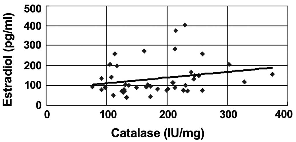
Catalase activity in the RBC during
the menstrual cycle
The average level of catalase in group A (210±72
IU/mg) was significantly higher than that in group B (168±62 IU/mg)
(P<0.05). In the Spearman's rank correlation analysis, a
significant correlation (P=0.04) was found between the estrogen
levels in the blood and the catalase expression in the RBC
(Fig. 2).
Discussion
Oxidative stress is responsible for aging and
coronary heart diseases (10,
11). Rapid induction of
intracellular GSH synthesis occurs in response to various oxidative
stress parameters, which is a critical determinant of cellular
tolerance to oxidative stress (12). Damy et al (13) reported that cardiac and systemic GSH
deficiency is associated with the functional status and structural
cardiac abnormalities of patients with cardiac diseases. As a
result, GSH serves an important function in fighting oxidative
damage and helping prevent heart diseases. Additionally, the GSH
level is consistently increased following various types of
treatments and in different situations, such as amenorrhea,
post-menopause or treatments with oral contraceptive drugs
(14–17). In the present study, blood samples
were collected from females who were not taking contraceptive
drugs, between ages 22 and 51 years and have regular menstrual
cycles. The antioxidant levels of the samples were determined on
the same day to avoid hemolysis. In a regular menstrual cycle, the
GSH level increased when the estrogen concentration was
significantly increased.
An antioxidant-like catalase is also inversely
associated with coronary heart disease (18). However, Yang et al (19) reported that high levels of catalase
activity were associated with a low risk of coronary heart diseases
in females with diabetes but not in those without diabetes.
Pre-menopausal females have a lower risk of heart diseases compared
to males, but the risk becomes the same post-menopause (20). Estrogen levels exist in a high
concentration pre-menopause, which decreases oxidative stress in
the cardiovascular system. In the present data, estrogen levels
increased and decreased according to the menstrual cycle and the
catalase level shifted according to the cycle. This process may
protect females from cardiovascular diseases.
The effect of estrogen on catalase in the condition
of elevation in previous studies differed from the results of the
present study. Massafra et al (21, 22)
reported that a significant positive correlation existed between
physiological estrogen and antioxidant expression during the
menstrual cycle, but no significant changes occurred in catalase.
However, females in different situations, such as multiple blood
sampling, which thus have different effects, may take different
mechanism routes. Therefore, the present study underlined a
significant correlation between estrogen and catalase/GSH and
suggested that estradiol upregulated the expression of the
antioxidants, which may also be mediated through the same
mechanism. Certain unknown factors involved in the whole pathway
should be further investigated.
Acknowledgements
The present study was supported by the grant (no.
9818) from Tsaotun Psychiatric Center of Ministry of Health and
Welfare, Taiwan.
References
|
1
|
Southorn PA and Powis G: Free radicals in
medicine. I. Chemical nature and biologic reactions. Mayo Clin
Proc. 63:381–389. 1988. View Article : Google Scholar : PubMed/NCBI
|
|
2
|
Southorn PA and Powis G: Free radicals in
medicine. II. Involvement in human disease. Mayo Clin Proc.
63:390–408. 1988. View Article : Google Scholar : PubMed/NCBI
|
|
3
|
Sakac V and Sakac M: Free oxygen radiacals
and kidney diseases - part I. Med Pregl. 53:463–474. 2000.(In
Croatian). PubMed/NCBI
|
|
4
|
Agar NS, Sadrzadeh SM, Hallaway PE, et al:
Erythrocyte catalase. A somatic oxidant defense? J Clin Invest.
77:319–321. 1986. View Article : Google Scholar : PubMed/NCBI
|
|
5
|
van Asbeck BS, Hoidal J, Vercellotti GM,
et al: Protection against lethal hyperoxia by tracheal insufflation
of erythrocytes: role of red cell glutathione. Science.
227:756–759. 1985. View Article : Google Scholar : PubMed/NCBI
|
|
6
|
Dabrosin C and Ollinger K: Variability of
glutathione during the menstrual cycle-due to estrogen effects on
hepatocytes? Free Radic Biol Med. 36:145–151. 2004. View Article : Google Scholar : PubMed/NCBI
|
|
7
|
Nishikawa M, Hashida M and Takakura Y:
Catalase delivery for inhibiting ROS-mediated tissue injury and
tumor metastasis. Adv Drug Deliv Rev. 61:319–326. 2009. View Article : Google Scholar : PubMed/NCBI
|
|
8
|
Kolberg C, Horst A, Kolberg A, et al:
Effects of high-velocity, low-amplitude manipulation on catalase
activity in men with neck pain. J Manipulative Physiol Ther.
33:300–307. 2010. View Article : Google Scholar : PubMed/NCBI
|
|
9
|
Owens CW and Belcher RV: A colorimetric
micro-method for the determination of glutathione. Biochem J.
94:705–711. 1965.PubMed/NCBI
|
|
10
|
Cencioni C, Spallotta F, Martelli F, et
al: Oxidative stress and epigenetic regulation in ageing and
age-related diseases. Int J Mol Sci. 14:17643–17663. 2013.
View Article : Google Scholar : PubMed/NCBI
|
|
11
|
Halliwell B: The role of oxygen radicals
in human disease, with particular reference to the vascular system.
Haemostasis. 23 (Suppl 1):118–126. 1993.PubMed/NCBI
|
|
12
|
Rahman I, Antonicelli F and MacNee W:
Molecular mechanism of the regulation of glutathione synthesis by
tumor necrosis factor-alpha and dexamethasone in human alveolar
epithelial cells. J Biol Chem. 274:5088–5096. 1999. View Article : Google Scholar : PubMed/NCBI
|
|
13
|
Damy T, Kirsch M, Khouzami L, et al:
Glutathione deficiency in cardiac patients is related to the
functional status and structural cardiac abnormalities. PLoS One.
4:e48712009. View Article : Google Scholar : PubMed/NCBI
|
|
14
|
Massafra C, Buonocore G, Berni S, et al:
Antioxidant erythrocyte enzyme activities during oral
contraception. Contraception. 47:590–596. 1993. View Article : Google Scholar : PubMed/NCBI
|
|
15
|
Massafra C, Buonocore G, Gioia D, et al:
Changes in the erythrocyte antioxidant enzyme system during
transdermal estradiol therapy for secondary amenorrhea. Gynecol
Endocrinol. 10:155–158. 1996. View Article : Google Scholar : PubMed/NCBI
|
|
16
|
Massafra C, Buonocore G, Gioia D, et al:
Effects of estradiol and medroxyprogesterone-acetate treatment on
erythrocyte antioxidant enzyme activities and malondialdehyde
plasma levels in amenorrhoic women. J Clin Endocrinol Metab.
82:173–175. 1997. View Article : Google Scholar : PubMed/NCBI
|
|
17
|
Bednarek-Tupikowska G, Tworowska U,
Jedrychowska I, et al: Effects of oestradiol and oestroprogestin on
erythrocyte antioxidative enzyme system activity in postmenopausal
women. Clin Endocrinol (Oxf). 64:463–468. 2006.PubMed/NCBI
|
|
18
|
Flores-Mateo G, Carrillo-Santisteve P,
Elosua R, et al: Antioxidant enzyme activity and coronary heart
disease: meta-analyses of observational studies. Am J Epidemiol.
170:135–147. 2009. View Article : Google Scholar : PubMed/NCBI
|
|
19
|
Yang S, Jensen MK, Rimm EB, et al:
Erythrocyte superoxide dismutase, glutathione peroxidase and
catalase activities and risk of coronary heart disease in generally
healthy women: a prospective study. Am J Epidemiol. 180:901–908.
2014. View Article : Google Scholar : PubMed/NCBI
|
|
20
|
Yang XP and Reckelhoff JF: Estrogen,
hormonal replacement therapy and cardiovascular disease. Curr Opin
Nephrol Hypertens. 20:133–138. 2011. View Article : Google Scholar : PubMed/NCBI
|
|
21
|
Massafra C, De Felice C, Gioia D, et al:
Variations in erythrocyte antioxidant glutathione peroxidase
activity during the menstrual cycle. Clin Endocrinol (Oxf).
49:63–67. 1998. View Article : Google Scholar : PubMed/NCBI
|
|
22
|
Massafra C, Gioia D, De Felice C, et al:
Effects of estrogens and androgens on erythrocyte antioxidant
superoxide dismutase, catalase and glutathione peroxidase
activities during the menstrual cycle. J Endocrinol. 167:447–452.
2000. View Article : Google Scholar : PubMed/NCBI
|