Introduction
Acute respiratory distress syndrome (ARDS), an
indication of acute lung injury (ALI), is highly associated with
sepsis, multiple transfusions and trauma (1). ARDS is an inflammatory condition,
manifested by a diffused alveolar damage, formation of hyaline
membranes, protein-rich edema fluid in the alveolar spaces,
capillary injury and disruption of the alveolar epithelium
(2). Lipopolysaccharides (LPS) are
thought to play a major role in initiating the inflammatory
processes that result in ALI, mainly by dysfunction of the
pulmonary surfactants (3). Exposure of
LPS, derived from the cell wall of Gram-negative bacteria, is a
well-known method to introduce acute lung inflammation and ARDS.
LPS activates alveolar macrophages and causes neutrophils to
infiltrate and damage the lungs.
Numerous studies have suggested that oxidant injury
to the pulmonary microvasculature is an important mechanism in the
pathogenesis of ARDS (4,5). There are animal models to prove that
pretreatment with scavengers of reactive oxygen species
significantly reduces the pulmonary hypertension, hypoxia and
increased microvascular permeability to proteins that otherwise
characterizes ALI following infusion of endotoxin (6).
Auricularia auricular-judae is a medicinal
edible fungus, which belongs to the basidiomycotina fungi, mainly
distributed in China, Taiwan, Thailand and Indonesia. The fruit of
Auricularia auricular-judae is rich in
hetero-polysaccharides that consist of a D-glucose residue backbone
with various chains of β-1,3-branch residues, such as mannose,
glucose, xylose and glucuronic acid. It has been proved that black
fungus polysaccharide not only exhibits an extremely high
nutritional value, but also has various pharmacological functions
in humans and animals. Black fungus polysaccharide has antioxidant
(7,8),
blood lipid lowering (9), antitumor
(10,11)
and anti-radiation (12) activity.
Therefore, the present study aimed to determine whether
pretreatment of AAP on LPS-induced ARDS in rats could ameliorate
the ALI.
Materials and methods
Chemicals and reagents
LPS (Escherichia coli LPS, 055:B5) was
purchased from Sigma Aldrich (St. Louis, MO, USA). The
myeloperoxidase (MPO) and malondialdehyde (MDA) kits were purchased
from Jiancheng Bioengineering Institute of Nanjing (Nanjing,
China). Tumor necrosis factor-α (TNF-α) and interleukin (IL)-6
ELISA kits were purchased from USCN Life Science Inc. (Wuhan,
China). The fruit body of Auricularia auricular-judae was
cultured in the Daxinganling region, Heilongjiang province,
China.
Extraction of AAP
AAP was extracted by hot water and
ultrasonic-assisted extraction. The concentrated supernatants were
subsequently precipitated with 3 volumes of absolute ethanol (95%)
and maintained at 4°C overnight. The resulting precipitate was
separated by centrifugation, dissolved in deionized water and
subsequently dialyzed. The non-dialyzed portion was, in addition,
lyophilized to result in a crude polysaccharide extract. The AAP
was decolorized by hydrogen peroxide (13).
Animals and modeling
Adult Sprague-Dawley rats provided by the Laboratory
Animal Center of Xinjiang Medical University (Xinjiang, China) were
used in all the experiments. All the animal care and experimental
procedures were approved by the Animal Care Committee of Xinjiang
Medical University. Adult Sprague-Dawley rats were randomly
assigned into the control, AAP, LPS and LPS plus AAP groups. The
control animals received an equal volume of normal saline at the
same time. The LPS group was induced by intraperitoneal injection
of 10 mg/kg LPS. Rats in the LPS plus AAP group were treated with
AAP for 7 days before LPS administration. Post-LPS infusion (12 h),
animals were sacrificed by overdose of ethyl carbamate and blood
samples were collected from the abdominal aorta. Blood samples were
anticoagulated with EDTA and centrifuged at 3,000 × g for 10 min at
4°C and the plasma was stored at −20°C until measurements were
performed. Following death at the end of the protocol, the left
lung was lavaged using 500 µl of saline 3 times (total volume, 1.5
ml) for protein leakage. The right lung tissues were divided into 3
pieces, one immersed in 10% formalin solution for histopathological
examination, one frozen in liquid nitrogen for quantitative
analysis and the remaining part for measurement of the wet/dry
(W/D) weight ratio.
Bronchoalveolar lavage
The lungs were lavaged with 500 µl of saline 3 times
(total volume, 1.5 ml). Retrieval volume was maximized by
compression of the thorax following the last lavage. The protein
concentration was determined using a protein kit.
Lung W/D weight ratio
The trachea and esophagus were separated from the
lungs by blunt dissection and the wet weight of the latter was
determined. Subsequently, the lungs were incubated at 60°C for 3–4
days to remove all moisture, the dry weight was measured and the
ratio of wet-to-dry weight calculated.
MPO and MDA activities in lung
homogenates
The MPO and MDA activities in the lung tissue were
assayed by MPO and MDA kits, respectively, following the
manufacturer's instructions.
Cytokines in blood
TNF-α and IL-6 in blood samples were determined with
ELISA kits from USCN Life Science Inc., performed according to the
manufacturer's instructions. All the measurements were performed in
duplicate.
Histological assessment
A section of the right lung was removed and put into
10% formaldehyde solution followed by dehydration, paraffin
embedding, sectioning and hematoxylin and eosin staining
sequentially. The changes of pathology were observed.
Statistical analysis
Results are presented as mean ± standard deviation.
For tests of significance between the groups, one-way analysis of
variance was performed. Comparisons between two groups were
performed using unpaired Student's t-test. P<0.05 was considered
to indicate a statistically significant difference. All the data
were performed in ≥3 independent experiments.
Results
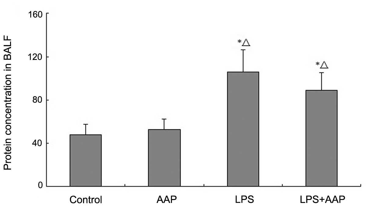
Effects of AAP on protein
concentration in bronchoalveolar lavage fluid (BALF)
To confirm the efficacy of LPS exposure, the protein
concentration in BALF was observed. As shown in Fig. 1, protein concentration in BALF markedly
increased in the LPS group compared with the control and AAP
groups. However, pretreatment with AAP caused the protein
concentration in BALF to decrease compared with the LPS group.
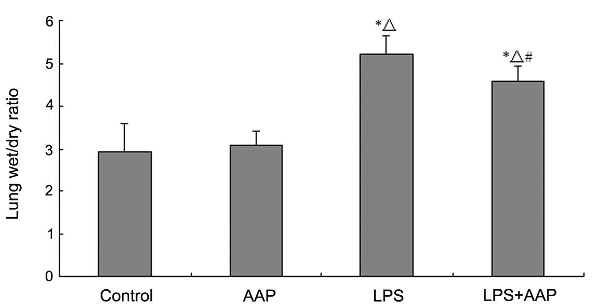
Effects of AAP on lung W/D weight
ratio
To investigate the effect of AAP on LPS-induced lung
edema, W/D weight ratios were detected. As shown in Fig. 2, there were no significant differences
between the control and AAP groups, which indicated that AAP had no
effect on lung edema in normal rats. LPS injected for 12 h caused a
significant increase in the lung W/D weight ratio compared with the
control group (P<0.01). As shown in Fig. 3, in the AAP pretreated group the lung
W/D weight ratios decreased compared with the LPS groups.
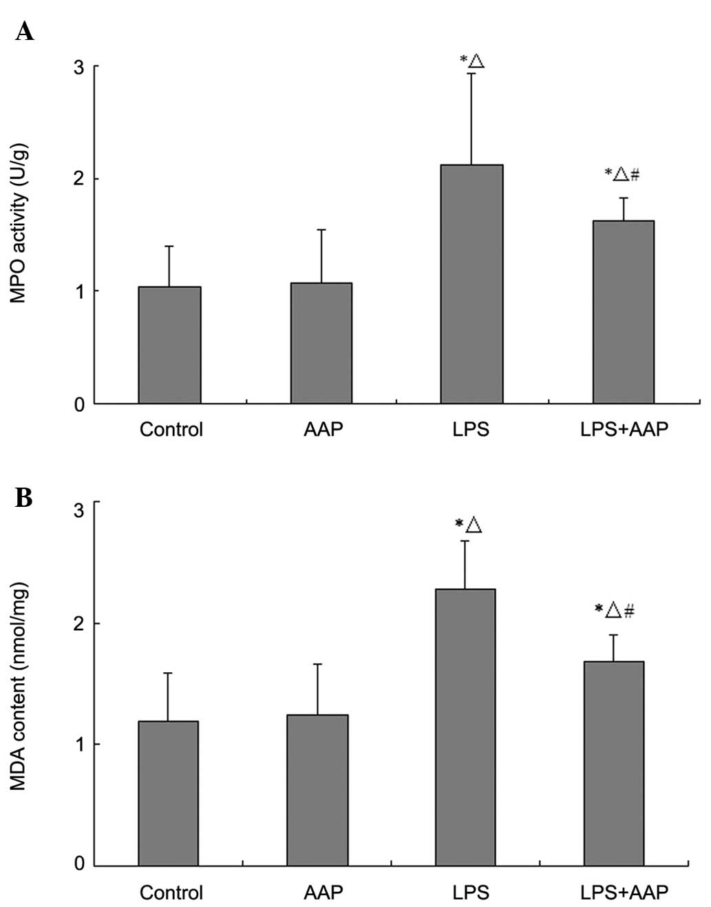
Effect of AAP on MDA level and MPO
activity in lung tissues of LPS-treated rats
To assess the lung neutrophil burden within
pulmonary tissues, lung MPO activity was measured. As shown in
Fig. 3, MPO activity increased
significantly compared with the control and AAP groups, however,
the MPO activity deceased in the LPS plus AAP group. In addition,
LPS induced an increase in the MDA level in lung tissues and AAP
significantly inhibited the MDA level.
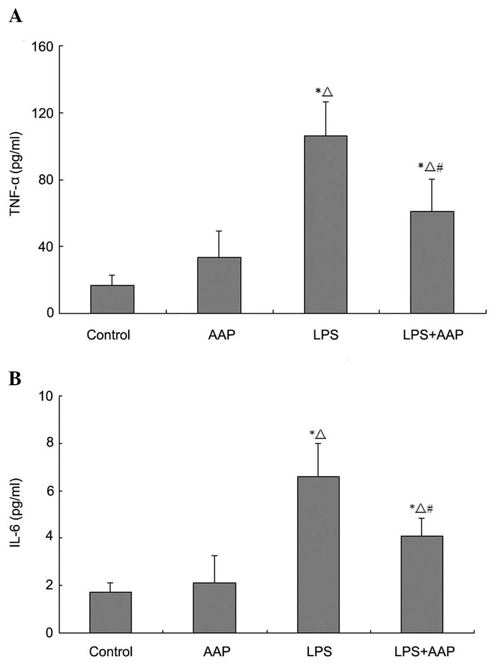
Effect of AAP on TNF-α and IL-6 in the
blood of LPS-treated rats
The concentration of TNF-α and IL-6 in the blood
represents pro-inflammatory mediators, which were thought to play
crucial roles in the development of ALI. As shown in Fig. 4, TNF-α and IL-6 levels increased
markedly in the LPS group compared with the control and AAP groups,
whereas these levels were decreased by AAP pretreatment.
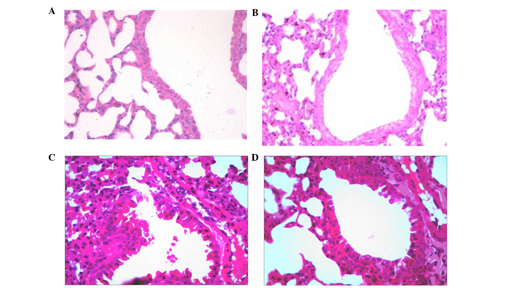
Effect of AAP on LPS-mediated lung
histopathological changes
In order to study the effects of AAP on ALI, the
histological changes were determined following AAP treatment in
LPS-treated rats. As shown in Fig. 5,
in the control and heparin groups, lung tissue showed a normal
structure and clear pulmonary alveoli under a light microscope. The
changes in the LPS group, such as a large number of neutrophil
sequestration and infiltration around the pulmonary vessel and
airway, distributed in the alveolar and interstitial were observed.
The LPS group pretreated with AAP markedly alleviated the
LPS-induced pathological changes of the lung. These results
indicated that AAP could protect rats from LPS-induced lung
damage.
Discussion
ARDS is a life-threatening respiratory failure due
to lung injury from a variety of precipitants (14). Despite advances in supportive
treatment, mainly associated with protective ventilation (15) and a fluid conservative strategy, the
morbidity and mortality of patients remains high (16). The lung pathogenesis of ALI/ARDS is
characterized by the diffuse alveolar damage, alveolar capillary
leakage and protein-rich pulmonary edema leading to the clinical
manifestation of poor lung compliance, severe hypoxemia and
bilateral infiltrates on chest radiograph (17). Neutrophils play a critical role in the
pathogenesis of ALI/ARDS and when activated release harmful
mediators, including cytokines, proteases, reactive oxygen species
and matrix metalloproteinases, leading to further damage (18). Numerous studies have suggested that
oxidative stress is an important mechanism in the pathogenesis of
ARDS (4,5). In a previous study, pretreatment with
scavengers of reactive oxygen species in an animal model has been
shown to significantly reduce the damage, such as pulmonary
hypertension, hypoxia and increased microvascular permeability, to
protein (19).
LPS is a principal component of the outer membrane
of Gram-negative bacteria and can enter the blood stream and elicit
inflammatory responses that may lead to shock and ultimately to
death (20). LPS-induced lung injury
in the rat is frequently used as a model for studying ALI (21). Thus, this model was used in the present
study to investigate the prevention of AAP on LPS-induced ALI in
mice.
AAP has shown a variety of pharmacological
properties. Black fungus polysaccharide has antioxidant (7,8), blood lipid
lowering (9), antitumor (10,11) and
anti-radiation (12) activity. In our
previous study, we proved that AAP has anti-inflammation (22) and antioxidant functions (23). MPO is an enzyme located mainly in the
primary granules of neutrophils and its main function is to kill
microorganisms, but under certain conditions, it produces excess
oxidants leading to tissue damage (24). In the present study, we found that MPO
activity increased significantly following LPS administration. By
contrast, pretreatment of AAP significantly decreased MPO activity
and reduced neutrophil infiltration. MDA is a lipid peroxidation
marker used to assess lipid peroxidation due to increased oxidative
stress (25). The blood levels of MDA
were markedly increased in LPS-induced mice, which could be
significantly reversed by the pretreatment of AAP. The lung W/D
weight ratio was evaluated as an index of pulmonary edema. It was
found that AAP decreased the LPS-induced lung W/D ratio. The
protein concentration in BALF markedly increased in the LPS group.
However, following pretreatment with AAP, the protein concentration
in BALF decreased compared with the LPS group. These results
suggested that AAP has a protective effect on LPS-induced ALI.
Excessive cytokine-mediated inflammation was thought
to play crucial roles in the development of ALI. In the present
study, LPS caused a significant increase in the level of TNF-α and
IL-6 in blood compared with the control group. By contrast, AAP
treatment significantly reduced TNF-α and IL-6 secretion. These
results suggested that the protective effects of AAP on LPS-induced
ALI are partly attributed to inhibition of TNF-α and IL-6
production.
In conclusion, the present study demonstrated that
AAP significantly ameliorated the lung injury induced by LPS in
rats via the inhibition of pro-inflammatory cytokine expression and
antioxidation. These results may provide a theoretical foundation
for treating ALI in the future.
Acknowledgements
The present study was supported by the National
Natural Science Foundation of China (grant no. 81260454) and the
Students Research Training Program of Xinjiang Medical University
(grant no. CX2014021).
References
|
1
|
Frutos-Vivar F, Ferguson ND and Esteban A:
Epidemiology of acute lung injury and acute respiratory distress
syndrome. Semin Respir Crit Care Med. 27:327–336. 2006. View Article : Google Scholar : PubMed/NCBI
|
|
2
|
Matuschak GM and Lechner AJ: Acute lung
injury and the acute respiratory distress syndrome: Pathophysiology
and treatment. Mo Med. 107:252–258. 2010.PubMed/NCBI
|
|
3
|
Kaplan RL, Sahn SA and Petty TL: Incidence
and outcome of the respiratory distress syndrome in gram-negative
sepsis. Arch Intern Med. 139:867–869. 1979. View Article : Google Scholar : PubMed/NCBI
|
|
4
|
Cross CE, Forte T, Stocker R, Louie S,
Yamamoto Y, Ames BN and Frei B: Oxidative stress and abnormal
cholesterol metabolism in patients with adult respiratory distress
syndrome. J Lab Clin Med. 115:396–404. 1990.PubMed/NCBI
|
|
5
|
Sabarirajan J, Vijayaraj P and Nachiappan
V: Induction of acute respiratory distress syndrome in rats by
lipopolysaccharide and its effect on oxidative stress and
antioxidant status in lung. Indian J Biochem Biophys. 47:278–284.
2010.PubMed/NCBI
|
|
6
|
Blackwell TS, Blackwell TR, Holden EP,
Christman BW and Christman JW: In vivo antioxidant treatment
suppresses nuclear factor-kappa B activation and neutrophilic lung
inflammation. J Immunol. 157:1630–1637. 1996.PubMed/NCBI
|
|
7
|
Ma H, Xu X and Feng L: Responses of
antioxidant defenses and membrane damage to drought stress in fruit
bodies of Auricularia auricula-judae. World J Microbiol Biotechnol.
30:119–124. 2014. View Article : Google Scholar : PubMed/NCBI
|
|
8
|
Xu L, Ma Q, Yao XP, et al: The effect of
extract from black funguson liver and small intestine's MDA, GSH-Px
in the septic shock rats. J XinJiang Med Univ. 36:450–455. 2013.(In
Chinese).
|
|
9
|
Han CR and Xu LP: Black fungus
polysaccharide extraction, purification and antihypelipidemic
effect. J Chin Inst Food Sci Technol. 15:54–58. 2007.(In
Chinese).
|
|
10
|
Reza MA, Hossain MA, Lee SJ, Yohannes SB,
Damte D, Rhee MH, Jo WS, Suh JW and Park SC: Dichlormethane extract
of the jelly ear mushroom Auricularia auricula-judae (higher
Basidiomycetes) inhibits tumor cell growth in vitro. Int J Med
Mushrooms. 16:37–47. 2014. View Article : Google Scholar : PubMed/NCBI
|
|
11
|
Cai R, Zhu XS, Qu HY, et al: Extraction of
Black Fungus Polysaccharide and Its Inhibitory Effect on Glioma.
Acta Med Univ Sci Technol Huazhong. 42:578–581. 2013.(In
Chinese).
|
|
12
|
Bai H, Wang Z, Cui J, Yun K, Zhang H, Liu
RH, Fan Z and Cheng C: Synergistic radiation protective effect of
purified Auricularia auricular-judae polysaccharide (AAP IV)
with grape seed procyanidins. Molecules. 19:20675–20694. 2014.
View Article : Google Scholar : PubMed/NCBI
|
|
13
|
Zhang H, Wang ZY, Yang L, Yang X, Wang X
and Zhang Z: In vitro antioxidant activities of sulfated
derivatives of polysaccharides extracted from Auricularia
auricular. Int J Mol Sci. 12:3288–3302. 2011. View Article : Google Scholar : PubMed/NCBI
|
|
14
|
Dushianthan A, Grocott MP, Postle AD and
Cusack R: Acute respiratory distress syndrome and acute lung
injury. Postgrad Med J. 87:612–622. 2011. View Article : Google Scholar : PubMed/NCBI
|
|
15
|
Delong P, Murray JA and Cook CK:
Mechanical ventilation in the management of acute respiratory
distress syndrome. Semin Dial. 19:517–524. 2006. View Article : Google Scholar : PubMed/NCBI
|
|
16
|
Buregeya E, Fowler RA, Talmor DS,
Twagirumugabe T, Kiviri W and Riviello ED: Acute respiratory
distress syndrome in the global context. Glob Heart. 9:289–295.
2014. View Article : Google Scholar : PubMed/NCBI
|
|
17
|
Gattinoni L, Bombino M, Pelosi P, Lissoni
A, Pesenti A, Fumagalli R and Tagliabue M: Lung structure and
function in different stages of severe adult respiratory distress
syndrome. JAMA. 271:1772–1779. 1994. View Article : Google Scholar : PubMed/NCBI
|
|
18
|
Zemans RL, Colgan SP and Downey GP:
Transepithelial migration of neutrophils: Mechanisms and
implications for acute lung injury. Am J Respir Cell Mol Biol.
40:519–535. 2009. View Article : Google Scholar : PubMed/NCBI
|
|
19
|
Weng TI, Wu HY, Kuo CW and Liu SH:
Honokiol rescues sepsis-associated acute lung injury and lethality
via the inhibition of oxidative stress and inflammation. Intensive
Care Med. 37:533–541. 2011. View Article : Google Scholar : PubMed/NCBI
|
|
20
|
Mu E, Ding R, An X, Li X, Chen S and Ma X:
Heparin attenuates lipopolysaccharide-induced acute lung injury by
inhibiting nitric oxide synthase and TGF-β/Smad signaling pathway.
Thromb Res. 129:479–485. 2012. View Article : Google Scholar : PubMed/NCBI
|
|
21
|
Ni YF, Tian F, Lu ZF, Yang GD, Fu HY, Wang
J, Yan XL, Zhao YC, Wang YJ and Jiang T: Protective effect of
nicotine on lipopolysaccharide-induced acute lung injury in mice.
Respiration. 81:39–46. 2011. View Article : Google Scholar : PubMed/NCBI
|
|
22
|
Sun Z, Yao XP, Yu WY, et al: Protective
effect of auricula polysaccharide on liver function of rats with
obstructive jaudice. J Trad Chin Med Pharm. 29:3985–3987. 2014.(In
Chinese).
|
|
23
|
Yao XP, Ma Q, Zhang JL, et al: The effects
of Auricularia Auricula Extract on NF-κB and SOD in Liver of Rats
with Obstructive Jaundice. Res Pract Chin Medicines. 27:35–37.
2013.(In Chinese).
|
|
24
|
Ma Z, Ji W, Fu Q and Ma S: Formononetin
inhibited the inflammation of LPS-induced acute lung injury in mice
associated with induction of PPAR gamma expression. Inflammation.
36:1560–1566. 2013. View Article : Google Scholar : PubMed/NCBI
|
|
25
|
Torun AN, Kulaksizoglu S, Kulaksizoglu M,
Pamuk BO, Isbilen E and Tutuncu NB: Serum total antioxidant status
and lipid peroxidation marker malondialdehyde levels in overt and
subclinical hypothyroidism. Clin Endocrinol (Oxf). 70:469–474.
2009. View Article : Google Scholar : PubMed/NCBI
|