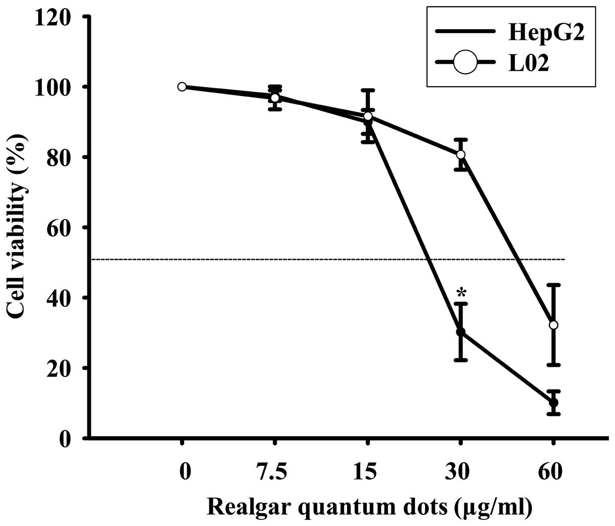
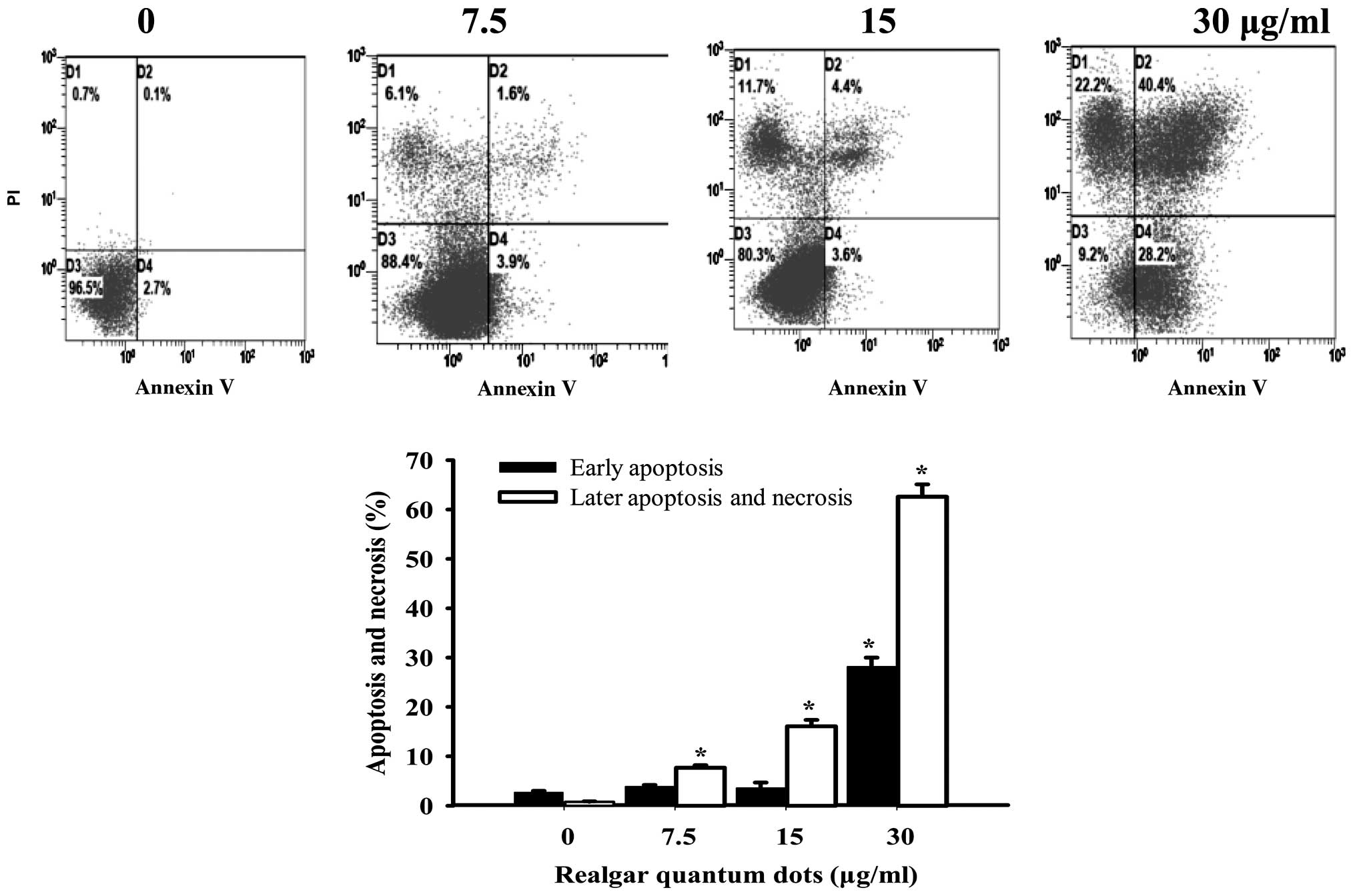
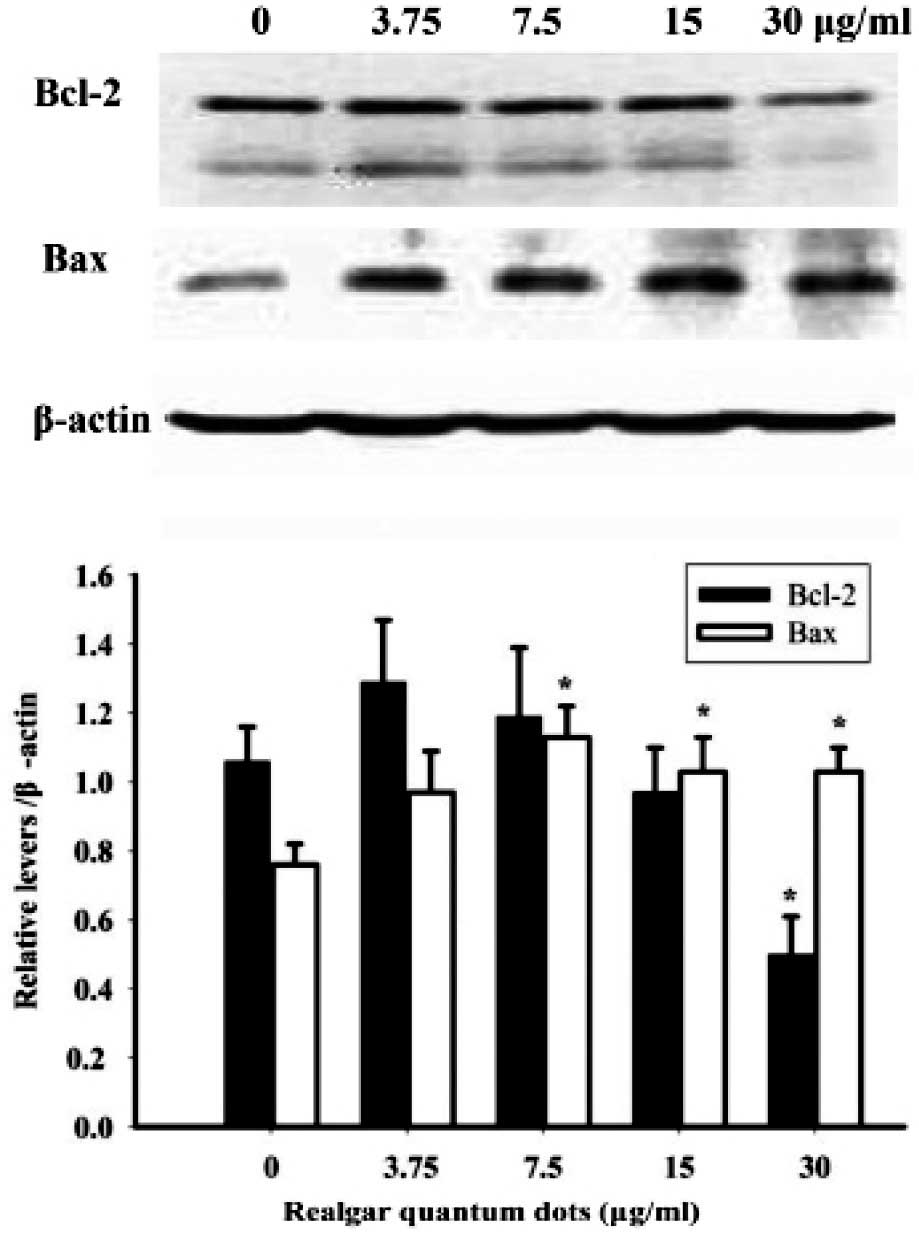
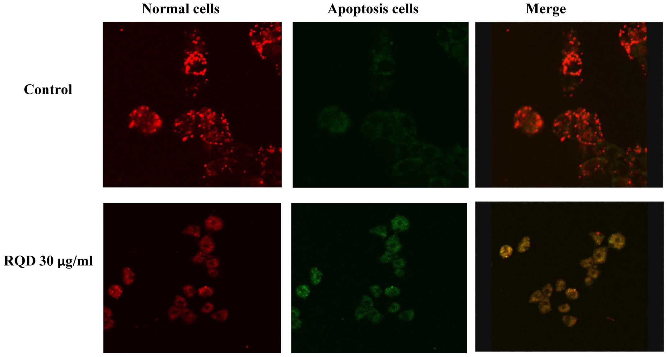
|
1
|
Liu J, Lu Y, Wu Q, Goyer RA and Waalkes
MP: Mineral arsenicals in traditional medicines: Orpiment, realgar
and arsenolite. J Pharmacol Exp Ther. 326:363–368. 2008. View Article : Google Scholar : PubMed/NCBI
|
|
2
|
Hu XM, Liu F and Ma R: Application and
assessment of Chinese arsenic drugs in treating malignant
hematopathy in China. Chin J Integr Med. 16:368–377. 2010.
View Article : Google Scholar : PubMed/NCBI
|
|
3
|
Ding W, Zhang L, Kim S, Tian W, Tong Y,
Liu J, Ma Y and Chen S: Arsenic sulfide as a potential anti cancer
drug. Mol Med Rep. 11:968–974. 2015.PubMed/NCBI
|
|
4
|
Liu X, Li X, Wang L, Lv X, Chen N, Li P,
Lu K and Wang X: Realgar induces apoptosis in the chronic
lymphocytic leukemia cell line MEC 1. Mol Med Rep. 8:1866–1870.
2013.PubMed/NCBI
|
|
5
|
Wang L, Zhou GB, Liu P, Song JH, Liang Y,
Yan XJ, Xu F, Wang BS, Mao JH, Shen ZX, et al: Dissection of
mechanisms of Chinese medicinal formula Realgar-Indigo naturalis as
an effective treatment for promyelocytic leukemia. Proc Natl Acad
Sci USA. 105:4826–4831. 2008. View Article : Google Scholar : PubMed/NCBI
|
|
6
|
Mao JH, Sun XY, Liu JX, Zhang QY, Liu P,
Huang QH, Li KK, Chen Q, Chen Z and Chen SJ: As4S4 targets
RING-type E3 ligase c-CBL to induce degradation of BCR-ABL in
chronic myelogenous leukemia. Proc Natl Acad Sci USA.
107:21683–21688. 2010. View Article : Google Scholar : PubMed/NCBI
|
|
7
|
Zhang QY, Mao JH, Liu P, Huang QH, Lu J,
Xie YY, Weng L, Zhang Y, Chen Q, Chen SJ, et al: A systems biology
understanding of the synergistic effects of arsenic sulfide and
Imatinib in BCR/ABL-associated leukemia. Proc Natl Acad Sci USA.
106:3378–3383. 2009. View Article : Google Scholar : PubMed/NCBI
|
|
8
|
Zhu HH, Wu DP, Jin J, Li JY, Ma J, Wang
JX, Jiang H, Chen SJ and Huang XJ: Oral tetra-arsenic tetra-sulfide
formula versus intravenous arsenic trioxide as first-line treatment
of acute promyelocytic leukemia: A multicenter randomized
controlled trial. J Clin Oncol. 31:4215–4221. 2013. View Article : Google Scholar : PubMed/NCBI
|
|
9
|
Wu J, Shao Y, Liu J, Chen G and Ho PC: The
medicinal use of realgar (As4S4) and its
recent development as an anticancer agent. J Ethnopharmacol.
135:595–602. 2011. View Article : Google Scholar : PubMed/NCBI
|
|
10
|
Baláž P and Sedlák J: Arsenic in cancer
treatment: Challenges for application of realgar nanoparticles (a
minireview). Toxins (Basel). 2:1568–1581. 2010. View Article : Google Scholar : PubMed/NCBI
|
|
11
|
Wu JZ and Ho PC: Evaluation of the in
vitro activity and in vivo bioavailability of realgar nanoparticles
prepared by cryo-grinding. Eur J Pharm Sci. 29:35–44. 2006.
View Article : Google Scholar : PubMed/NCBI
|
|
12
|
Zhao QH, Zhang Y, Liu Y, Wang HL, Shen YY,
Yang WJ and Wen LP: Anticancer effect of realgar nanoparticles on
mouse melanoma skin cancer in vivo via transdermal drug delivery.
Med Oncol. 27:203–212. 2010. View Article : Google Scholar : PubMed/NCBI
|
|
13
|
Tian Y, Wang X, Xi R, Pan W, Jiang S, Li
Z, Zhao Y, Gao G and Liu D: Enhanced antitumor activity of realgar
mediated by milling it to nanosize. Int J Nanomedicine. 9:745–757.
2014.PubMed/NCBI
|
|
14
|
Xie QJ, Cao XL, Bai L, Wu ZR, Ma YP and Li
HY: Anti-tumor effects and apoptosis induction by Realgar
bioleaching solution in Sarcoma-180 cells in vitro and transplanted
tumors in mice in vivo. Asian Pac J Cancer Prev. 15:2883–2888.
2014. View Article : Google Scholar : PubMed/NCBI
|
|
15
|
Wang J, Lin M, Zhang T, Yan Y, Ho PC, Xu
QH and Loh KP: Arsenic(II) sulfide quantum dots prepared by a wet
process from its bulk. J Am Chem Soc. 130:11596–11597. 2008.
View Article : Google Scholar : PubMed/NCBI
|
|
16
|
Subbarayan PR and Ardalan B: In the war
against solid tumors arsenic trioxide needs partners. J
Gastrointest Cancer. 45:363–371. 2014. View Article : Google Scholar : PubMed/NCBI
|
|
17
|
Qin Y, Jing F, Hai J, et al: Anti-tumor
effects of realgar quantum dots and Liushen pills in cervical
tumor-bearing mice. Chin J N Drugs Clin Remedies. 34:64–67.
2015.
|
|
18
|
Wei S, Cao H, Zhou X, Wu H and Yang J:
Prokaryotically and eukaryotically expressed interleukin-24 induces
breast cancer growth suppression via activation of apoptosis and
inhibition of tumor angiogenesis. Mol Med Rep. 11:3673–3681.
2015.PubMed/NCBI
|
|
19
|
Al-Fatlawi AA, Al-Fatlawi AA, Irshad M,
Zafaryab M, Rizvi MM and Ahmad A: Rice bran phytic acid induced
apoptosis through regulation of Bcl-2/Bax and p53 genes in HepG2
human hepatocellular carcinoma cells. Asian Pac J Cancer Prev.
15:3731–3736. 2014. View Article : Google Scholar : PubMed/NCBI
|
|
20
|
Yang HB, Song W, Cheng MD, Fan HF, Gu X,
Qiao Y, Lu X, Yu RH and Chen LY: Deoxycholic acid inhibits the
growth of BGC-823 gastric carcinoma cells via a p53 mediated
pathway. Mol Med Rep. 11:2749–2754. 2015.PubMed/NCBI
|
|
21
|
Il Jung H, Jo MJ, Kim HR, Choi YH and Kim
GD: Extract of Saccharina japonica induces apoptosis companied by
cell cycle arrest and endoplasmic reticulum stress in SK-Hep1 human
hepatocellular carcinoma cells. Asian Pac J Cancer Prev.
15:2993–2999. 2014. View Article : Google Scholar : PubMed/NCBI
|
|
22
|
Zhang B, Peng X, Li G, Xu Y, Xia X and
Wang Q: Oxidative stress is involved in Patulin induced apoptosis
in HEK293 cells. Toxicon. 94:1–7. 2015. View Article : Google Scholar : PubMed/NCBI
|
|
23
|
Verfaillie T, Garg AD and Agostinis P:
Targeting ER stress induced apoptosis and inflammation in cancer.
Cancer Lett. 332:249–264. 2013. View Article : Google Scholar : PubMed/NCBI
|