Introduction
Type 1 diabetes mellitus (T1D) is a chronic
condition in which the pancreas produces little or no insulin due
to autoimmune destruction of insulin-producing β cells in the
islets. As diabetes is caused by the loss of a single cell type,
cellular replacement therapy for diabetes mellitus (DM), especially
T1D, has received much attention. More recently, additional efforts
have been focused on the use of both autologous and allogeneic stem
cells as sources of new islets (1–5).
Bone marrow-derived mesenchymal stem cells (BMSCs)
exhibit considerable developmental plasticity that can be induced
to differentiate into insulin-producing cells in vitro.
Although it is possible to induce β-cell-specific gene expression
in murine and human BMSCs in vitro, insulin secretion of
these differentiated cells is extremely low (2). Findings of previous studies have
demonstrated that rodent BMSCs can adopt insulin-expressing
phenotype (6), drive the
phenotype of human BMSCs by the forced expression of β-cell
transcription factors, and generate cells capable of glucose
responsive insulin secretion (7).
BMSCs transplanted into diabetic mice contribute to endocrine
pancreatic regeneration (7–9).
BMSCs reversed experimental diabetes in vivo by enhancing
the regeneration and survival of endogenous β cells rather than
repopulating the islets with transdifferentiated β cells (10,11).
The potential mechanisms of these beneficial animal
and clinical results from stem cell transplantation remain to be
determined. However, the final destination of the cells and their
fate following their transplantation in vivo has not been
clearly determined. In this study, we used streptozotocin (STZ) to
induce diabetes in miniature pigs, transplanted autologous BMSCs
(ABMSCs) into the pancreas through targeted intervention,
identified them in vivo by MRI scanning and monitored
glycemic status and pancreatic islet β-cell function.
Materials and methods
Experimental animals
Tibetan miniature male pigs (3 months old) were
purchased from the Guangdong Experimental Animal Center (Guangzhou,
China). The animals were housed in a controlled temperature of
22–25°C, with a humidity of 50–70%, as well as air change and
lighting, The animals were fed twice daily according to the Larsen
protocol revised for the requirements of the present study
(commercial miniature pigs mixed fodder 30 g/kg body weight)
(14), and allowed access to
water ad libitum.
Central venous catheter implantation
A dual lumen venous catheter (5F; Arrow-Howes™
Multi-Lumen Catheter; Teleflex, Inc., Limerick, PA, USA) and ear
vein indwelling needle (24G; BD Biosciences, Franklin Lakes, NJ,
USA) were surgically inserted into the internal jugular vein and
ear vein under general anesthesia. All the procedures were
performed in accordance with the principles of Laboratory Animal
Care (National Institutes of Health publication no. 85–23, revised
1985) and the animal protocol was approved by the Sun Yat-sen
University, Institutional Animal Care and Use Committee (IACUC,
Approval ID no.: 2008020102).
T1D model
The T1D model was induced by intravenous
administration of STZ (Sigma-Aldrich, St. Louis, MO, USA) with
sodium citrate as the buffer through indwelling catheters, at a
dose of 120 mg/kg body weight after 24-h fasting in conscious
animals, while the normal control miniature pigs received an
injection of the same amount of citric buffer (30). Seventy two hours after the
injection of STZ, the fasting blood glucose (FBG) was detected in
the subsequent days. Diabetes was confirmed following three
consecutive readings of blood glucose ≥16.7 mmol/l (300 mg/dl).
Animal grouping and treatment
Experimental animals were randomly divided into
three groups with 5 miniature pigs in each group: normal controls
(NC) group without DM, diabetic minipig control group (DMC) and
diabetic minipigs with autologous BMSC transplantation group
(DMAB). DMC and DMAB animals received subcutaneous injection of
Humulin N (Eli Lilly and Company, Indianapolis, IN, USA) after T1D
was confirmed in order to maintain the blood glucose levels of
animals at a the range of 5.0–10.0 mmol/l. After 4 weeks of DM,
miniature pigs in the DMAB group were transplanted with ABMSCs
labeled with superparamagnetic iron oxide (SPIO) (Feridex; Advanced
Magnetics, Inc. Cambridge, MA, USA). Miniature pigs in the DMC
group were transplanted with the same volume of PBS. The experiment
procedure is shown in Fig. 1.
BMSC isolation, culture and labeling
BMSCs of animals were isolated, cultured and labeled
as previously described (31).
Flow cytometry was applied to detect the expression
of the following cell markers: CD34-FITC, CD45-FITC, CD29-FITC and
CD44-FITC (BD Biosciences) (32).
Third passage BMSCs were labeled with a SPIO
nanoparticle contrast agent prior to transplantation. SPIO
suspensions with an iron concentration of 50 mg/l were mixed with
an equal volume of Poly-L-lysine (PLL) (Sigma-Aldrich) at 1.5 mg/ml
for 1 h under gentle agitation. The above ferumoxides-PLL complex
was then added to the culture medium and incubated for 48 h. After
cells were fixed with 4% ethanol and stained with Prussian blue
(PB), light microscopy was performed to assess the iron labeling
efficiency. By way of regular fixation, dehydration, embedding,
ultrathin section (with the section thickness of 50–80 nm) and
staining in turn, cellular ultrastructure and localization of iron
in BMSCs were observed under a transmission electron microscope
(JEM-1200EX; JEOL Ltd., Tokyo, Japan).
Transplantation of ABMSCs
Transplantation was carried out after 4 weeks of
diabetes in the DMAB group (33).
Animals underwent 12-h fasting. Following successful general
anesthesia, the animals were fixed to the operating table of
digital subtraction angiography (DSA) in the supine position. The
right femoral artery was punctured and a micro catheter was
catheterized into the dorsal pancreatic artery. The ABMSCs
according to their body weight (5×10⁵ cells/kg) were
directly injected into the dorsal pancreatic artery (11,34).
In vivo tracing of ABMSCs following
transplantation
The animals were fixed in an experimental special
holder and anesthetized as described above. Magnetic resonance
imaging (MRI) (3.0T MRI system) was carried out in different
sequences 1 week prior to transplantation, and 3 and 6 weeks after
transplantation to observe the local signal change of the pancreas
(35,36).
Intravenous glucose tolerance testing
(IVGTT)
IVGTTs were carried out prior to establishment of
the DM model, transplantation of ABMSCs and 6 weeks after
transplantation. Miniature pigs were fasted overnight (12 h),
weighed and injected intravenously with a bolus of 50% glucose (0.5
g/kg of body weight) at ear margin venipuncture. Blood was then
drawn from an indwelling needle puncture of the external jugular
vein at 0, 3, 5, 7, 10, 20, 30, 60 and 120 min after the glucose
challenge.
Oral glucose tolerance testing
(OGTT)
At the second day of the IVGTT, miniature pigs were
fasted overnight (12 h), weighed and administered glucose (2 g/kg
weight) and 25 g mixed feed (dissolved in 100 ml water). Blood was
then drawn from an indwelling needle puncture of the external
jugular vein at 0, 30, 60, 120 and 180 min after the glucose
challenge.
Measurement of biochemical indices
Blood samples were obtained from an indwelling
needle puncture of the external jugular vein and measured for blood
glucose (glucose oxidase method). Serum insulin (PI-12K porcine
nsulin RIA kit; Millipore Corp., St. Charles, MO, USA) and
C-peptide (PCP-22K Porcine C-peptide RIA kit; Millipore Corp.) was
quantified by porcine insulin and the C-peptide assay (RIA)
kit.
Histology and immunohistochemistry
Paraffin-embedded pancreatic samples were reviewed
histologically using hematoxylin and eosin staining. PB staining
was performed to determine intracellular iron using Perls’ reaction
method.
Specimens were then deparaffinized with xylene and
rehydrated. To block non-specific staining, the sections were
incubated in a buffer containing 5% goat serum for 30 min at 37°C.
Primary monoclonal antibody of mouse anti-human insulin, glucagon
and factor VIII-R (OriGene Technologies, Inc., Beijing, China) were
used at dilutions of 1:400, 1:300 and 1:300, respectively, and
incubated overnight at 4°C. IgG-conjugated horseradish peroxidase
(HRP) and 3,3′-diaminobenzidine tetrahydrochloride (DAB) (Vector
Laboratories, Burlingame, CA, USA) were employed to visualize
antibody binding.
Statistical analysis
Statistical data were processed by SPSS 12.0
software. Data with normal distribution were expressed as mean ±
SD. One-way analysis of variance (ANOVA) or the unpaired Student’s
t-test was performed to determine statistical difference, while the
χ2 test was used for qualitative data. Two-way analysis
of variance (ANOVA) was used to determine the effects of treatement
and period of time, followed by Bonferroni group comparisons.
P<0.05 was considered to indicate statistical significance.
Results
Hyperglycemia was controlled and insulin
use was reduced following transplantation of ABMSCs
No significant difference was observed in blood
glucose during 1–4 weeks after establishment of the type 1 diabetes
model between the DMC and DMAB groups (Fig. 1A). However, the FBG level was
gradually reduced, and maintained between 2.3 and 7.5 mmol/l after
ABMSC transplantation (Fig. 2A).
Moreover, the insulin dose was gradually decreased and discontinued
during the 17–28th day post-operation (Fig. 2B).
β-cell function was partially restored in
the DMAB group
IVGTT showed that there was no significance among
the three groups in terms of blood glucose, serum insulin and
C-peptide level at the baseline (Fig.
3A–C). The blood glucose, serum insulin and C-peptide levels in
the DMC and DMAB groups at the time point of pre-ABMSCs were in
line with the characteristics of T1D (Fig. 3D–F). Insulin and C-peptide curves
showed a flat form. At the end-point of our experiment, there was
no obvious change of blood glucose, serum insulin and C-peptide
level in the NC and DMC groups compared to those prior to ABMSC
transplantation (Fig. 3G–I). The
peak of blood glucose in the DMAB group was significantly reduced,
and the curve was close to the normal glucose tolerance (NGT) curve
(Fig. 3G). Insulin and C-peptide
secretion levels prior to and after glucose load were significantly
higher than those of DMC group, which presented a small secretion
peak between 3 and 10 min, and was gradually reduced to basic level
(Fig. 3H and I).
For the area under the curve (AUC) of blood glucose,
serum insulin and C-peptide level in IVGTT, there was no
significance among the three groups at the baseline (Fig. 3J–L). Prior to ABMSC
transplantation, AUC of blood glucose in the DMC and DMAB groups
was significantly greater than that of the NC group (Fig. 3J), whereas AUC of serum insulin
and C-peptide in the DMC and DMAB groups was significantly less
than those of the NC group, respectively (Fig. 3K and L). There was no significant
difference of AUC between the DMC and DMAB groups (Fig. 3K and L). At the end-point of our
experiment, AUC of blood glucose in the DMAB group was less than
that prior to ABMSC transplantation, although it was not
significantly different compared to that of the baseline level
(Fig. 3J). The AUCs of insulin
and C-peptide in the DMAB group were significantly more than those
of the DMC group (Fig. 3K and L).
OGTT showed a similar trend to IVGTT for the three groups (Fig. 4A–D).
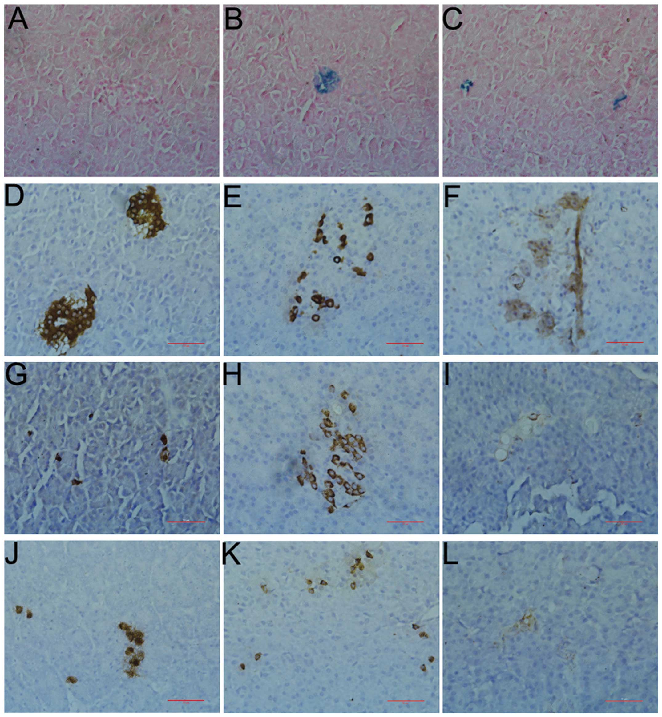
PB staining was positive in pancreas with
transplanted ABMSCs
The positive PB staining cells in the pancreas of
miniature pigs showed that the islet and pancreatic ductal
epithelial cells derived from the SPIO-labeled BMSCs. The cells
were positive in the DMAB group (Fig.
5B–C) but not in the NC group (Fig. 5A), suggesting that SPIO-labeled
BMSCs were implanted in the islet following transplantation,
differentiated into islet cell or formed new islet, or implanted in
exocrine pancreas and differentiated into pancreatic ductal
epithelial cells.
Small islets were regenerated in pancreas
after transplantation of ABMSCs
As shown in Fig.
5D–F, there was an abundance of islets; the boundary was clear;
the cell mass shape was circular, oval, or irregular, and present
in alignment, with plump cytoplasm, and a circular nucleus in the
NC groups. Capillary was rich around the islet or penetrating the
internal islet. By contrast, the islets were scarce, the boundary
was not clear, and the contour was incomplete or collapsed in the
DMC groups. Additionally, the cells were arranged in a disorderly
manner, the nuclear size and shape were irregular, and some of the
islet cells appeared to have vacuolar degeneration, i.e., few β
cells remained while the α cells became the main pancreatic islet
cells. Capillaries were significantly reduced and unevenly
distributed (Fig. 5G–I).
New small islets were identified in the majority of
islets that were distributed in different parts of the pancreas in
the DMAB groups. The new islets and survival islets coexisted. The
size and number of the new islets were obviously fewer than those
of the NC groups. The contour of the cells was regular, and cells
were distributed uniformly, with the cytoplasm being much richer
and light dyed lighter compared with the NC groups. The nuclear
size and form were normal. There were more β cells present as
compared with α cells, and capillaries were more abundant than
those of the DMC group (Fig.
5J–L).
Successful real-time tracing of
transplanted ABMSCs by MRI
At 1 week before transplantation, 3 and 6 weeks
after transplantation, there was no significant change among MRI
TFL T1WI signals of the pancreases in the DMAB group
(Fig. 6A–C). However, the
sporadic mottled low-signal area emerged distinctly in the
pancreases of DMAB animals in MRI TFL T2WI at 1 week
prior to transplantation, and 3 and 6 weeks after ABMSC
transplantation (Fig. 6D–F).
PB staining of BMSCs
SPIO-labeled BMSCs showed numerous blue particles in
the cytoplasm of the labeled cells and nano-iron particles near the
nucleus (Fig. 7A and B).
Discussion
T1D is obtained from the specific autoimmune
reaction of β cells induced by a variety of pathogenic factors,
with 20–30% of viable β cells prior to onset of typical clinical
symptoms. However, autoimmune reaction begins to accelerate after
typical clinical manifestations, similar to β-cell damage (12). Thus protection of the remaining β
cells and promotion of intrinsic β-cell regeneration and new islet
cell formation has become the focus of study in T1D.
Islet function is restored following
ABMSC transplantation
A deep vein path was established in order for OGTT
and IVGTT to be evaluated repeatedly (13–15). Three different time-points were
selected, including the baseline before T1D model, and time-points
pre- and post-ABMSCs. The results showed that the OGTT and IVGTT
glucose curve was significantly lower than that in the DMC group,
albeit similar to that of the NC group at the end-point of the
experiments. Moreover, there was improvement in the basal and
post-glucose-challenge insulin and C-peptide secretion in the DMAB
group, with the insulin and C-peptide secretion peaks evident 3–10
min after intravenous injection of 50% glucose, which suggests that
the initial phase insulin release of β cells in the DMAB group was
significantly improved. In addition, the exogenous insulin use
gradually decreased after operation until the discontinuation of
insulin in the DMAB group, with the FBG being maintained at normal
and relatively stable levels. The results of this study indicate
that ABMSCs can effectively restore the islet, improve β-cell
function and glucose tolerance, and reverse the hyperglycemic
conditions in early STZ-induced T1D.
Results of previous animal studies have shown that
peripheral vein infusion or pancreatic local injection of
autologous HSC or BMSCs can improve the islet function of mice
(16,17), and miniature pigs (18), and reverse hyperglycemic
conditions 15–30 days after transplantation. A follow-up clinical
trial lasting almost 5 years (average 29.8 months) showed that
peripheral venous transplantation of autologous HSC joint with
immune intervention therapy can improve the islet function of new
developed T1D patients; 20 cases (23 cases in total) stopped
exogenous insulin treatment, and 12 patients had discontinued
insulin for ≥31 months (19,20). The abovementioned data suggest
that stem cell transplantation is expected to become an effective
means for the early treatment of T1D. However, the abovementioned
studies have certain limitations such as non-autologous cell
transplantation, lack of different time window of cell
transplantation, non-directional pancreatic transplantation, more
severe trauma, or lack of standardization and quantitation in
grafts (21,22). In the present study, we
transplanted autologous and quantitative BMSCs into pancreas by
super microcatheter targeting into pancreas dorsal artery in the
early stage T1D pigs, which has solved problems including time
window selection, optimization of transplantation procedure, graft
standardization and quantification in the treatment of T1D with
autologous stem cell transplantation.
In vivo tracing of transplanted
autologous cells
SPIO is a negative contrast agent used in magnetic
resonance and has been approved for clinical diagnosis by FDA. It
is identified by the reticuloendothelial system after intravenous
injection, and is absorbed by phagocytes. The corresponding
regional signal is reduced following phagocytosis of SPIO
particles. In this study, we have established a safe, real-time,
dynamic, and non-invasive MRI tracing technique in T1D miniature
pigs. We observed uneven pancreas signal in T2WI at
post-operative 3 and 6 weeks, an obvious scattered low-signal area,
and local low-signal pancreas presented s gradually decreasing
trend with the passage of time. Thus MRI may be used for short-term
tracing and observing the distribution and survival of SPIO-labeled
ABMSCs in the pancreas of miniature pigs.
SPIO-labeled islet is clearly observed by MRI
imaging 18 weeks after transplantation (23). The 1.5T MRI system shows the body
distribution of SPIO-labeled BMSCs transplanted into brain
(24), and heart (25) in rats, mice and miniature pigs.
The real-time tracing time of SPIO-labeled BMSCs is relatively
shorter compared to that of real-time islet transplantation
tracing; however, it may still be used for short-term tracing in
the clinic.
The role of ABMSCs in islet repairing and
remodeling
Immunohistochemistry of pancreas showed that PB
staining of some islet and pancreatic ductal epithelial cells was
positive, while insulin, glucagon and vascular endothelial VIII
factor staining were visible. The islets were mainly new small
islets in the DMAB group, which coexisted with survival islets. The
area of new islets and cell number were significantly less than
those of normal islets. The majority of cells in the new islets
were β cells, with α cells a minority. This result suggests that
directional transplanted ABMSCs may be implanted in the pancreas
and differentiated into islet and pancreatic ductal epithelial
cells in miniature pigs. However, whether ABMSCs restore islets
hyperplasia by promoting pancreatic inherent stem cell
differentiation into islet cells, self-replicate into inherent
islet cells in a paracrine manner should be investigated.
BMSCs are capable of repairing and remodeling the
islets by promoting intrinsic islet cell replication (26) and the formation of new islets in
type 1 diabetic mice (10,27).
Transplanted BMSCs differentiate into endothelial cells, improve
local microcirculation (28) and
promote the repair of diabetic neuropathy by secreting vascular
endothelial growth factor (VEGF) and basic fibroblast growth factor
(bFGF) in in a paracrine manner (28,29).
In summary, quantitative ABMSC transplantation
targeted into the pancreas can effectively improve the islet
function of miniature pigs with early T1D, reverse high blood
glucose state, discontinue exogenous insulin therapy in the short
term and maintain normal blood glucose. MRI scanning can be used
for dynamic, non-invasive and real-time tracing of SPIO-labeled
ABMSCs in the short term after transplantation.
Acknowledgements
This study was funded by grants from the National
Science Fund for Distinguished Young Scholars (81025005), China
National Natural Science Foundation Projects for Young Scientists
(No. 81100520), 5010 project of Sun Yat-sen University (2007030)
and the Natural Science Foundation of Shandong Province Government
(No. Y2007C036). We would like to thank Professor Wenhua Ling from
the School of Public Health, Sun Yat-sen University for providing
his cell laboratory for this study.
References
|
1
|
Haller MJ, Viener HL, Wasserfall C, Brusko
T, Atkinson MA and Schatz DA: Autologous umbilical cord blood
infusion for type 1 diabetes. Exp Hematol. 36:710–715. 2008.
View Article : Google Scholar : PubMed/NCBI
|
|
2
|
Limbert C, Päth G, Jakob F and Seufert J:
Beta-cell replacement and regeneration: strategies of cell-based
therapy for type 1 diabetes mellitus. Diabetes Res Clin Pract.
79:389–399. 2008. View Article : Google Scholar : PubMed/NCBI
|
|
3
|
Hussain MA and Theise ND: Stem-cell
therapy for diabetes mellitus. Lancet. 364:203–205. 2004.
View Article : Google Scholar
|
|
4
|
Jurewicz M, Yang S, Augello A, et al:
Congenic mesenchymal stem cell therapy reverses hyperglycemia in
experimental type 1 diabetes. Diabetes. 59:3139–3147. 2010.
View Article : Google Scholar : PubMed/NCBI
|
|
5
|
Zhao Y, Jiang Z, Zhao T, et al: Reversal
of type 1 diabetes via islet β cell regeneration following immune
modulation by cord blood-derived multipotent stem cells. BMC Med.
10:32012.
|
|
6
|
Tang DQ, Cao LZ, Burkhardt BR, Xia CQ,
Litherland SA, Atkinson MA and Yang LJ: In vivo and in vitro
characterization of insulin-producing cells obtained from murine
bone marrow. Diabetes. 53:1721–1732. 2004. View Article : Google Scholar : PubMed/NCBI
|
|
7
|
Ianus A, Holz GG, Theise ND and Hussain
MA: In vivo derivation of glucose-competent pancreatic endocrine
cells from bone marrow without evidence of cell fusion. J Clin
Invest. 111:843–850. 2003. View Article : Google Scholar : PubMed/NCBI
|
|
8
|
Lee RH, Seo MJ, Reger RL, Spees JL, Pulin
AA, Olson SD and Prockop DJ: Multipotent stromal cells from human
marrow home to and promote repair of pancreatic islets and renal
glomeruli in diabetic NOD/scid mice. Proc Natl Acad Sci USA.
103:17438–17443. 2006. View Article : Google Scholar : PubMed/NCBI
|
|
9
|
Gao X, Song L, Shen K, Wang H, Niu W and
Qin X: Transplantation of bone marrow derived cells promotes
pancreatic islet repair in diabetic mice. Biochem Biophys Res
Commun. 371:132–137. 2008. View Article : Google Scholar : PubMed/NCBI
|
|
10
|
Hasegawa Y, Ogihara T, Yamada T, et al:
Bone marrow (BM) transplantation promotes beta-cell regeneration
after acute injury through BM cell mobilization. Endocrinology.
148:2006–2015. 2007. View Article : Google Scholar : PubMed/NCBI
|
|
11
|
Urbán VS, Kiss J, Kovács J, Gócza E, Vas
V, Monostori E and Uher F: Mesenchymal stem cells cooperate with
bone marrow cells in therapy of diabetes. Stem Cells. 26:244–253.
2008.PubMed/NCBI
|
|
12
|
Couri CE and Voltarelli JC: Autologous
stem cell transplantation for early type 1 diabetes mellitus.
Autoimmunity. 41:666–672. 2008. View Article : Google Scholar : PubMed/NCBI
|
|
13
|
Larsen MO, Wilken M, Gotfredsen CF, Carr
RD, Svendsen O and Rolin B: Mild streptozotocin diabetes in the
Göttingen minipig. A novel model of moderate insulin deficiency and
diabetes. Am J Physiol Endocrinol Metab. 282:E1342–E1351. 2002.
|
|
14
|
Larsen MO, Rolin B, Wilken M, Carr RD and
Gotfredsen CF: Measurements of insulin secretory capacity and
glucose tolerance to predict pancreatic beta-cell mass in vivo in
the nicotinamide/streptozotocin Göttingen minipig, a model of
moderate insulin deficiency and diabetes. Diabetes. 52:118–123.
2003.PubMed/NCBI
|
|
15
|
Hara H, Lin YJ, Zhu X, et al: Safe
induction of diabetes by high-dose streptozotocin in pigs.
Pancreas. 36:31–38. 2008. View Article : Google Scholar : PubMed/NCBI
|
|
16
|
Wen Y, Ouyang J, Yang R, Chen J, Liu Y,
Zhou X and Burt RK: Reversal of new-onset type 1 diabetes in mice
by syngeneic bone marrow transplantation. Biochem Biophys Res
Commun. 374:282–287. 2008. View Article : Google Scholar : PubMed/NCBI
|
|
17
|
Ezquer FE, Ezquer ME, Parrau DB, Carpio D,
Yañez AJ and Conget PA: Systemic administration of multipotent
mesenchymal stromal cells reverts hyperglycemia and prevents
nephropathy in type 1 diabetic mice. Biol Blood Marrow Transplant.
14:631–640. 2008. View Article : Google Scholar : PubMed/NCBI
|
|
18
|
Chang C, Niu D, Zhou H, Zhang Y, Li F and
Gong F: Mesenchymal stroma cells improve hyperglycemia and insulin
deficiency in the diabetic porcine pancreatic microenvironment.
Cytotherapy. 10:796–805. 2008. View Article : Google Scholar : PubMed/NCBI
|
|
19
|
Olmos PR and Borzone G: Autologous
nonmyeloablative hematopoietic stem cell transplantation in newly
diagnosed type 1 diabetes mellitus. JAMA. 302:624author reply
624–625. 2009. View Article : Google Scholar
|
|
20
|
Couri CE, Oliveira MC, Stracieri AB, et
al: C-peptide levels and insulin independence following autologous
nonmyeloablative hematopoietic stem cell transplantation in newly
diagnosed type 1 diabetes mellitus. JAMA. 301:1573–1579. 2009.
View Article : Google Scholar
|
|
21
|
Scolding N, Marks D and Rice C: Autologous
mesenchymal bone marrow stem cells: practical considerations. J
Neurol Sci. 265:111–115. 2008. View Article : Google Scholar : PubMed/NCBI
|
|
22
|
Yamout B, Hourani R, Salti H, et al: Bone
marrow mesenchymal stem cell transplantation in patients with
multiple sclerosis: a pilot study. J Neuroimmunol. 227:185–189.
2010. View Article : Google Scholar : PubMed/NCBI
|
|
23
|
Jirák D1, Kríz J, Herynek V, et al: MRI of
transplanted pancreatic islets. Magn Reson Med. 52:1228–1233.
2004.
|
|
24
|
Neri M, Maderna C, Cavazzin C, et al:
Efficient in vitro labeling of human neural precursor cells with
superparamagnetic iron oxide particles: relevance for in vivo cell
tracking. Stem Cells. 26:505–516. 2008. View Article : Google Scholar : PubMed/NCBI
|
|
25
|
Amsalem Y, Mardor Y, Feinberg MS, et al:
Iron-oxide labeling and outcome of transplanted mesenchymal stem
cells in the infarcted myocardium. Circulation. 116(Suppl 11):
I38–I45. 2007. View Article : Google Scholar : PubMed/NCBI
|
|
26
|
Murray HE, Paget MB, Bailey CJ and Downing
R: Sustained insulin secretory response in human islets co-cultured
with pancreatic duct-derived epithelial cells within a rotational
cell culture system. Diabetologia. 52:477–485. 2009. View Article : Google Scholar
|
|
27
|
Lipsett M, Aikin R, Castellarin M, Hanley
S, Jamal AM, Laganiere S and Rosenberg L: Islet neogenesis: a
potential therapeutic tool in type 1 diabetes. Int J Biochem Cell
Biol. 38:498–503. 2006. View Article : Google Scholar : PubMed/NCBI
|
|
28
|
Jeong JO, Kim MO, Kim H, et al: Dual
angiogenic and neurotrophic effects of bone marrow-derived
endothelial progenitor cells on diabetic neuropathy. Circulation.
119:699–708. 2009. View Article : Google Scholar : PubMed/NCBI
|
|
29
|
Shibata T, Naruse K, Kamiya H, et al:
Transplantation of bone marrow-derived mesenchymal stem cells
improves diabetic polyneuropathy in rats. Diabetes. 57:3099–3107.
2008. View Article : Google Scholar : PubMed/NCBI
|
|
30
|
Kin T, Korbutt GS, Kobayashi T, Dufour JM
and Rajotte RV: Reversal of diabetes in pancreatectomized pigs
after transplantation of neonatal porcine islets. Diabetes.
54:1032–1039. 2005. View Article : Google Scholar
|
|
31
|
Tang KX, Yan JH, Shen YF, et al: Tracing
type 1 diabetic Tibet miniature pig’s bone marrow mesenchymal stem
cells in vitro by magnetic resonance imaging. J Diabetes.
6:123–131. 2014.PubMed/NCBI
|
|
32
|
Layton DS, Strom AD, O’Neil TE, et al:
Development of an anti-porcine CD34 monoclonal antibody that
identifies hematopoietic stem cells. Exp Hematol. 35:171–178. 2007.
View Article : Google Scholar : PubMed/NCBI
|
|
33
|
Nir T, Melton DA and Dor Y: Recovery from
diabetes in mice by beta cell regeneration. J Clin Invest.
117:2553–2561. 2007. View
Article : Google Scholar : PubMed/NCBI
|
|
34
|
Alvarez SS, Jimenez LM, Murillo AZ, et al:
A new approach for bone marrow-derived stem cells intrapancreatic
autotransplantation in diabetic rats. Microsurgery. 26:539–542.
2006. View Article : Google Scholar : PubMed/NCBI
|
|
35
|
Frank JA, Miller BR, Arbab AS, et al:
Clinically applicable labeling of mammalian and stem cells by
combining superparamagnetic iron oxides and transfection agents.
Radiology. 228:480–487. 2003. View Article : Google Scholar
|
|
36
|
Barnett BP, Arepally A, Karmarkar PV, et
al: Magnetic resonance-guided, real-time targeted delivery and
imaging of magnetocapsules immunoprotecting pancreatic islet cells.
Nat Med. 13:986–991. 2007. View
Article : Google Scholar
|