Introduction
Intrahepatic cholangiocarcinoma (ICC) has become the
second most common primary liver cancer, representing 10–25% of
cases, with poor responsiveness to existing drug therapies
(1). Liver transplantation is
useful in the treatment of cholangiocarcinoma. However, recurrence
of the disease is common due to the unique biological
characteristics involved, such as cholangiocyte differentiation and
abundant stromal desmoplasia. Due to the lack of an early
diagnosis, most patients are not eligible for surgical resection
(2–3). Therefore, novel treatment strategies
against ICC are needed to improve survival, particularly in
high-risk subgroups.
Although many frequently mutated genes have been
identified in cholangiocarcinoma, such as TP53 (37–44%) and
KRAS (17–54%) (4), none of
these signature genes have become targets of therapy. Sequencing
efforts are continuously conducted in order to generate in-depth
information with regard to the somatic alterations in ICC. Receptor
tyrosine kinases (RTKs), the important mediators of extracellular
signals, regulate key cell growth, survival, and motility pathways.
In various types of cancer, dysregulated RTK activation was found
in the process of initiation and progression. Recently, the
oncogenic mutations of the orphan RTK c-ros oncogene (ROS) fusion
genes was found in almost 9% of cholangiocarcinoma patients
(5). Several ROS kinase fusion
proteins have been identified, including the
fused-in-glioblastoma-ROS1 (FIG-ROS), SLC34A2-ROS1 (SLC-ROS),
CD74-ROS1, EZR-ROS1, LRIG3-ROS1, SDC4-ROS1, and TPM3-ROS1 (5). FIG-ROS was first identified in a
human glioblastoma cell line (6)
and more recently in patients with ICC (5). In animal models, FIG-ROS has been
validated as a potent oncoprotein in ICC (7). In clinical application, anaplastic
lymphoma kinase (ALK) kinase is mostly homologous with ROS. Phase
I/II clinical trials have focused on the ALK inhibitor crizotinib
for its efficacy in ROS1-driven lung cancer patients, leading to
its approval by the Food and Drug Administration (FDA) (8). Thus, ROS kinase fusion proteins
present a potential and promising drug target for patients with
ICC. However, few studies have demonstrated the effects and precise
molecular mechanisms of FIG-ROS underlying ICC.
The aim of this study was to investigate the role of
FIG-ROS in ICC via different serial shRNA sequence transfections.
Although FIG shRNA transfection showed a marginal effect on HUCCT1
cells, the co-transfection of ROS and FIG shRNA exhibited a
stronger effect on HUCCT1 cell proliferation, apoptosis, cell cycle
progression, migration and invasion compared to ROS shRNA treated
alone. Thus, we confirmed that FIG-ROS serves as a potent
oncoprotein in ICC and that ROS1-6290 and FIG-363 segments may
serve as therapeutic targets for ICC harboring ROS1 fusion
proteins.
Materials and methods
Tissue specimen collection
Study protocols were approved by the Ethics
Committee of the Third Xiangya Hospital, Central South University
(Hunan, China). Four ICC tissues and three normal tissues were
obtained at the Department of General Surgery of the Third Xiangya
Hospital of Central South University. Informed consent was obtained
from patients. Tissues were immediately frozen in liquid nitrogen
following surgical removal.
Immunohistochemistry
Tissues were fixed in formalin, sectioned and
mounted on poly-l-lysine-coated glass slides. Paraffin sections
were deparaffinized, and incubated in antigen retrieval buffer for
2 min at 95°C and then for 10 min at room temperature. The sections
were then treated in 3% hydrogen peroxide for 5 min. Non-specific
antibody binding was blocked with 5% BSA in TBST. The sections were
treated with mouse anti-ROS1 monoclonal antibody (Abcam, Cambridge,
UK) overnight at 4°C in PBS, rinsed, and subsequently incubated for
1 h with biotinylated HRP-conjugated goat anti-mouse secondary
antibody (Abcam), followed by the avidin-biotin complex (Dako,
Copenhagen, Denmark). The sections were developed with DAB,
counterstained with hematoxylin, and examined under a microscope
(DM1750M; Leica, Solms, Germany) to assess the
immunoreactivity.
Cell lines and cell culture
Human ICC cell lines, HUCCT1, RBC, and QBC939, were
purchased from ATCC. Cells were cultured in DMEM and 10% fetal
bovine serum (FBS) was added at 37°C in a humidified incubator
containing 5% CO2.
Plasmid construction and
transfection
The plasmids pGPU6/GFP/Neo-ROS1-homo-6191,
pGPU6/GFP/Neo-ROS1-homo-6290, pGPU6/GFP/Neo-ROS1-homo-6443,
pGPU6/GFP/Neo-ROS1-homo-6976, pGPU6/GFP/Neo-FIG-homo-363,
pGPU6/GFP/Neo-FIG-homo-475, pGPU6/GFP/Neo-FIG-homo-504,
pGPU6/GFP/Neo-FIG-homo-675 were purchased from GenePharma
(Shanghai, China). The plasmid pGPU6/GFP/Neo-shNC (GenePharma) was
used as a negative control (NC). The targeting sequences of each
shRNA are shown in Table I.
HUCCT1 cells were transfected with these plasmids, respectively,
using Lipofectamine 2000 (Invitrogen Life Technologies, Shanghai,
China) according to the manufacturer’s instructions. Subsequently,
the cells were incubated at 37°C with 5% CO2 for 72 h
using MTT assay.
 | Table ITarget sequence of shRNA. |
Table I
Target sequence of shRNA.
| shRNA name | Target sequence |
|---|
| shNC |
5′-GTTCTCCGAACGTGTCACGT-3′ |
| ROS1-homo-6290 |
5′-GAGGAGACCTTCTTACTTAT-3′ |
| ROS1-homo-6443 |
5′-GCTAGAAATTGCCTTGTTTCC-3′ |
| ROS1-homo-6976 |
5′-GCCAGTTGCTTTAATGGAAAC-3′ |
| ROS1-homo-6191 |
5′-GCACATCTGATGAGCAAATTT-3′ |
| FIG-homo-504 |
5′-GCCCAGTCTGTGTCTCAAATC-3′ |
| FIG-homo-475 |
5′-GCTCCTGCTTTGCACAGCTTT-3′ |
| FIG-homo-363 |
5′-CTGGAGAAGGAGTTCGACAAA-3′ |
| FIG-homo-675 |
5′-GCTGACTCTGGTACCATTAAG-3′ |
Western blotting
Tissues or cells were solubilized in cold RIPA lysis
buffer. Proteins were separated with 12% SDS-PAGE, and transferred
onto a polyvinylidene difluoride (PVDF) membrane. The membrane was
incubated with TBST containing 5% skimmed milk at 37°C for 2 h. The
membrane was then incubated with rabbit anti-ROS, rabbit anti-FIG,
and mouse anti-GAPDH primary antibodies (all from Santa Cruz
Biotechnology, Inc., Santa Cruz, USA), respectively, at room
temperature for 2 h. After washing with PBST 4 times for 10 min
each time, the membrane was incubated with the goat anti-rabbit and
goat anti-mouse secondary antibodies (Santa Cruz Biotechnology,
Inc.) at 4°C overnight. After washing with PBST 4 times for 10 min
each time, an ECL kit (Pierce Chemical, Rockford, IL, USA) was used
to perform chemiluminent detection. Image-Pro plus software 6.0 was
used to analyze the relative protein expression, represented as the
density ratio versus GAPDH. GAPDH was used as an internal
reference.
Cell proliferation assay
MTT assay was used to measure cell proliferation. At
72 h post-transfection, 100 μl cell suspension (1×10⁵
cells/ml) was seeded into 96-well plates, and incubated at 37°C
with 5% CO2 for 0, 1, 3, 5 and 7 days, respectively. For
the MTT assay, the transfection medium in each well was replaced by
100 μl of fresh serum-free medium with 0.5 g/l MTT. After
incubation at 37°C for 4 h, the MTT medium was removed by
aspiration and 50 μl of DMSO was added to each well. After reacting
for 10 min at room temperature, formazan production was detected by
measurement of the optical density (OD) at 570 nm using a Bio-Tek
ELx-800 type ELISA reader (BioTek, Winooski, VT, USA). This assay
was repeated 3 times.
Cell apoptosis assay
For cell apoptosis assay, 1×10⁶ cells
were collected, washed twice with cold phosphate-buffered saline
(PBS), and then resuspended in 500 μl 1× binding buffer. Annexin
V-FITC (5 μl) and propidium iodide (PI; 5 μl) were added to the
solution and mixed well. After incubation for 15 min at room
temperature in the dark, the cells were analyzed using FACSCalibur
flow cytometry (BD Biosciences, Franklin Lakes, NJ, USA).
Cell cycle assay
For all groups, 1×10⁶ cells were
collected in PBS and fixed in 70% ethanol overnight at −20°C. The
cells were pelleted at 1,000 rpm/min for 5 min, washed in PBS, and
then pelleted at 1,000 rpm/min for 5 min. Cells were resuspended in
300 μl propidium iodide staining buffer and incubated for 30 min at
room temperature. DNA content analyses were performed using
FACSCalibur flow cytometry (BD Biosciences). This assay was
repeated 3 times.
Colony formation assay
For each group, 4 ml complete medium containing 200
cells was added to a 60-mm dish. Following cell culture at 37°C
with 5% CO2 for 14 days, the supernatant was discarded,
and the cells were washed with PBS 3 times. The cells were then
fixed with 4% paraformaldehyde for 15 min, and stained with GIMSA
(Solarbio, Beijing, China) for 20 min. Colonies were counted under
an inverted microscope (Nikon, Tokyo, Japan). This assay was
repeated 3 times.
Cell migration assay
Corning-Costar 3494 Transwell (Corning Life
Sciences, Oneonta, NY, USA) was used to perform cell migration
assay, according to the manufacturer’s instructions. In brief, cell
suspension (5×10⁵ cells/ml) was prepared in serum-free
DMEM. For each group, 500 μl of DMEM with 10% FBS was added into
the lower chamber, and 300 μl of cell suspension was added into the
upper chamber. After incubation at 37°C with 5% CO2 for
24 h, the cells that did not migrate through the membrane were
gently removed. Cells that migrated through the membrane were
stained for 20 min, rinsed in water, and air dried. Six fields were
randomly selected under the microscope (Nikon), and the stained
cell number was counted. This assay was repeated 3 times.
Cell invasion assay
For the cell invasion assay, 24-well Transwell
chambers (Chemicon, Temecula, CA, USA) with a layer of matrix gel
were used. For each group, cell suspension (5×10⁵
cells/ml) was prepared in serum-free DMEM, and 500 μl of DMEM with
10% FBS was added into the lower chamber, and 300 μl of cell
suspension was added into the upper chamber. After incubation at
37°C with 5% CO2 for 24 h, the non-invading cells and
matrix gel were removed, and the same procedure described above was
performed.
Statistical analysis
Data are expressed as mean ± SD of three independent
experiments. SPSS.13.0 software was used to perform statistical
analysis. Differences were analyzed using one-way analysis of
variance (ANOVA) or two-way ANOVA. P<0.05 was considered
statistically significant.
Results
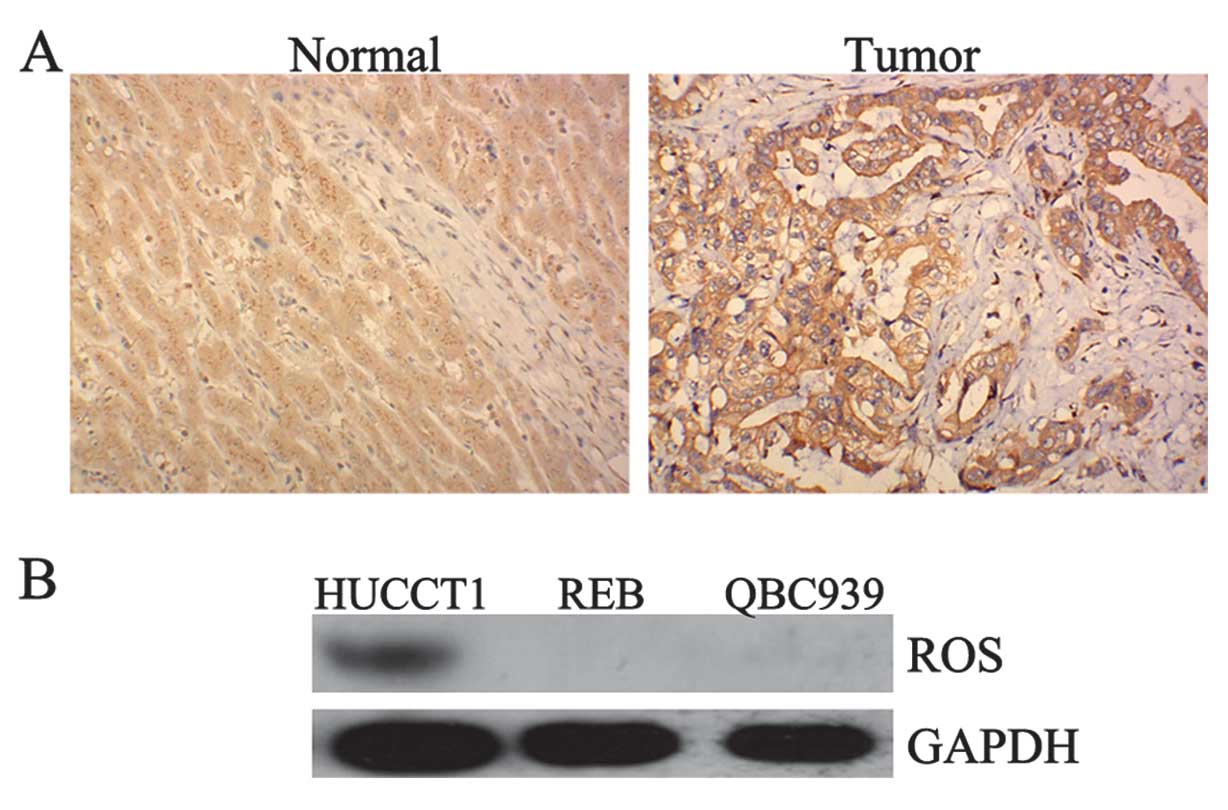
Positive expression of ROS in ICC tissues
and HUCCT1 cells
To determine the role of ROS in ICC, we firstly
performed immunohistochemistry and western blotting to determine
the protein expression of ROS in ICC tissues as well as in the ICC
cell lines, HUCCT1, REB, and QBC939. As demonstrated in Fig. 1A, one ICC sample showed a positive
expression of ROS, while none of the normal samples showed any
positive expression of ROS. Furthermore, we found that ROS was
positively expressed in HUCCT1 cells. However, we did not detect
ROS expression in the REB and QBC939 cell lines (Fig. 1B). Accordingly, the HUCCT1 cell
line was used in subsequent experiments.
shRNA-mediated downregulation of ROS-FIG
protein expression
To investigate the role of ROS-FIG fusion protein in
ICC, HUCCT1 cells were transfected with plasmids expressing ROS-
and FIG-specific shRNA, respectively. After confirming the
transfection efficiency by observing GFP fluorescence (data not
shown), we examined the protein level of ROS and FIG in each group,
respectively, by using western blotting. As shown in Fig. 2A, ROS1-6191, ROS1-6290, and
ROS1-6976 shRNAs effectively inhibited the protein level of ROS in
HUCCT1 cells, and ROS1-6290 shRNA had the strongest inhibitory
effect. As shown in Fig. 2B, the
expression of FIG protein was significantly downregulated in HUCCT1
cells transfected with plasmids expressing FIG-363, FIG-475, and
FIG-504 shRNAs, respectively, compared to the control HUCCT1 cells
without any transfection. FIG-363 shRNA had the strongest
inhibitory effect. Based on these findings, the plasmids expressing
ROS1-6290 and FIG-363 shRNA, respectively, were used in subsequent
experiments.
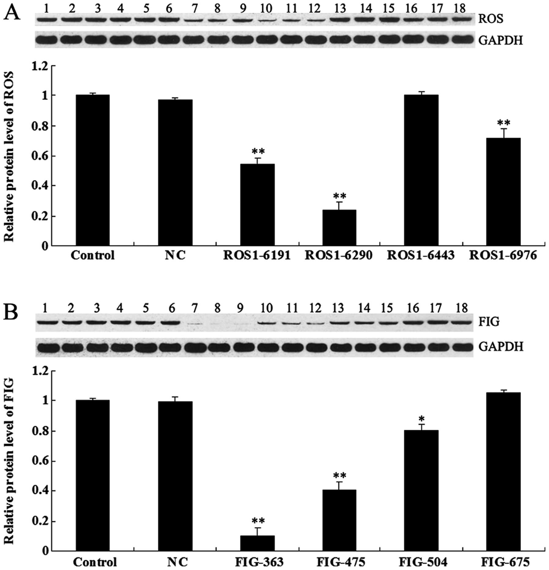 | Figure 2(A) Western blotting was performed to
examine the protein level of c-ros-oncogene (ROS) in HUCCT1 cells
transfected with ROS-specific shRNA. GAPDH was used as an internal
reference. Control (lanes 1–3), cells without any transfection;
negative control (NC) (lanes 4–6), cells transfected with blank
vector; ROS1-6191 (lanes 7–9), cells transfected with ROS1-6191
shRNA; ROS1-6290 (lanes 10–12), cells transfected with ROS1-6290
shRNA; ROS1-6443 (lanes 13–15), cells transfected with ROS1-6443
shRNA and ROS1-6976 (lanes 16–18), cells transfected with ROS1-6976
shRNA. **P<0.01 vs. control. (B) Western blotting was
performed to examine the protein level of fused-in-glioblastoma
(FIG) in HUCCT1 cells transfected with FIG-specific shRNA. GAPDH
was used as an internal reference. Control (lanes lanes 1–3), cells
without any transfection. NC (lanes 4–6), cells transfected with
blank vector. FIG-363 (lanes 7–9), cells transfected with FIG-363
shRNA. FIG-475 (lanes 10–12), cells transfected with FIG-475 shRNA.
FIG-504 (lanes 13–15), cells transfected with FIG-504 shRNA.
FIG-675 (lanes 16–18), cells transfected with FIG-6975 shRNA.
*P<0.05 and **P<0.01 vs. control. |
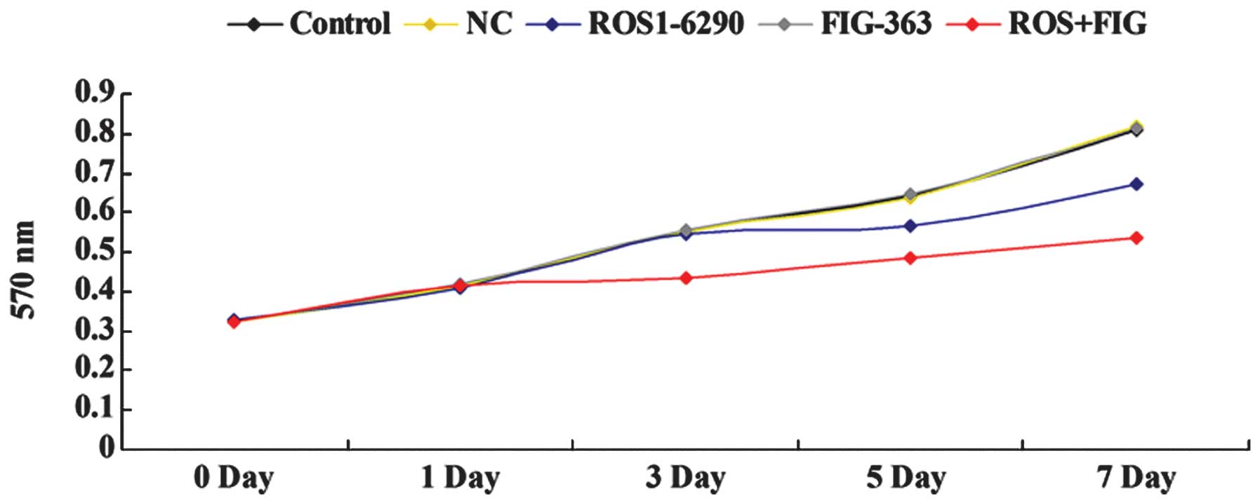
Effects of shRNA-mediated ROS-FIG
downregulation on HUCCT1 cell proliferation
An MTT assay was performed to investigate the role
of ROS-FIG in ICC cell proliferation. HUCCT1 cells were transfected
with ROS1-6290, or FIG-363 shRNA plasmid, or both. As shown in
Fig. 3, single downregulation of
FIG had no effect on HUCCT1 cell proliferation; however, the
shRNA-mediated downregulation of ROS or ROS+FIG effectively
inhibited the proliferation of HUCCT1 cells. Moreover, our findings
showed that co-downregulation of ROS and FIG had an improved
inhibitory effect on HUCCT1 cell proliferation, compared to the
single downregulation of ROS (Fig.
3).
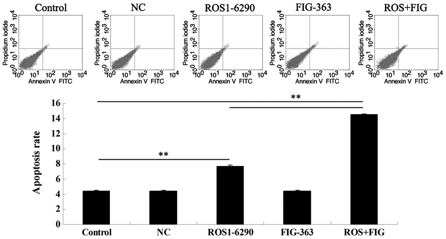
Effects of shRNA-mediated ROS-FIG
downregulation on HUCCT1 cell apoptosis
Since the downregulated cell proliferation induced
by ROS- and FIG-specific shRNAs may be attributed to the occurrence
of cell apoptosis, we determined the cell apoptotic rate by using
PI/Annexin V staining and flow cytometry. Fig. 4 shows that the shRNA-mediated
downregulation of FIG had no effect on HUCCT1 cell apoptosis.
However, single downregulation of ROS or co-inhibition of ROS and
FIG induced HUCCT1 cell apoptosis. Additionally, the apoptotic rate
in HUCCT1 cells co-transfected with ROS1-6290 and FIG-363 shRNA
plasmids was much higher, compared with that in ROS1-6290 group
(Fig. 4). These findings suggest
that ROS- and FIG-specific shRNA-mediated downregulation of HUCCT1
cell proliferation was partially at least due to the induction of
cell apoptosis.
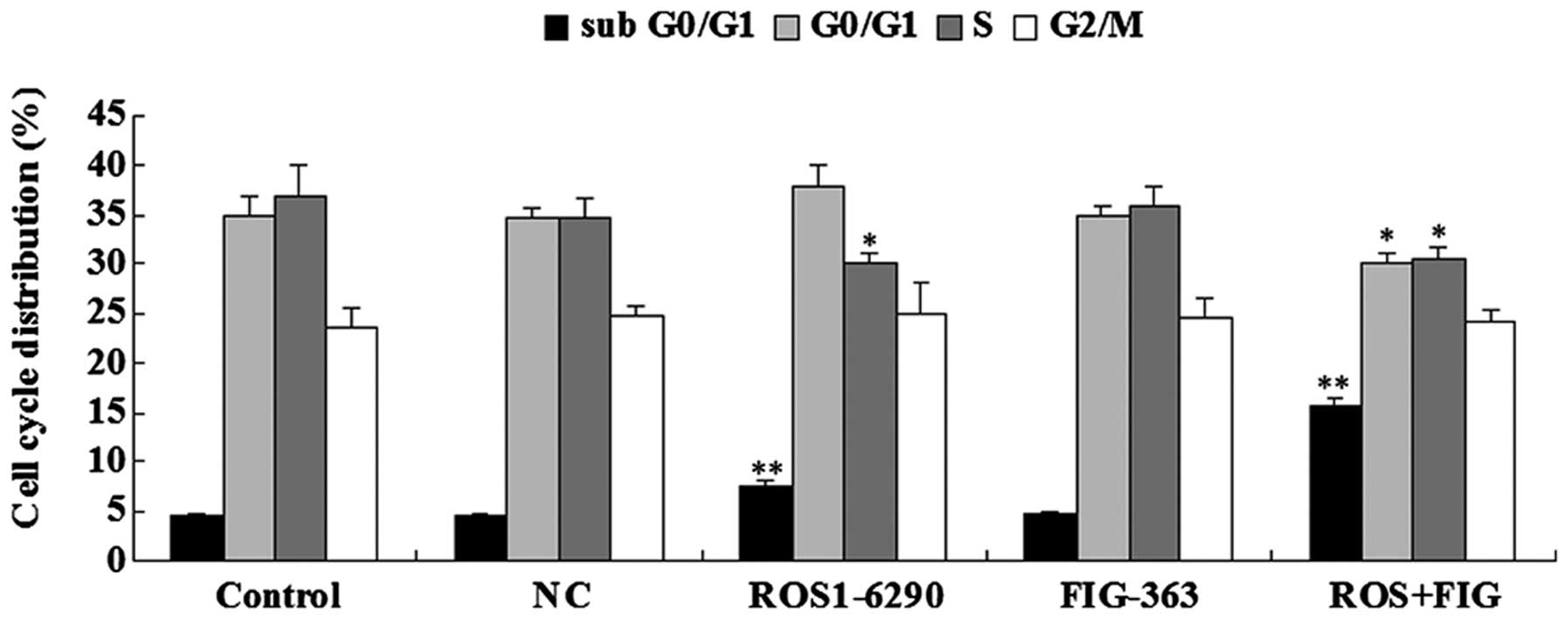
Effects of shRNA-mediated ROS-FIG
downregulation on cell the cycle progression of HUCCT1 cells
As the shRNA-mediated downregulation of HUCCT1 cell
proliferation may also be due to the abnormal cell cycle
progression, we examined the cell cycle distribution in each group
using PI staining and flow cytometry. As shown in Fig. 5, HUCCT1 cells transfected with
ROS1-6290 shRNA plasmid or co-transfected with ROS1-6290 and
FIG-363 shRNA plasmids showed a higher percentage in sub G0/G1
stage, when compared with the control group. Since sub G0/G1 stage
is an index for cell apoptosis, these findings were consistent with
the previous apoptotic assay data. By contrast, HUCCT1 cells
transfected with ROS1-6290 shRNA plasmid or co-transfected with
ROS1-6290 and FIG-363 shRNA plasmids showed a different cell cycle
distribution compared to the control group, indicating that the
inhibition of ROS or ROS-FIG suppressed HUCCT1 cell proliferation
partially at least by inducing an abnormal cell cycle progression
(Fig. 5).
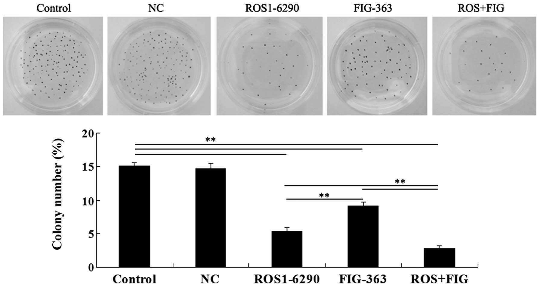
Effects of shRNA-mediated ROS-FIG
downregulation on colony-formation ability of HUCCT1 cells
The role of ROS-FIG in the regulation of
colony-formation ability of HUCCT1 cells was investigated. Single
inhibition of ROS or FIG, or the co-inhibition of ROS and FIG
significantly suppressed the colony-formation ability of HUCCT1
cells (Fig. 6). The data
demonstrated that the suppressive rate was ROS+FIG shRNA > ROS
shRNA > FIG shRNA (Fig.
6).
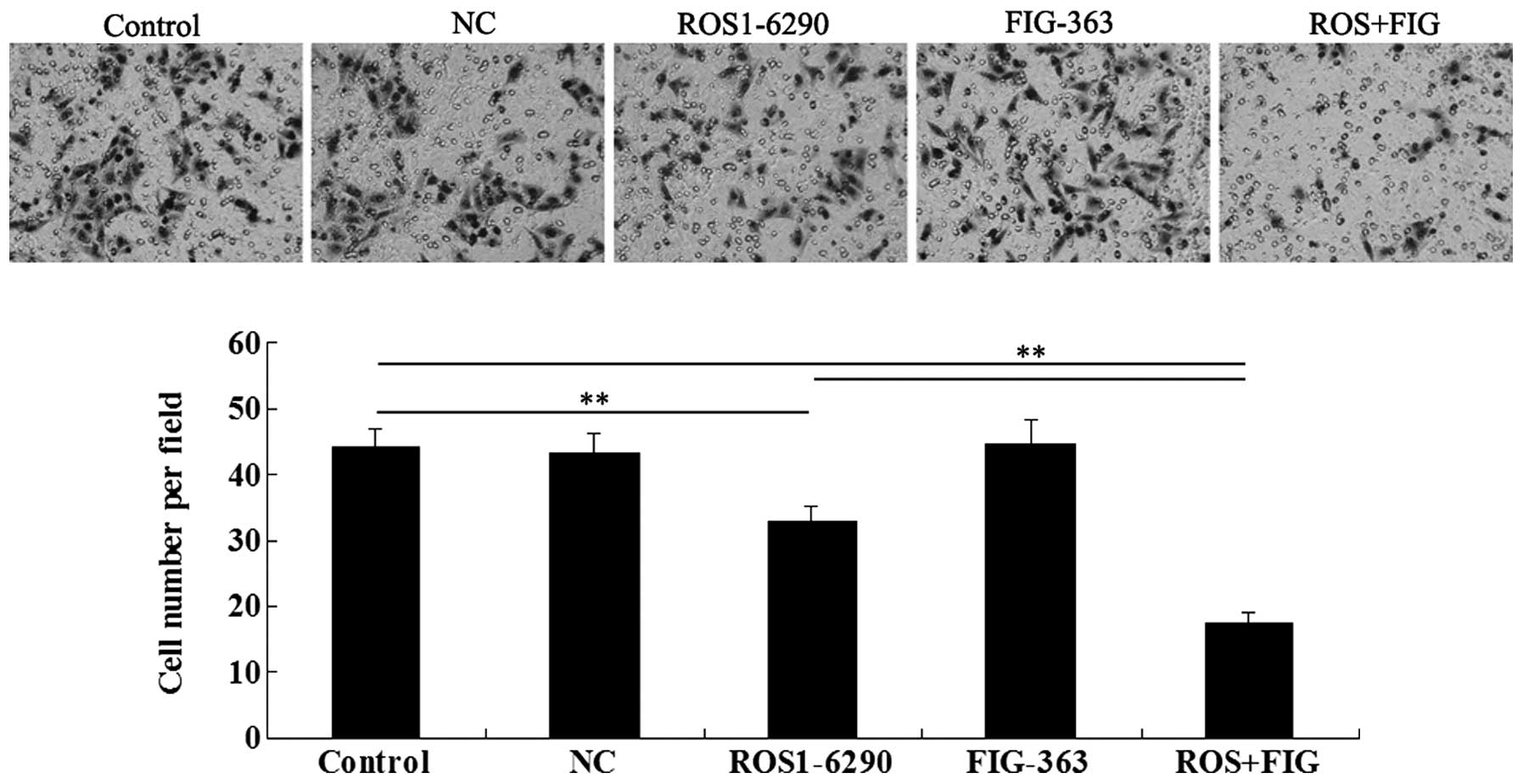
Effects of shRNA-mediated ROS-FIG
downregulation on HUCCT1 cell migration
We investigated the effect of shRNA-mediated ROS-FIG
inhibition on HUCCT1 cell migration. Single inhibition of ROS or
co-inhibition of ROS and FIG notably downregulated HUCCT1 cell
migration, while single inhibition of FIG had no effect on cell
migration (Fig. 7). Co-inhibition
of ROS and FIG showed a stronger inhibitory effect on HUCCT1 cell
migration than that of single inhibition of ROS.
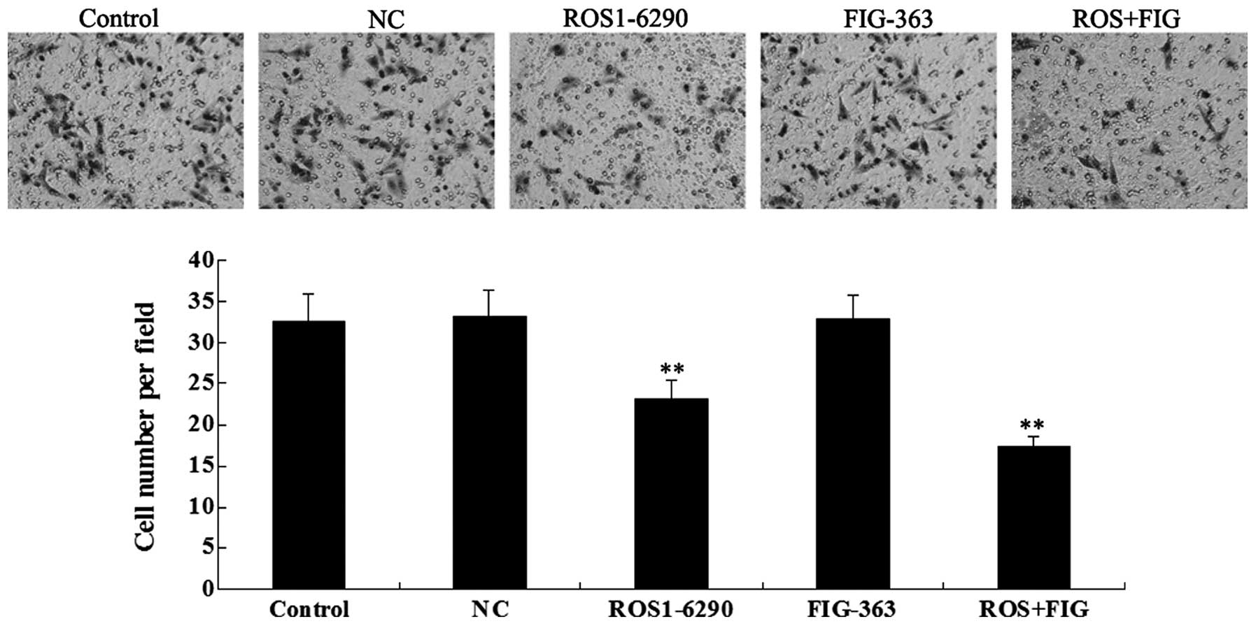
Effects of shRNA-mediated ROS-FIG
downregulation on HUCCT1 cell invasion
As tumor cell invasion is a key index for tumor
malignancy, we determined the effect of ROS-FIG downregulation on
HUCCT1 cell invasion by performing a Transwell assay.
Downregulation of ROS or the co-inhibition of ROS and FIG
significantly suppressed HUCCT1 cell invasion, compared with the
control cells without any transfection (Fig. 8). However, single inhibition of
FIG showed no effect. In addition, unlike the cell migration data,
the co-inhibition of ROS and FIG did not show a stronger
suppressive effect on cell invasion, compared with single
inhibition of ROS.
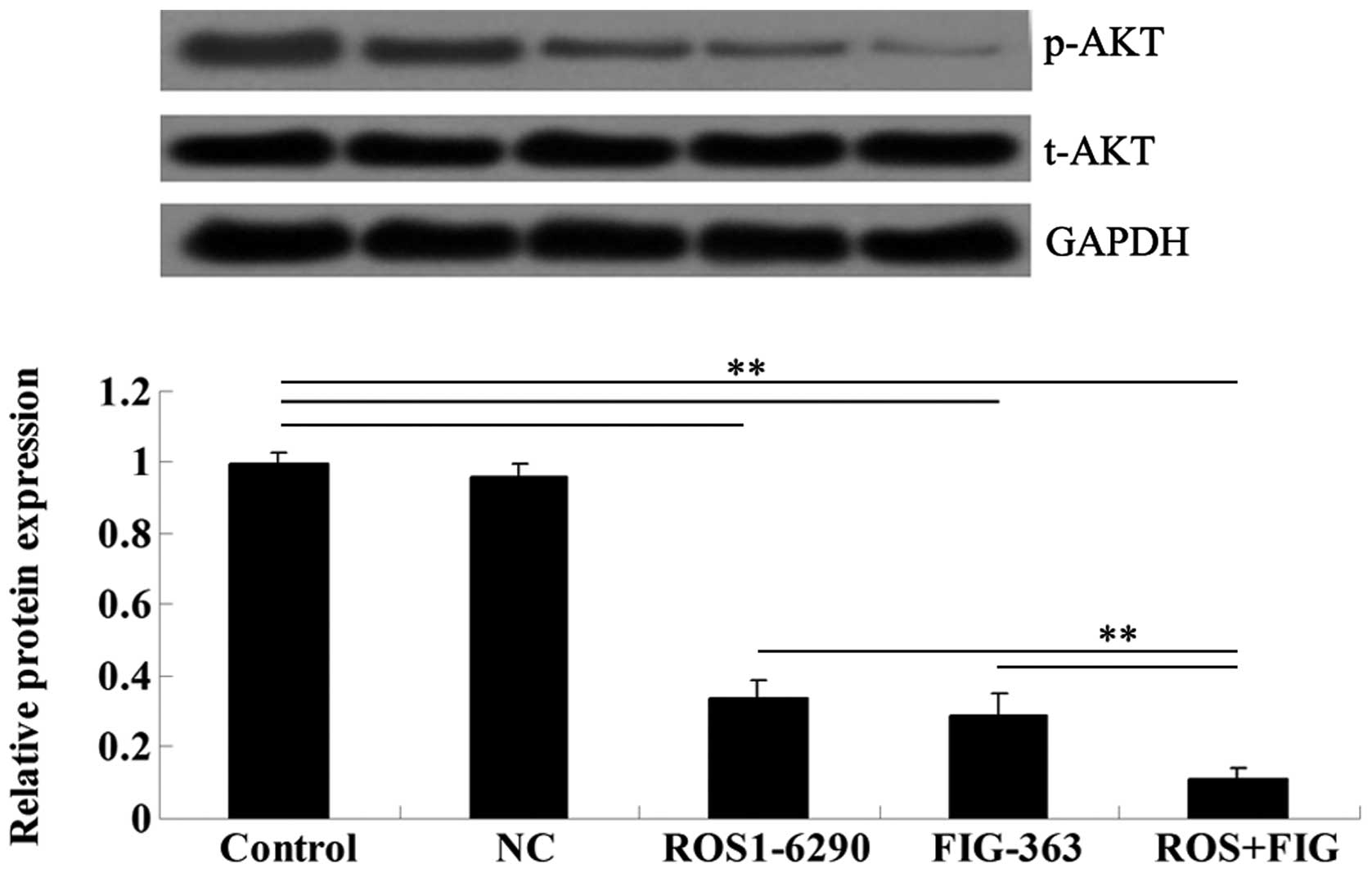
ROS-FIG downregulation inhibited the
activity of Akt signaling
To assess the molecular mechanism by which ROS-FIG
downregulation affected HUCCT1 cell proliferation, apoptosis, and
cell cycle progression, we examined the activity of Akt signaling,
which has been demonstrated to be upregulated in various types of
cancer including ICC (9). Single
inhibition of ROS or co-inhibition of ROS and FIG notably
suppressed the activity of Akt signaling in HUCCT1 cells (Fig. 9). Moreover, the co-inhibition of
ROS and FIG showed a stronger inhibitory effect when compared to
single inhibition of ROS.
Discussion
Cholangiocarcinoma, an aggressive and lethal cancer,
originates from the neoplastic transformation of the intra- and
extra-hepatic bile ducts epithelial cells (10). Morphologically, ICCs were
classified as mass-forming, periductular infiltrating, or
intraductal growth pattern (11–12). Numerous risk factors promote the
development of ICC. ROS fusions were identified originally in
glioblastoma and non-small cell lung cancer (NSCLC) to promote
their progression (13–14). The oncogenic activation of ROS1
was observed in a subset of patients with cholangiocarcinoma,
glioblastoma and lung cancer (5–6,15).
Wild-type full-length ROS1 is a 2,347 amino acid transmembrane
tyrosine kinase (TK) receptor, consisting of an extracellular
ligand-binding domain composed of nine repeated fibronectin-like
motifs, an intracellular TK domain, and a short transmembrane
domain (16). Dysregulated ROS1
may occur in different types, including ROS1 gene fusion,
overexpression, or mutations. In many cases, the ROS1 pathway was
activated by interchromosomal translocation or intrachromosomal
deletion, which resulted in N-terminal ROS1 fusion genes.
Increasing evidence (20) has
shown that ROS1 fusions as a distinct subgroup within various types
of cancer promoted the development of ROS1-directed therapeutic
strategies.
Of the four ICC samples we collected from our
hospital, only one showed a positive protein expression for ROS. Gu
et al detected FIG-ROS fusions in only 2 of 23 Chinese
cholangiocarcinoma patients (5).
Similarly, a positive rate of only 0.7% (11/1,476) of ROS fusions
among lung cancer patients was observed in Japan (13). A low expression of FIG-ROS fusions
in Chinese ICC patients was identified in our study, which is in
accordance with previous studies (5). This low expression should be
considered as important. Although rare, emerging evidence supports
ROS fusions as a valid therapeutic target in molecularly defined
patients. In malignant gliomas, the demethylation of ROS promoter
enhanced the elevated expression of ROS kinase (17). Chromosomal rearrangements involve
ROS kinase in glioblastoma and NSCLC (6,18).
Expression of FIG-ROS in the central nervous system induces
glioblastoma formation in vivo (19). Additionally, the inhibition of ROS
fusions induced growth inhibition and cell death in BaF3 cells
expressing this fusion protein (5). Thus, specific ROS inhibitors, such
as crizotinib and foretinib (20), may provide approaches for the
treatment of patients with liver cancer harboring ROS fusions.
The ICC cell lines HUCCT1, REB, and QBC939 were used
to determine whether they contain FIG-ROS fusions. Results showed
that only HUCCT1 cells showed a positive expression of ROS. These
FIG-ROS-expressed HUCCT1 cells were suitable for screening ROS
inhibitors in in vitro models. In a recent study, we
constructed different serial sequences of FIG and ROS1 shRNA to
downregulate their expression. A marked inhibitory effect was
observed in the FIG-363 and ROS1-6290 shRNA groups, indicating that
these segments are potentially efficient and specific targets for
FIG-ROS fusion inhibitors. By the loss of function study, we found
that downregulation of FIG showed a marginal effect on HUCCT1
cells; however, simultaneously decreased ROS and FIG exhibited a
stronger effect on HUCCT1 cell proliferation, apoptosis, cell cycle
progression, migration and invasion than single downregulation of
ROS. These data suggest that FIG contributes to the role of ROS in
HUCCT1 cells, but not required.
Apoptosis is a type of cell death that is
characterized by a series of morphologic changes, and plays an
important role in the development and tissue homeostasis of cells.
Promotion of apoptosis is the ultimate goal of preventive
strategies for cancer (21). The
data in this study show that the treatment of HUCCT1 cells with
FIG-ROS shRNA resulted in the inhibition of cell growth. There was
significant suppression on the colony-formation ability of HUCCT1
cells after co-inhibition of ROS and FIG. Subsequent experiments
using PI staining and flow cytometry showed that apoptosis
induction and cell-cycle progression blockage were equally
responsible for the inhibition of tumor cell proliferation. The
control of cell-cycle progression in cancer cells is considered an
effective method to stop or arrest tumor growth. Several
anti-cancer drugs arrest the tumor cell cycle at the G1, S, and
G2/M phase (22). The findings of
our study (?) showed that FIG-ROS shRNA mediated the cell-cycle
arrest in the sub G0/G1 phase. Moreover, inhibition of FIG-ROS
fusion caused the reduction of migration and invasion in HUCCT1
cells.
Recently, Davies et al suggested that EGFR
pathway activation mediated resistance to ROS1 inhibition in NSCLC
(23). Aberrant ROS1 kinase
activity resulted in the activated downstream signaling of several
oncogenic pathways, including AKT/mTOR, RAS-MAPK/ERK, and
Src-homology 2 domain-containing phosphatase (SHP)-1 and -2
pathways (19,24). Moreover, Akt signaling has been
shown to control cell proliferation, survival, and cell cycle
progression and is aberrantly upregulated in various types of
cancers including ICC (25,26). Accordingly, we examined the
activity of Akt signaling, and showed that the co-inhibition of ROS
and FIG exerted a stronger inhibitory effect on Akt signaling
activity when compared to single inhibition of ROS.
In conclusion, this study confirmed that FIG-ROS
fusion as a potent oncoprotein in ICC. Specifically, downregulated
ROS1-6290 segment mediated by shRNA exhibited a stronger inhibitory
effect on HUCCT1 cell proliferation. Downregulation of FIG showed a
synergistic effect with ROS1. Therefore, the ROS1 inhibitors
directed to ROS1-6290 segment may be an effective strategy for a
subset of human ICC harboring ROS1 fusion proteins.
References
|
1
|
McLean L and Patel T: Racial and ethnic
variations in the epidemiology of intrahepatic cholangiocarcinoma
in the United States. Liver Int. 26:1047–1053. 2006. View Article : Google Scholar
|
|
2
|
Khan SA, Davidson BR, Goldin R, Pereira
SP, Rosenberg WM, et al: Guidelines for the diagnosis and treatment
of cholangiocarcinoma: consensus document. Gut. 51(Suppl 6):
VI1–VI9. 2002.PubMed/NCBI
|
|
3
|
Endo I, Gonen M, Yopp AC, Dalal KM, Zhou
Q, et al: Intrahepatic cholangiocarcinoma: rising frequency,
improved survival, and determinants of outcome after resection. Ann
Surg. 248:84–96. 2008. View Article : Google Scholar : PubMed/NCBI
|
|
4
|
Ong CK, Subimerb C, Pairojkul C, Wongkham
S, Cutcutache I, et al: Exome sequencing of liver fluke-associated
cholangiocarcinoma. Nat Genet. 44:690–693. 2012. View Article : Google Scholar : PubMed/NCBI
|
|
5
|
Gu TL, Deng X, Huang F, Tucker M, Crosby
K, et al: Survey of tyrosine kinase signaling reveals ROS kinase
fusions in human cholangiocarcinoma. PLoS One. 6:e156402011.
View Article : Google Scholar : PubMed/NCBI
|
|
6
|
Charest A, Lane K, McMahon K, Park J,
Preisinger E, et al: Fusion of FIG to the receptor tyrosine kinase
ROS in a glioblastoma with an interstitial del(6) (q21q21). Genes
Chromosomes Cancer. 37:58–71. 2003. View Article : Google Scholar : PubMed/NCBI
|
|
7
|
Saborowski A, Saborowski M, Davare MA,
Druker BJ, Klimstra DS and Lowe SW: Mouse model of intrahepatic
cholangiocarcinoma validates FIG-ROS as a potent fusion oncogene
and therapeutic target. Proc Natl Acad Sci USA. 110:19513–19518.
2013. View Article : Google Scholar : PubMed/NCBI
|
|
8
|
Shaw AT, Camidge DR, Engelman JA, et al:
Clinical activity of crizotinib in advanced non-small cell lung
cancer (NSCLC) harboring ROS1 gene rearrangement. J Clin Oncol.
30:abstr 7508. 2012.
|
|
9
|
Sirica AE, Zhang Z, Lai GH, Asano T, Shen
XN, et al: A novel ‘patient-like’ model of cholangiocarcinoma
progression based on bile duct inoculation of tumorigenic rat
cholangiocyte cell lines. Hepatology. 47:1178–1190. 2008.
|
|
10
|
Malhi H and Gores GJ: Cholangiocarcinoma:
modern advances in understanding a deadly old disease. J Hepatol.
45:856–867. 2006. View Article : Google Scholar : PubMed/NCBI
|
|
11
|
Sirica AE: Cholangiocarcinoma: molecular
targeting strategies for chemoprevention and therapy. Hepatology.
41:5–15. 2005. View Article : Google Scholar
|
|
12
|
Malhi H and Gores GJ: Cholangiocarcinoma:
modern advances in understanding a deadly old disease. J Hepatol.
45:856–867. 2006. View Article : Google Scholar : PubMed/NCBI
|
|
13
|
Takeuchi K, Soda M, Togashi Y, Suzuki R,
Sakata S, et al: RET, ROS1 and ALK fusions in lung cancer. Nat Med.
18:378–381. 2012. View
Article : Google Scholar : PubMed/NCBI
|
|
14
|
Lira ME, Choi YL, Lim SM, Deng S, Huang D,
et al: A Single-Tube Multiplexed Assay for Detecting ALK, ROS1, and
RET Fusions in Lung Cancer. J Mol Diagn. 16:229–243. 2014.
View Article : Google Scholar : PubMed/NCBI
|
|
15
|
Bergethon K, Shaw AT, Ou SH, Katayama R,
Lovly CM, et al: ROS1 rearrangements define a unique molecular
class of lung cancers. J Clin Oncol. 30:863–870. 2012. View Article : Google Scholar : PubMed/NCBI
|
|
16
|
Nagarajan L, Louie E, Tsujimoto Y,
Balduzzi PC, Huebner K and Croce CM: The human c-ros gene (ROS) is
located at chromosome region 6q16–6q22. Proc Natl Acad Sci USA.
83:6568–6572. 1986.
|
|
17
|
Jun HJ, Woolfenden S, Coven S, Lane K,
Bronson R, et al: Epigenetic regulation of c-ROS receptor tyrosine
kinase expression in malignant gliomas. Cancer Res. 69:2180–2184.
2009. View Article : Google Scholar : PubMed/NCBI
|
|
18
|
Rikova K, Guo A, Zeng Q, Possemato A, Yu
J, et al: Global survey of phosphotyrosine signaling identifies
oncogenic kinases in lung cancer. Cell. 131:1190–1203. 2007.
View Article : Google Scholar : PubMed/NCBI
|
|
19
|
Charest A, Wilker EW, McLaughlin ME, Lane
K, Gowda R, et al: ROS fusion tyrosine kinase activates a SH2
domain-containing phosphatase-2/phosphatidylinositol
3-kinase/mammalian target of rapamycin signaling axis to form
glioblastoma in mice. Cancer Res. 66:7473–7481. 2006. View Article : Google Scholar : PubMed/NCBI
|
|
20
|
Davare MA, Saborowski A, Eide CA, Tognon
C, Smith RL, et al: Foretinib is a potent inhibitor of oncogenic
ROS1 fusion proteins. Proc Natl Acad Sci USA. 110:19519–19524.
2013. View Article : Google Scholar : PubMed/NCBI
|
|
21
|
Farnebo M, Bykov VJ and Wiman KG: The p53
tumor suppressor: a master regulator of diverse cellular processes
and therapeutic target in cancer. Biochem Biophys Res Commun.
396:85–89. 2010. View Article : Google Scholar : PubMed/NCBI
|
|
22
|
Mork CN, Faller DV and Spanjaard RA: A
mechanistic approach to anticancer therapy: targeting the cell
cycle with histone deacetylase inhibitors. Curr Pharm Des.
11:1091–1104. 2005. View Article : Google Scholar : PubMed/NCBI
|
|
23
|
Davies KD, Mahale S, Astling DP, Aisner
DL, Le AT, et al: Resistance to ROS1 inhibition mediated by EGFR
pathway activation in non-small cell lung cancer. PLoS One.
8:e822362013. View Article : Google Scholar : PubMed/NCBI
|
|
24
|
Acquaviva J, Wong R and Charest A: The
multifaceted roles of the receptor tyrosine kinase ROS in
development and cancer. Biochim Biophys Acta. 1795:37–52.
2009.PubMed/NCBI
|
|
25
|
Fava G, Alpini G, Rychlicki C, Saccomanno
S, DeMorrow S, et al: Leptin enhances cholangiocarcinoma cell
growth. Cancer Res. 68:6752–6761. 2008. View Article : Google Scholar : PubMed/NCBI
|
|
26
|
Yoon H, Min JK, Lee JW, Kim DG and Hong
HJ: Acquisition of chemoresistance in intrahepatic
cholangiocarcinoma cells by activation of AKT and extracellular
signal-regulated kinase (ERK)1/2. Biochem Biophys Res Commun.
405:333–337. 2011. View Article : Google Scholar : PubMed/NCBI
|