|
1
|
Sarks SH: Drusen patterns predisposing to
geographic atrophy of the retinal pigment epithelium. Aust J
Ophthalmol. 10:91–97. 1982. View Article : Google Scholar : PubMed/NCBI
|
|
2
|
Bressler NM, Silva JC, Bressler SB, Fine
SL and Green WR: Clinicopathologic correlation of drusen and
retinal pigment epithelial abnormalities in age-related macular
degeneration. 1994. Retina. 25:130–142. 2005. View Article : Google Scholar : PubMed/NCBI
|
|
3
|
Dentchev T, Milam AH, Lee VM, Trojanowski
JQ and Dunaief JL: Amyloid-beta is found in drusen from some
age-related macular degeneration retinas, but not in drusen from
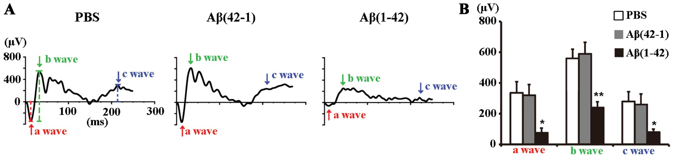
normal retinas. Mol Vis. 9:184–190. 2003.PubMed/NCBI
|
|
4
|
Johnson LV, Leitner WP, Rivest AJ, Staples
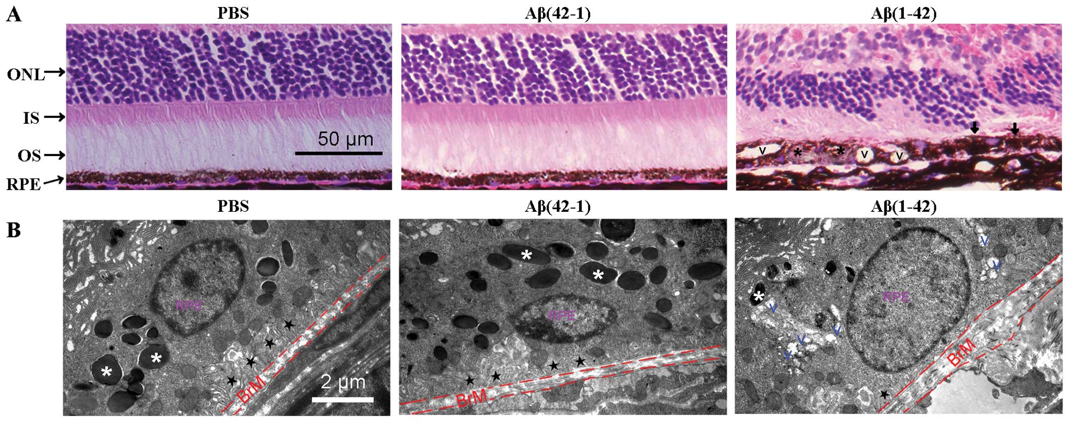
MK, Radeke MJ and Anderson DH: The Alzheimer’s A beta -peptide is
deposited at sites of complement activation in pathologic deposits
associated with aging and age-related macular degeneration. Proc
Natl Acad Sci USA. 99:11830–11835. 2002. View Article : Google Scholar
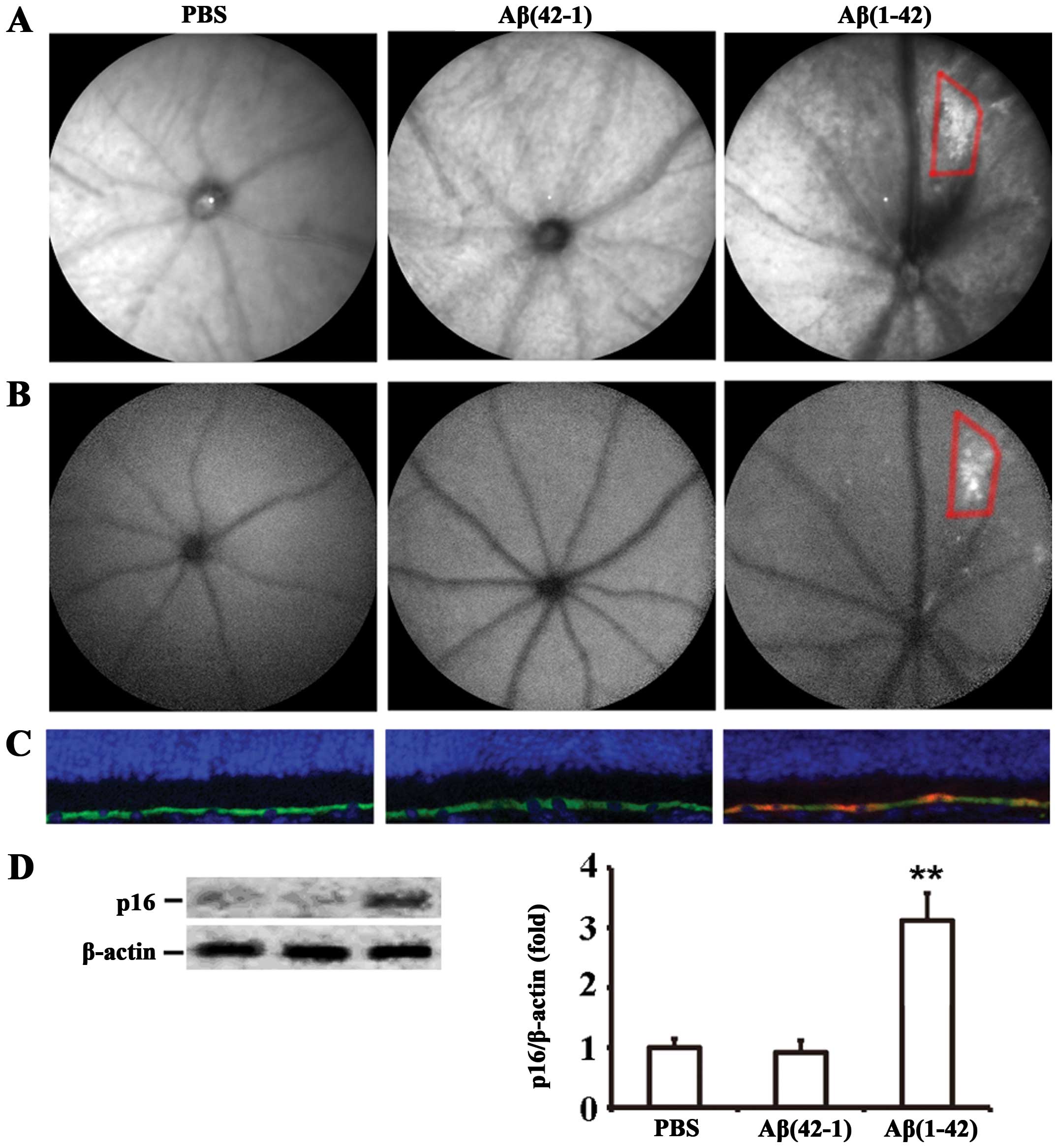
|
|
5
|
Kurji KH, Cui JZ, Lin T, et al: Microarray
analysis identifies changes in inflammatory gene expression in
response to amyloid-beta stimulation of cultured human retinal
pigment epithelial cells. Invest Ophthalmol Vis Sci. 51:1151–1163.
2010. View Article : Google Scholar
|
|
6
|
Cao L, Liu C, Wang F and Wang H: SIRT1
negatively regulates amyloid-beta-induced inflammation via the
NF-kappaB pathway. Braz J Med Biol Res. 46:659–669. 2013.
View Article : Google Scholar : PubMed/NCBI
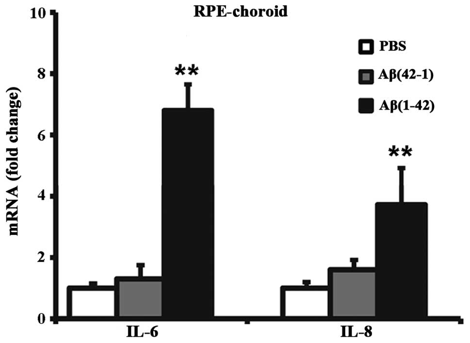
|
|
7
|
Liu RT, Gao J, Cao S, et al: Inflammatory
mediators induced by amyloid-beta in the retina and RPE in vivo:
implications for inflammasome activation in age-related macular
degeneration. Invest Ophthalmol Vis Sci. 54:2225–2237. 2013.
View Article : Google Scholar : PubMed/NCBI
|
|
8
|
Ding JD, Johnson LV, Herrmann R, et al:
Anti-amyloid therapy protects against retinal pigmented epithelium
damage and vision loss in a model of age-related macular
degeneration. Proc Natl Acad Sci USA. 108:E279–E287. 2011.
View Article : Google Scholar : PubMed/NCBI
|
|
9
|
Buschini E, Piras A, Nuzzi R and Vercelli
A: Age related macular degeneration and drusen: neuroinflammation
in the retina. Prog Neurobiol. 95:14–25. 2011. View Article : Google Scholar : PubMed/NCBI
|
|
10
|
Coppé JP, Patil CK, Rodier F, et al:
Senescence-associated secretory phenotypes reveal
cell-nonautonomous functions of oncogenic RAS and the p53 tumor
suppressor. PLoS Biol. 6:2853–2868. 2008. View Article : Google Scholar : PubMed/NCBI
|
|
11
|
Kumar S, Millis AJ and Baglioni C:
Expression of interleukin 1-inducible genes and production of
interleukin 1 by aging human fibroblasts. Proc Natl Acad Sci USA.
89:4683–4687. 1992. View Article : Google Scholar : PubMed/NCBI
|
|
12
|
Campisi J: Senescent cells, tumor
suppression, and organismal aging: good citizens, bad neighbors.
Cell. 120:513–522. 2005. View Article : Google Scholar : PubMed/NCBI
|
|
13
|
Ben-Porath I and Weinberg RA: The signals
and pathways activating cellular senescence. Int J Biochem Cell
Biol. 37:961–976. 2005. View Article : Google Scholar : PubMed/NCBI
|
|
14
|
Lombard DB, Chua KF, Mostoslavsky R,
Franco S, Gostissa M and Alt FW: DNA repair, genome stability, and
aging. Cell. 120:497–512. 2005. View Article : Google Scholar : PubMed/NCBI
|
|
15
|
He N, Jin WL, Lok KH, Wang Y, Yin M and
Wang ZJ: Amyloid-β(1–42) oligomer accelerates senescence in adult
hippocampal neural stem/progenitor cells via formylpeptide receptor
2. Cell Death Dis. 4:e9242013. View Article : Google Scholar
|
|
16
|
Golde TE and Miller VM:
Proteinopathy-induced neuronal senescence: a hypothesis for brain
failure in Alzheimer’s and other neurodegenerative diseases.
Alzheimers Res Ther. 1:52009. View
Article : Google Scholar
|
|
17
|
Bhat R, Crowe EP, Bitto A, et al:
Astrocyte senescence as a component of Alzheimer’s disease. PLoS
One. 7:e450692012. View Article : Google Scholar
|
|
18
|
Donnini S, Solito R, Cetti E, et al: Abeta
peptides accelerate the senescence of endothelial cells in vitro
and in vivo, impairing angiogenesis. FASEB J. 24:2385–2395. 2010.
View Article : Google Scholar : PubMed/NCBI
|
|
19
|
Cao L, Wang H, Wang F, Xu D, Liu F and Liu
C: Aβ-induced senescent retinal pigment epithelial cells create a
proinflammatory microenvironment in AMD. Invest Ophthalmol Vis Sci.
54:3738–3750. 2013. View Article : Google Scholar : PubMed/NCBI
|
|
20
|
Bruban J, Maoui A, Chalour N, et al:
CCR2/CCL2-mediated inflammation protects photoreceptor cells from
amyloid-beta-induced apoptosis. Neurobiol Dis. 42:55–72. 2011.
View Article : Google Scholar : PubMed/NCBI
|
|
21
|
Hood DC and Birch DG: A quantitative
measure of the electrical activity of human rod photoreceptors
using electroretinography. Vis Neurosci. 5:379–387. 1990.
View Article : Google Scholar : PubMed/NCBI
|
|
22
|
Penn RD and Hagins WA: Signal transmission
along retinal rods and the origin of the electroretinographic
a-wave. Nature. 223:201–204. 1969. View
Article : Google Scholar : PubMed/NCBI
|
|
23
|
Robson JG and Frishman LJ: Response
linearity and kinetics of the cat retina: the bipolar cell
component of the dark-adapted electroretinogram. Vis Neurosci.
12:837–850. 1995. View Article : Google Scholar : PubMed/NCBI
|
|
24
|
Tian N and Slaughter MM: Correlation of
dynamic responses in the ON bipolar neuron and the b-wave of the
electroretinogram. Vision Res. 35:1359–1364. 1995. View Article : Google Scholar : PubMed/NCBI
|
|
25
|
Robson JG, Maeda H, Saszik SM and Frishman
LJ: In vivo studies of signaling in rod pathways of the mouse using
the electroretinogram. Vision Res. 44:3253–3268. 2004. View Article : Google Scholar : PubMed/NCBI
|
|
26
|
Hanitzsch R and Lichtenberger T: Two
neuronal retinal components of the electroretinogram c-wave. Doc
Ophthalmol. 94:275–285. 1997. View Article : Google Scholar : PubMed/NCBI
|
|
27
|
Green WR and Enger C: Age-related macular
degeneration histopathologic studies. The 1992 Lorenz E Zimmerman
Lecture. Ophthalmology. 100:1519–1535. 1993. View Article : Google Scholar : PubMed/NCBI
|
|
28
|
Perez SE, Lumayag S, Kovacs B, Mufson EJ
and Xu S: Beta-amyloid deposition and functional impairment in the
retina of the APPswe/PS1DeltaE9 transgenic mouse model of
Alzheimer’s disease. Invest Ophthalmol Vis Sci. 50:793–800. 2009.
View Article : Google Scholar
|
|
29
|
Bruban J, Glotin AL, Dinet V, et al:
Amyloid-beta(1–42) alters structure and function of retinal
pigmented epithelial cells. Aging Cell. 8:162–177. 2009. View Article : Google Scholar : PubMed/NCBI
|
|
30
|
Drüeke T, Hennessen U, Nabarra B, et al:
Ultrastructural and functional abnormalities of intestinal and
renal epithelium in the SHR. Kidney Int. 37:1438–1448. 1990.
View Article : Google Scholar : PubMed/NCBI
|
|
31
|
Anderson DH, Mullins RF, Hageman GS and
Johnson LV: A role for local inflammation in the formation of
drusen in the aging eye. Am J Ophthalmol. 134:411–431. 2002.
View Article : Google Scholar : PubMed/NCBI
|
|
32
|
Cho YY, Kim DJ, Lee HS, et al: Autophagy
and cellular senescence mediated by Sox2 suppress malignancy of
cancer cells. PLoS One. 8:e571722013. View Article : Google Scholar : PubMed/NCBI
|
|
33
|
Butterfield DA and Boyd-Kimball D: Amyloid
beta-peptide(1–42) contributes to the oxidative stress and
neurodegeneration found in Alzheimer disease brain. Brain Pathol.
14:426–432. 2004. View Article : Google Scholar : PubMed/NCBI
|
|
34
|
Yu AL, Fuchshofer R, Kook D, Kampik A,
Bloemendal H and Welge-Lüssen U: Subtoxic oxidative stress induces
senescence in retinal pigment epithelial cells via TGF-beta
release. Invest Ophthalmol Vis Sci. 50:926–935. 2009. View Article : Google Scholar : PubMed/NCBI
|
|
35
|
Katz ML, Wendt KD and Sanders DN: RPE65
gene mutation prevents development of autofluorescence in retinal
pigment epithelial phagosomes. Mech Ageing Dev. 126:513–521. 2005.
View Article : Google Scholar : PubMed/NCBI
|
|
36
|
Rawes V, Kipling D, Kill IR and Faragher
RG: The kinetics of senescence in retinal pigmented epithelial
cells: a test for the telomere hypothesis of ageing? Biochemistry
(Mosc). 62:1291–1295. 1997.
|
|
37
|
Feeney-Burns L, Hilderbrand ES and
Eldridge S: Aging human RPE: morphometric analysis of macular,
equatorial, and peripheral cells. Invest Ophthalmol Vis Sci.
25:195–200. 1984.PubMed/NCBI
|
|
38
|
Katz ML and Robison WG Jr: Age-related
changes in the retinal pigment epithelium of pigmented rats. Exp
Eye Res. 38:137–151. 1984. View Article : Google Scholar : PubMed/NCBI
|
|
39
|
Wing GL, Blanchard GC and Weiter JJ: The
topography and age relationship of lipofuscin concentration in the
retinal pigment epithelium. Invest Ophthalmol Vis Sci. 17:601–607.
1978.PubMed/NCBI
|
|
40
|
Holz FG, Bindewald-Wittich A, Fleckenstein
M, Dreyhaupt J, Scholl HP and Schmitz-Valckenberg S; FAM-Study
Group. Progression of geographic atrophy and impact of fundus
autofluorescence patterns in age-related macular degeneration. Am J
Ophthalmol. 143:463–472. 2007. View Article : Google Scholar : PubMed/NCBI
|
|
41
|
Sparrow JR, Hicks D and Hamel CP: The
retinal pigment epithelium in health and disease. Curr Mol Med.
10:802–823. 2010. View Article : Google Scholar : PubMed/NCBI
|
|
42
|
Freund A, Orjalo AV, Desprez PY and
Campisi J: Inflammatory networks during cellular senescence: causes
and consequences. Trends Mol Med. 16:238–246. 2010. View Article : Google Scholar : PubMed/NCBI
|
|
43
|
Tsuji T, Aoshiba K and Nagai A: Alveolar
cell senescence exacerbates pulmonary inflammation in patients with
chronic obstructive pulmonary disease. Respiration. 80:59–70. 2010.
View Article : Google Scholar
|
|
44
|
Dagouassat M, Gagliolo JM, Chrusciel S, et
al: The cyclooxygenase-2-prostaglandin E2 pathway maintains
senescence of chronic obstructive pulmonary disease fibroblasts. Am
J Respir Crit Care Med. 187:703–714. 2013. View Article : Google Scholar : PubMed/NCBI
|
|
45
|
Sparmann A and Bar-Sagi D: Ras-induced
interleukin-8 expression plays a critical role in tumor growth and
angiogenesis. Cancer Cell. 6:447–458. 2004. View Article : Google Scholar : PubMed/NCBI
|
|
46
|
Acosta JC, O’Loghlen A, Banito A, et al:
Chemokine signaling via the CXCR2 receptor reinforces senescence.
Cell. 133:1006–1018. 2008. View Article : Google Scholar : PubMed/NCBI
|
|
47
|
Kuilman T, Michaloglou C, Vredeveld LC, et
al: Oncogene-induced senescence relayed by an interleukin-dependent
inflammatory network. Cell. 133:1019–1031. 2008. View Article : Google Scholar : PubMed/NCBI
|
|
48
|
Jonas JB, Tao Y, Neumaier M and Findeisen
P: Cytokine concentration in aqueous humour of eyes with exudative
age-related macular degeneration. Acta Ophthalmol. 90:e381–e388.
2012. View Article : Google Scholar : PubMed/NCBI
|
|
49
|
Hong DS, Angelo LS and Kurzrock R:
Interleukin-6 and its receptor in cancer: implications for
translational therapeutics. Cancer. 110:1911–1928. 2007. View Article : Google Scholar : PubMed/NCBI
|
|
50
|
Parmeggiani F, Romano MR, Costagliola C,
et al: Mechanism of inflammation in age-related macular
degeneration. Mediators Inflamm. 2012:5467862012. View Article : Google Scholar : PubMed/NCBI
|
|
51
|
Parmeggiani F, Sorrentino FS, Romano MR,
et al: Mechanism of inflammation in age-related macular
degeneration: an up-to-date on genetic landmarks. Mediators
Inflamm. 2013:4356072013. View Article : Google Scholar : PubMed/NCBI
|
|
52
|
Jeyapalan JC and Sedivy JM: Cellular
senescence and organismal aging. Mech Ageing Dev. 129:467–474.
2008. View Article : Google Scholar : PubMed/NCBI
|
|
53
|
Ksiazek K, Mikula-Pietrasik J, Olijslagers
S, Jörres A, von Zglinicki T and Witowski J: Vulnerability to
oxidative stress and different patterns of senescence in human
peritoneal mesothelial cell strains. Am J Physiol Regul Integr Comp
Physiol. 296:R374–R382. 2009. View Article : Google Scholar
|
|
54
|
Weyemi U, Lagente-Chevallier O, Boufraqech
M, et al: ROS-generating NADPH oxidase NOX4 is a critical mediator
in oncogenic H-Ras-induced DNA damage and subsequent senescence.
Oncogene. 31:1117–1129. 2012. View Article : Google Scholar :
|
|
55
|
Sitte N, Merker K, Von Zglinicki T, Grune
T and Davies KJ: Protein oxidation and degradation during cellular
senescence of human BJ fibroblasts: part I - effects of
proliferative senescence. FASEB J. 14:2495–2502. 2000. View Article : Google Scholar : PubMed/NCBI
|
|
56
|
Rayess H, Wang MB and Srivatsan ES:
Cellular senescence and tumor suppressor gene p16. Int J Cancer.
130:1715–1725. 2012. View Article : Google Scholar :
|
|
57
|
Campisi J and d’Adda di Fagagna F:
Cellular senescence: when bad things happen to good cells. Nat Rev
Mol Cell Biol. 8:729–740. 2007. View Article : Google Scholar : PubMed/NCBI
|
|
58
|
Baker DJ, Wijshake T, Tchkonia T, et al:
Clearance of p16Ink4a-positive senescent cells delays
ageing-associated disorders. Nature. 479:232–236. 2011. View Article : Google Scholar : PubMed/NCBI
|
|
59
|
Timmers AM, Zhang H, Squitieri A and
Gonzalez-Pola C: Subretinal injections in rodent eyes: effects on
electrophysiology and histology of rat retina. Mol Vis. 7:131–137.
2001.PubMed/NCBI
|