Introduction
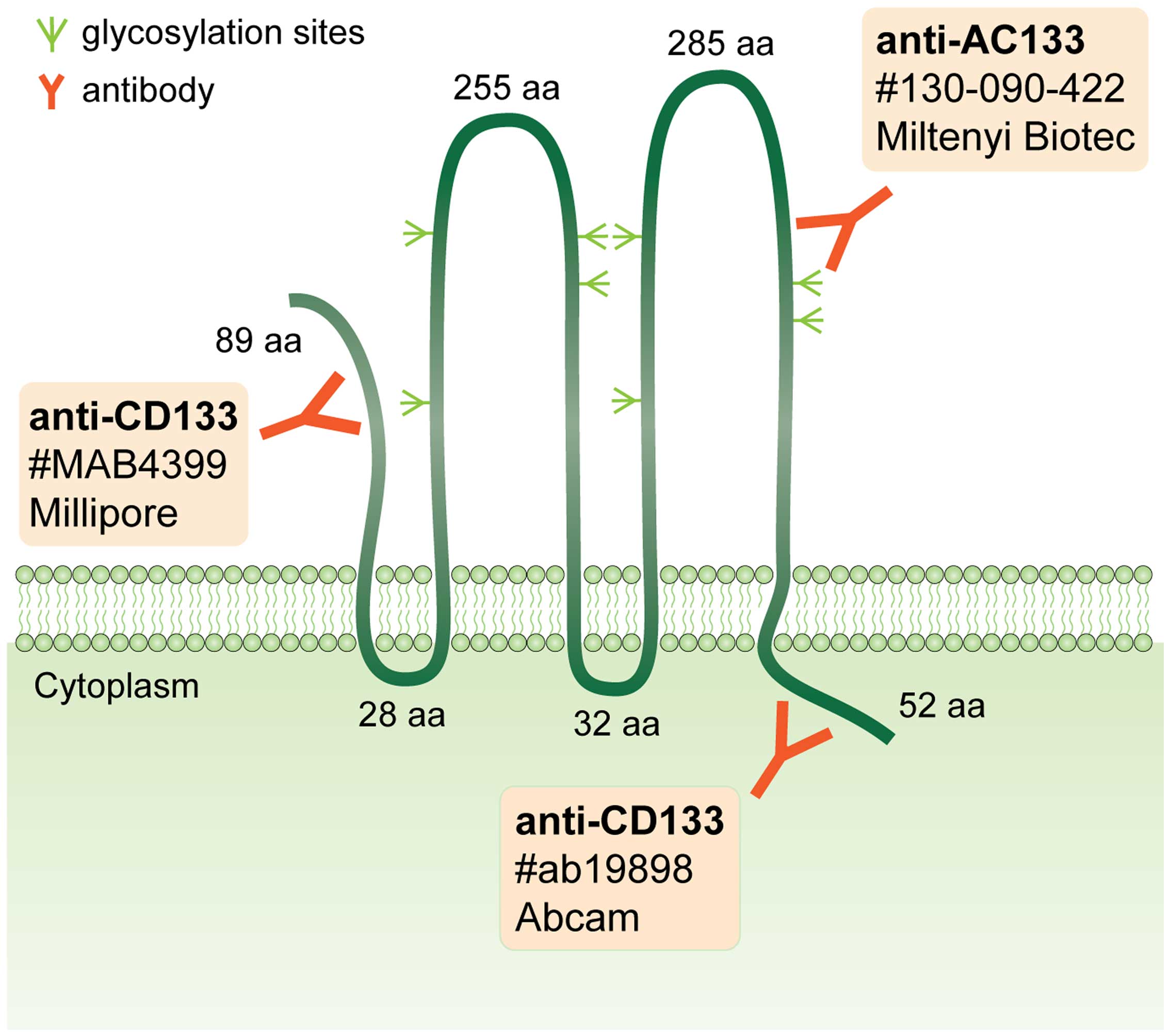
CD133 (also known as prominin-1) is a glycoprotein
that is typically localized to the plasma membrane. The molecule
consists of five transmembrane domains, two large extracellular
loops, an extracellular N-terminus and an intracellular C-terminus.
Eight potential glycosylation sites have been identified within the
extracellular domains, with four per loop. Human CD133 is encoded
by the PROM1 gene, which is located in chromosomal region
4p15.32. At least seven CD133 isoforms resulting from alternative
splicing have been described in humans (1,2).
CD133 is widely used to identify stem cells, and its
glycosylated epitope, AC133, has recently been discussed as a
marker of cancer stem cells (CSCs) in various human malignancies
(2–4). In our previous studies, we
identified CD133-positive cells that presented typical membrane
positivity in two of the most common types of pediatric sarcomas,
osteosarcoma (5) and
rhabdomyosarcoma (RMS) (6). The
expression of CD133 in these two solid tumors, as well as the
tumorigenicity of CD133-positive cells, has been confirmed by other
research groups (7–10). Therefore, CD133 is currently
accepted as one of the markers of a CSC phenotype in pediatric
sarcomas, including RMS (11–13).
During our recent study aimed at the analysis of CSC
markers in pediatric sarcomas, we noted a surprising result: a
stable subset of cells in each of five RMS cell lines examined
exhibited an exclusive nuclear localization of CD133 (these data
are published in this article). To date, a similar localization of
this antigen has been described only in one case report of breast
cancer (14) and in a large study
on lung cancer (15) using
immunohistochemical methods, nevertheless, without any verification
or systematic description. For this reason, in this study, we
sought to analyze this interesting phenomenon in detail using three
independent anti-CD133 commercial antibodies (Fig. 1).
Materials and methods
Cell culture
Five cell lines derived from pediatric patients with
RMS were included in this study: NSTS-8, NSTS-9, NSTS-11, NSTS-22
and NSTS-28. The first three cell lines were described in our
previous study (6), and the last
two were derived using the same procedure to generate primary
cultures (16). All cell lines
were authenticated by the immunodetection of MyoD, and the subtype
was distinguished using FKHR break detection by fluorescence
in situ hybridization (FISH). Authentication using MyoD
detection was performed in the same passages as the experiments;
FISH analysis of the FKHR break was completed up to passage
10. The cell lines were maintained in Dulbecco’s modified Eagle’s
medium (DMEM) supplemented with 20% fetal calf serum, 2 mM
glutamine, 100 IU/ml penicillin and 100 µg/ml streptomycin
(all purchased from GE Healthcare Europe GmbH, Freiburg, Germany).
The cells were maintained under standard conditions at 37°C in a
humified atmosphere containing 5% CO2 and were
subcultured once or twice per week. The Research Ethics Committee
of the School of Science (Masaryk University, Brno, Czech Republic)
approved the study protocol, and a written statement of informed
consent was obtained from each participant or his/her legal
guardian prior to participation in this study. A brief description
of the cohort of patients included in this study is provided in
Table I.
 | Table IDescription of patients from whom
tumor samples were obtained to establish the rhabdomyosarcoma cell
lines and the results concerning the mean percentage of cells with
an exclusive nuclear localization of CD133. |
Table I
Description of patients from whom
tumor samples were obtained to establish the rhabdomyosarcoma cell
lines and the results concerning the mean percentage of cells with
an exclusive nuclear localization of CD133.
| Cell line | Gender | Age (years) | RMS type | No. of passages
analyzed | Mean percentage of
cells with exclusive nuclear localization of CD133
|
|---|
| Anti-CD133 antibody
(rabbit polyclonal) | Anti-CD133 antibody
(mouse monoclonal) | Anti-AC133 antibody
(mouse monoclonal) |
|---|
| NSTS-8 | F | 21 | A | 15–18 | 4.6 | 6.1 | 3.4 |
| NSTS-9 | M | 17 | A | 13–18 | 4.6 | 5.2 | 4.1 |
| NSTS-11 | F | 16 | E | 11–19 | 3.8 | 4.6 | 6.6 |
| NSTS-22 | F | 5 | A | 11–15 | 4.0 | 6.0 | 6.4 |
| NSTS-28 | M | 8 | A | 12–16 | 4.1 | 5.2 | 7.5 |
Indirect immunofluorescence
The cells were cultivated on coverslips in Petri
dishes for one day and then rinsed with phosphate-buffered saline
(PBS) and fixed with 3% paraformaldehyde (Sigma-Aldrich, St. Louis,
MO, USA) at room temperature for 20 min. After washing again with
PBS, nonspecific binding was blocked with 3% bovine serum albumin
(BSA; Sigma-Aldrich) in PBS for 10 min. The cells were then
incubated with primary antibody at 37°C for 60 min, washed three
times in PBS and then incubated with the corresponding secondary
antibody at 37°C for 45 min. Rabbit polyclonal anti-CD133 (Cat. no.
ab19898, dilution 1:70; Abcam, Cambridge, UK), mouse monoclonal
anti-CD133 (clone 17A6.1, Cat. no. MAB4399, dilution 1:100;
Millipore, Billerica, MA, USA), mouse monoclonal anti-AC133 (clone
AC133, Cat. no. 130-090-422, dilution 1:4; Miltenyi Biotec,
Bergisch Gladbach, Germany), and mouse monoclonal anti-α-tubulin
antibody (clone: TU-01, Cat. no. 11-250, dilution 1:100; Exbio,
Vestec, Czech Republic), which served as a control, were used as
the primary antibodies. Anti-rabbit Alexa Fluor 488 (Cat. no.
A11008, dilution 1:200) and anti-mouse Alexa Fluor 488 antibody
(Cat. no. A11001, dilution 1:200) (both from Invitrogen, Paisley,
UK) were used as the secondary antibodies. After a final wash with
PBS, the cell nuclei were counterstained with 0.05% Hoechst 33342
(Life Technologies, Carlsbad, CA, USA) for 10 min, and the
coverslips were mounted using Dako fluorescence mounting medium
(Dako, Glostrup, Denmark). An Olympus BX-51 microscope was used for
sample evaluation; micrographs were captured using an Olympus DP72
CCD camera and analyzed using the Cell P imaging system (Olympus,
Tokyo, Japan). At least 200 cells were evaluated overall within
discrete areas of each sample, and the samples were prepared from
at least three independent passages of all examined cell lines. The
mean percentages of cells showing exclusive nuclear CD133
localization were determined for entire samples of individual cell
lines. For the detailed examination of CD133 nuclear localization,
the same protocol for indirect immunofluorescence was employed, and
the specimens were then examined using an Olympus FluoView-500
confocal imaging system combined with an inverted Olympus IX-81
microscope. The images were recorded using an Olympus DP70 CCD
camera and analyzed using analySIS FIVE software (Soft Imaging
System GmbH, Muenster, Germany) and an Olympus FluoView Confocal
Laser Scanning Microscope System 4.3.
Transmission electron microscopy
(TEM)
To perform the immunodetection of CD133 in ultrathin
sections, the cells grown on coverslips were rinsed with PBS and
fixed in 3% paraformaldehyde (Sigma-Aldrich) and 0.1%
glutaraldehyde (AppliChem GmbH, Darmstadt, Germany) in PBS at room
temperature for 60 min. Following a PBS rinse and dehydration, the
cells were embedded in LR White medium (Polysciences Inc., London,
UK). The labeling of the ultrathin sections was performed on grids.
CD133 was detected using mouse monoclonal anti-CD133 antibody
(dilution 1:25; Millipore) and a gold particle-conjugated secondary
antibody (anti-mouse IgG 20 nm gold, Cat. no. ab27242, dilution
1:40; Abcam). Ultrathin sections incubated without primary antibody
or with the TU-01 primary monoclonal antibody against α-tubulin
(dilution 1:200; Exbio) were used as controls. Following
immunodetection, the specimens were contrasted with 2.5% uranyl
acetate (PLIVA-Lachema, Brno, Czech Republic) for 10 min and with
Reynolds solution (Sigma-Aldrich) for 6 min at room temperature.
The specimens were then observed under a Morgagni 268(D)
transmission electron microscope (FEI Co., Hillsboro, OR, USA). The
images were captured using an Olympus Veleta TEM CCD camera and
analyzed using iTEM Olympus Soft Imaging Solution (Olympus).
Immunoblot analysis
To analyze the nuclear and cytoplasmic fractions, a
Nuclear Protein Extraction kit (Thermo Fisher Scientific, Rockford,
IL, USA) was used according to the manufacturer’s instructions. A
20 µl sample of protein extract was loaded onto an 8% sodium
dodecyl sulfate (SDS)-polyacrylamide gel and separated by
electrophoresis. Subsequently, the proteins were transferred onto
polyvinylidene difluoride (PVDF) membranes (Bio-Rad Laboratories,
Hercules, CA, USA), blocked in 5% non-fat milk at room temperature
for 60 min, and incubated with primary antibodies, rabbit
polyclonal anti-CD133 (Abcam), mouse monoclonal anti-α-tubulin
(Exbio), or rabbit monoclonal anti-lamin B2 antibody (clone: D8P3U,
Cat. no. 12255S; Cell Signaling Technology, Danvers, MA, USA) at a
1:1,000 dilution overnight at 4°C. Anti-α-tubulin and anti-lamin B2
served as the controls for the purity of the cytoplasmic and
nuclear cell fractions, respectively. After washing with
Tris-buffered saline (TBS)-Tween-20, the membranes were incubated
with the corresponding horseradish peroxidase (HRP)-conjugated
secondary antibodies anti-mouse IgG-HRP (cat. no. A9917, dilution
1:5,000; Sigma-Aldrich) and anti-rabbit IgG-HRP antibodies (cat.
no. 7074, dilution 1:5,000; Cell Signaling Technology) at room
temperature for 60 min. Signal detection was performed using ECL
Prime Western Blotting Detection Reagent (GE Healthcare) according
to the manufacturer’s instructions.
Results
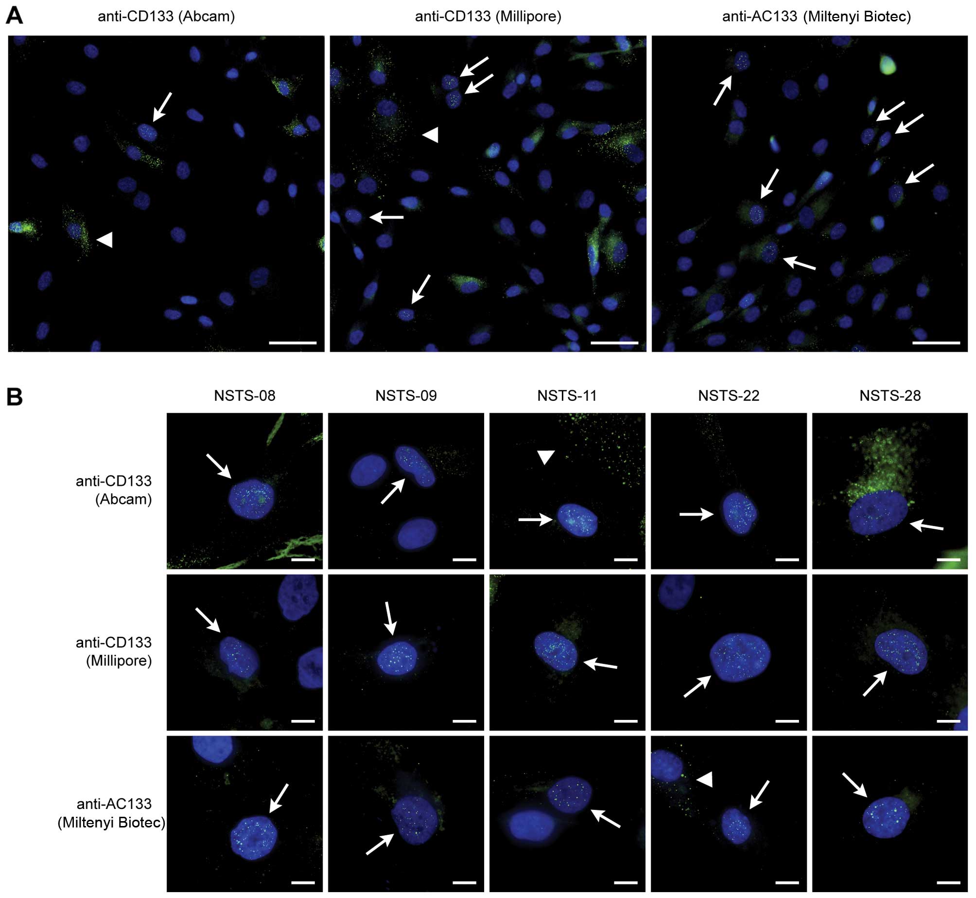
For all five RMS cell lines examined in this study,
we performed a detailed analysis of the presence of cells with
nuclear CD133 positivity using indirect immunofluorescence with
three independent anti-CD133 antibodies (Fig. 1). A subset of cells showed only
nuclear CD133 positivity, i.e., no detectable membrane or
cytoplasmic positive signal. The results were markedly similar in
all five cell lines analyzed, regardless of the primary antibody
utilized, and the proportion ranged from 3.4 to 7.5%, with only
minor differences observed among the individual anti-CD133
antibodies (Fig. 2A and Table I). We also performed a detailed
morphological analysis of the cells that exhibited exclusive
nuclear positivity for CD133 (Fig.
2B); as can be seen on these micrographs, the pattern of CD133
nuclear positivity was markedly similar in all of the cell
lines.
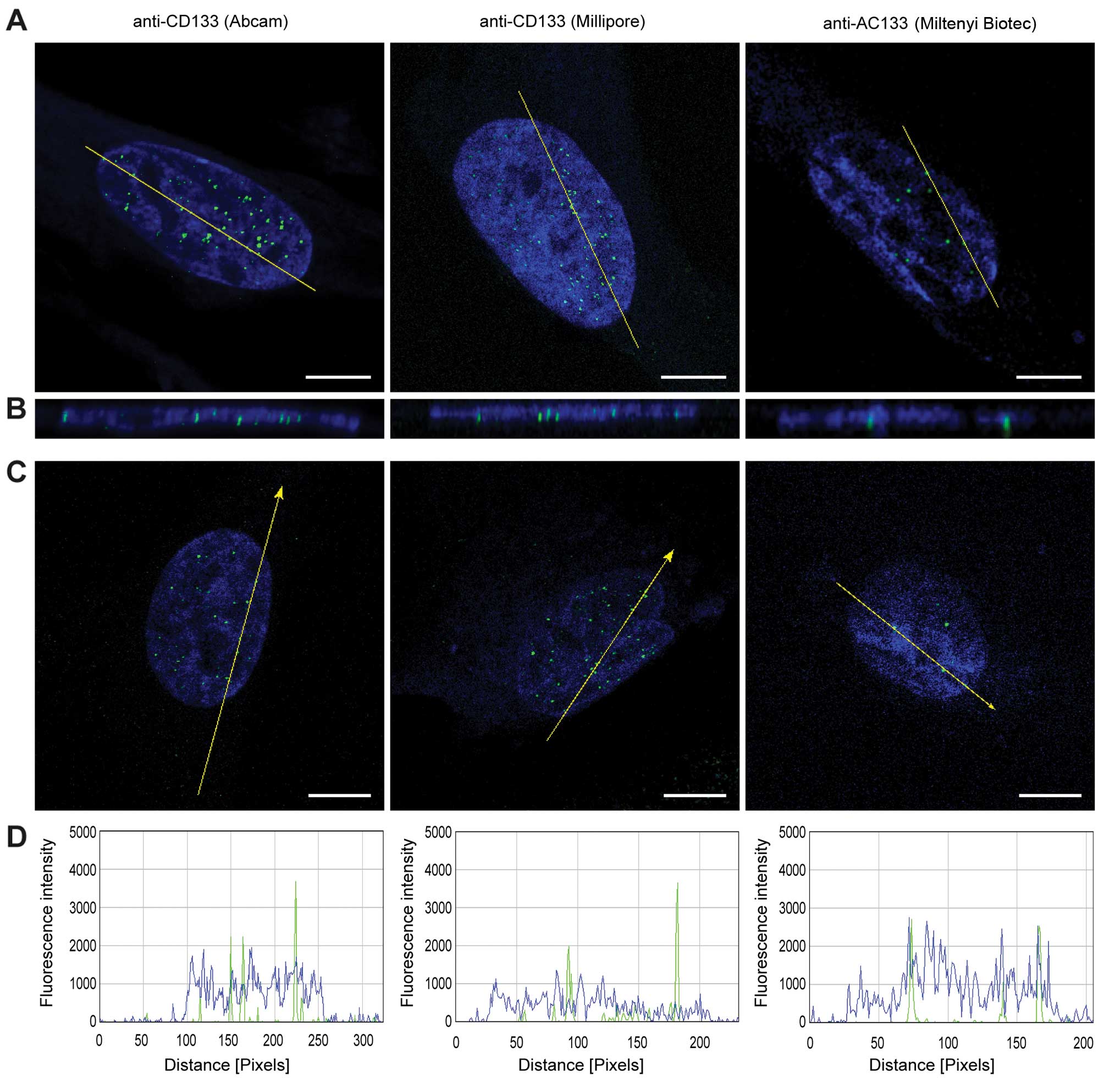
To confirm the presence of CD133 in the nuclei of
the RMS cells visualized using indirect immunofluorescence, we
employed confocal microscopy and software cross-section analysis
through these CD133-positive nuclei (Fig. 3). As is apparent from the results,
the localization of the fluorescence signal for CD133 was detected
within the cell nuclei both on the software cross-sections
(Fig. 3B) and on the plot
diagrams of the fluorescence intensity (Fig. 3D).
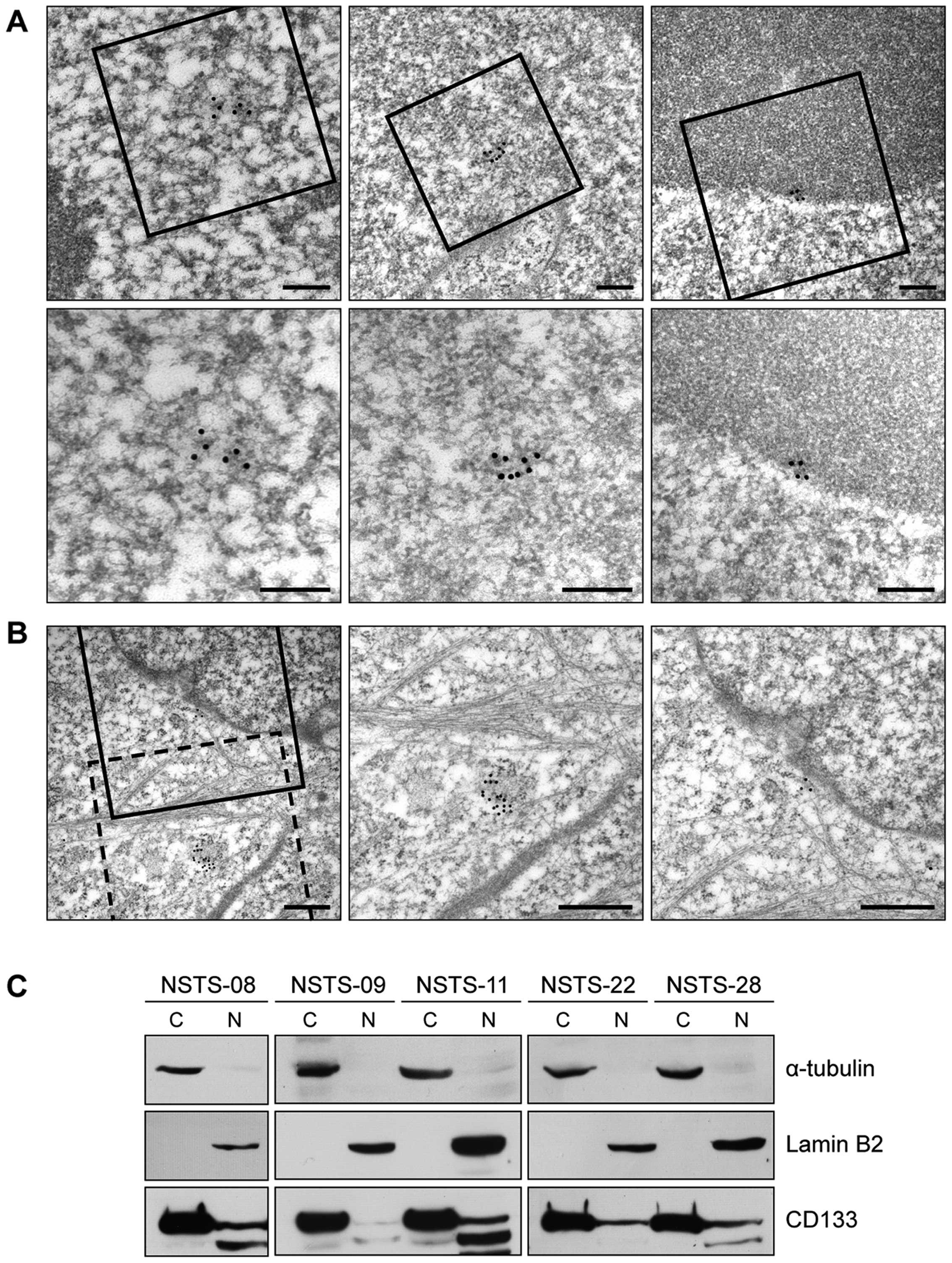
Furthermore, we also used immunogold labeling with
TEM to verify the localization of CD133 in the nuclei of the RMS
cells. To avoid any artifacts associated with this methodological
approach, the accumulation of three or more gold particles together
was considered to indicate a positive signal. The results clearly
indicated the presence of CD133 in both the nuclei and nucleoli
(Fig. 4A and B).
These results are all completely consistent with the
microscopic observations described above (Fig. 2): the software cross-sections also
showed clear, punctuate signals for CD133 within the nucleus
(Fig. 3B and D), i.e., no diffuse
positivity throughout the entire nucleus was observed.
Nevertheless, the TEM micrographs also showed the presence of CD133
in the nucleoli (Fig. 4B), and
this observation corresponds with the diffuse positivity for CD133
observed in some of the nucleoli (Fig. 2B). In addition to cells with the
typical membrane positivity or exclusive nuclear positivity for
CD133, we also sporadically noted clusters of positive signals in
the cytoplasm near the cell nucleus or very close to the nuclear
envelope (Fig. 4B).
Final confirmation of the results achieved through
microscopic methods was carried out by immunoblot analysis of the
cytoplasmic and nuclear fractions of all five RMS cell lines. The
presence of CD133-specific bands of various intensities was
detected in all nuclear fractions, in addition to the strong
CD133-specific bands in the cytoplasmic fractions (Fig. 4C). The purity of both subcellular
fractions was confirmed by the presence/absence of α-tubulin and
lamin B2. These results are completely in accordance with our other
findings (reported above) achieved by indirect immunofluorescence
and TEM, i.e., in all five RMS cell lines, the presence of a small
subpopulation of cells with CD133 in the nucleus was revealed.
Discussion
As described above, we unexpectedly identified an
atypical nuclear localization of CD133 in a relatively stable
subset of cells in five RMS cell lines established in our
laboratory. To date, this atypical localization of CD133 was
described in one case report on breast cancer (14) and in a large study of prognostic
markers on lung cancer (15)
using immunohistochemical methods. Nevertheless, published data on
CD133 expression in human cancer cells are partly inconsistent,
possibly due to different analytical tools, as well as
methodological limitations and pitfalls (2). For this reason, results obtained by
immunohistochemistry or flow cytometry must be confirmed with
alternative antibodies and should be complemented by the
utilization of different detection methods of either protein or
transcript (2). Furthermore, the
glycosylation of CD133 epitopes in relation to the CSC phenotype
should be also taken into account, particularly if the antibodies
against AC133 epitope are commercially available (17).
In this study, we verified the nuclear localization
of CD133 in RMS cells using three independent anti-CD133
antibodies, including both rabbit polyclonal and mouse monoclonal
antibodies (Fig. 1). Indirect
immunofluorescence and confocal microscopy followed by software
cross-section analysis, TEM and cell fractionation with
immunoblotting were also employed, and all the results undeniably
confirmed the presence of CD133 in the nuclei of stable minor
subpopulations of all five RMS cell lines.
These results strongly support the hypothesis that a
stable subpopulation of cells with nuclear positivity for CD133 is
a common phenomenon in RMS cell lines. Surprisingly, and to the
best of our knowledge, similar results have not been reported to
date for RMS cells. Although certain micrographs from our previous
study showed cells with an accumulation of fluorescent signal for
CD133 in the nucleus (6), we
assumed that this finding was an artifact resulting from the use of
a rabbit polyclonal antibody against CD133, (which was the only
anti-CD133 antibody available at the time), although we had never
detected similar nuclear positivity for CD133 in osteosarcoma or
glioblastoma cell lines using the same antibody (5,18).
Other authors investigating CD133 expression in RMS and RMS cell
lines have not described the pattern of CD133 positivity in detail,
and no micrographs of the individual cells are available in their
published articles (9,10).
Very recently, one publication has mentioned the
nuclear localization of CD133 in triple-negative breast cancer
cells as revealed by immunohistochemistry; nevertheless, this study
is a case report based on only one simple descriptive method and
therefore does not include any continuing systematic analysis of
this apparently interesting finding. Moreover, two of three methods
listed in this article, quantitative RT-PCR and flow cytometry, are
not suitable for identifying the cell surface, cytoplasmic or
nuclear localization of any protein (14).
To date, another study concerning the possible value
of CD133 as a prognostic indicator of survival in patients with
non-small cell lung cancer (NSCLC) was just published. These
interesting results suggest that CD133 expression in the nucleus of
NSCLC cells was related to tumor diameter, tumor differentiation
and the TNM stage. Kaplan-Meier survival and Cox regression
analyses revealed that a high CD133 expression in the nucleus, as
well as in the cytoplasm also predicted the poor prognosis of NSCLC
(15).
As mentioned above, in addition to cells with the
typical membrane positivity or exclusive nuclear positivity for
CD133, clusters of positive signals in the cytoplasm near the cell
nucleus or very close to the nuclear envelope were also
sporadically noted. This finding is in accordance with our
previously published observations of sporadic cytoplasmic
positivity for CD133 in RMS cells (6), as well as with the deposition of
CD133 in cytoplasmic vesicles that has been described in
osteosarcoma (19) and the
recently suggested mechanism of CD133 internalization and
trafficking into lysosomes through interactions between CD133 and
the histone deacetylase HDAC6 (20).
Taken together, our results undeniably confirmed the
presence of CD133 in the cell nuclei of stable minor subpopulations
in RMS cell lines. These results, although surprising and novel,
were achieved through three independent methods using three
independent antibodies purchased from three separate suppliers.
Nevertheless, the main question of what is the exact
role of CD133 in the nucleus of RMS cells remains unanswered. In a
previous study on breast cancer, the authors suggested that CD133
in the nucleus may act as transcriptional regulator and is most
likely associated with a poor prognosis; however, this conclusion
is largely speculative in this case report (14). By contrast, the most recent
findings on NSCLC undoubtedly proved the association of nuclear
positivity for CD133 poor prognosis in these patients (15).
Although a similar function of another type of
surface molecule internalized into the cell nucleus, receptor
tyrosine kinases, has been reported (21–23), CD133 belongs to a distinct class
of cell membrane proteins, and the analogies to this process are
therefore limited. Furthermore, recent studies also discuss the
involvement of internalized CD133 in cell signaling pathways, such
as the canonical Wnt pathway (20), or report an association between
CD133 and the PI3K/Akt pathway (24–26). Regardless, the elucidation of the
possible role of CD133 in the nucleus of cancer cells should be
based on detailed descriptions of the localization and interactions
of CD133 with other molecules in the cell nucleus. These
experiments will be the focus of our upcoming study on this
interesting phenomenon.
Acknowledgments
This study was supported by grant IGA NT13443-4 of
the Ministry of Healthcare of the Czech Republic. We thank Johana
Maresova and Dobromila Klemova for their skilful technical
assistance.
Abbreviations:
|
BSA
|
bovine serum albumin
|
|
CSCs
|
cancer stem cells
|
|
DMEM
|
Dulbecco’s modified Eagle’s medium
|
|
FISH
|
fluorescence in situ
hybridization
|
|
HRP
|
horseradish peroxidase
|
|
NSCLC
|
non-small cell lung cancer
|
|
PBS
|
phosphate-buffered saline
|
|
PVDF
|
polyvinylidene difluoride
|
|
RMS
|
rhabdomyosarcoma
|
|
SDS
|
sodium dodecyl sulfate
|
|
TBS
|
Tris-buffered saline
|
|
TEM
|
transmission electron microscopy
|
References
|
1
|
Corbeil D, Karbanová J, Fargeas CA and
Jászai J: Prominin-1 (CD133): Molecular and cellular features
across species. Adv Exp Med Biol. 777:3–24. 2013. View Article : Google Scholar
|
|
2
|
Grosse-Gehling P, Fargeas CA, Dittfeld C,
Garbe Y, Alison MR, Corbeil D and Kunz-Schughart LA: CD133 as a
biomarker for putative cancer stem cells in solid tumours:
Limitations, problems and challenges. J Pathol. 229:355–378. 2013.
View Article : Google Scholar
|
|
3
|
Friedman GK and Gillespie GY: Cancer stem
cells and pediatric solid tumors. Cancers (Basel). 3:298–318. 2011.
View Article : Google Scholar
|
|
4
|
Xia P: Surface markers of cancer stem
cells in solid tumors. Curr Stem Cell Res Ther. 9:102–111. 2014.
View Article : Google Scholar
|
|
5
|
Veselska R, Hermanova M, Loja T, Chlapek
P, Zambo I, Vesely K, Zitterbart K and Sterba J: Nestin expression
in osteosarcomas and derivation of nestin/CD133 positive
osteosarcoma cell lines. BMC Cancer. 8:3002008. View Article : Google Scholar : PubMed/NCBI
|
|
6
|
Sana J, Zambo I, Skoda J, Neradil J,
Chlapek P, Hermanova M, Mudry P, Vasikova A, Zitterbart K, Hampl A,
et al: CD133 expression and identification of CD133/nestin positive
cells in rhabdomyosarcomas and rhabdomyosarcoma cell lines. Anal
Cell Pathol (Amst). 34:303–318. 2011. View Article : Google Scholar
|
|
7
|
Di Fiore R, Santulli A, Ferrante RD,
Giuliano M, De Blasio A, Messina C, Pirozzi G, Tirino V, Tesoriere
G and Vento R: Identification and expansion of human
osteosarcoma-cancer-stem cells by long-term 3-aminobenzamide
treatment. J Cell Physiol. 219:301–313. 2009. View Article : Google Scholar : PubMed/NCBI
|
|
8
|
Tirino V, Desiderio V, Paino F, De Rosa A,
Papaccio F, Fazioli F, Pirozzi G and Papaccio G: Human primary bone
sarcomas contain CD133+ cancer stem cells displaying
high tumorigenicity in vivo. FASEB J. 25:2022–2030. 2011.
View Article : Google Scholar : PubMed/NCBI
|
|
9
|
Walter D, Satheesha S, Albrecht P,
Bornhauser BC, D’Alessandro V, Oesch SM, Rehrauer H, Leuschner I,
Koscielniak E, Gengler C, et al: CWS Study Group: CD133 positive
embryonal rhabdomyosarcoma stem-like cell population is enriched in
rhabdospheres. PLoS One. 6:e195062011. View Article : Google Scholar
|
|
10
|
Pressey JG, Haas MC, Pressey CS, Kelly VM,
Parker JN, Gillespie GY and Friedman GK: CD133 marks a myogenically
primitive subpopulation in rhabdomyosarcoma cell lines that are
relatively chemoresistant but sensitive to mutant HSV. Pediatr
Blood Cancer. 60:45–52. 2013. View Article : Google Scholar
|
|
11
|
Veselska R, Skoda J and Neradil J:
Detection of cancer stem cell markers in sarcomas. Klin Onkol.
25:2S16–2S20. 2012.PubMed/NCBI
|
|
12
|
Dela Cruz FS: Cancer stem cells in
pediatric sarcomas. Front Oncol. 3:1682013. View Article : Google Scholar : PubMed/NCBI
|
|
13
|
Satheesha S and Schafer BW: Cancer stem
cells in pediatric sarcomas. Stem Cells and Cancer Stem Cells.
Hayat MA: Springer; Dordrecht: pp. 111–126. 2014, View Article : Google Scholar
|
|
14
|
Cantile M, Collina F, D’Aiuto M, Rinaldo
M, Pirozzi G, Borsellino C, Franco R, Botti G and Di Bonito M:
Nuclear localization of cancer stem cell marker CD133 in
triple-negative breast cancer: A case report. Tumori. 99:e245–e250.
2013.PubMed/NCBI
|
|
15
|
Huang M, Zhu H, Feng J, Ni S and Huang J:
High CD133 expression in the nucleus and cytoplasm predicts poor
prognosis in non-small cell lung cancer. Dis Markers.
2015:9860952015. View Article : Google Scholar : PubMed/NCBI
|
|
16
|
Veselska R, Kuglik P, Cejpek P, Svachova
H, Neradil J, Loja T and Relichova J: Nestin expression in the cell
lines derived from glioblastoma multiforme. BMC Cancer. 6:322006.
View Article : Google Scholar : PubMed/NCBI
|
|
17
|
Bidlingmaier S, Zhu X and Liu B: The
utility and limitations of glycosylated human CD133 epitopes in
defining cancer stem cells. J Mol Med Berl. 86:1025–1032. 2008.
View Article : Google Scholar : PubMed/NCBI
|
|
18
|
Loja T, Chlapek P, Kuglik P, Pesakova M,
Oltova A, Cejpek P and Veselska R: Characterization of a GM7
glioblastoma cell line showing CD133 positivity and both
cytoplasmic and nuclear localization of nestin. Oncol Rep.
21:119–127. 2009.
|
|
19
|
Tirino V, Desiderio V, d’Aquino R, De
Francesco F, Pirozzi G, Graziano A, Galderisi U, Cavaliere C, De
Rosa A, Papaccio G and Giordano A: Detection and characterization
of CD133+ cancer stem cells in human solid tumours. PLoS
One. 3:e34692008. View Article : Google Scholar
|
|
20
|
Mak AB, Nixon AM, Kittanakom S, Stewart
JM, Chen GI, Curak J, Gingras AC, Mazitschek R, Neel BG, Stagljar I
and Moffat J: Regulation of CD133 by HDAC6 promotes β-catenin
signaling to suppress cancer cell differentiation. Cell Rep.
2:951–963. 2012. View Article : Google Scholar : PubMed/NCBI
|
|
21
|
Wang SC and Hung MC: Nuclear translocation
of the epidermal growth factor receptor family membrane tyrosine
kinase receptors. Clin Cancer Res. 15:6484–6489. 2009. View Article : Google Scholar : PubMed/NCBI
|
|
22
|
Mills IG: Nuclear translocation and
functions of growth factor receptors. Semin Cell Dev Biol.
23:165–171. 2012. View Article : Google Scholar
|
|
23
|
Carpenter G and Liao HJ: Receptor tyrosine
kinases in the nucleus. Cold Spring Harb Perspect Biol.
5:a0089792013. View Article : Google Scholar : PubMed/NCBI
|
|
24
|
Takenobu H, Shimozato O, Nakamura T,
Ochiai H, Yamaguchi Y, Ohira M, Nakagawara A and Kamijo T: CD133
suppresses neuroblastoma cell differentiation via signal pathway
modification. Oncogene. 30:97–105. 2011. View Article : Google Scholar
|
|
25
|
Wei Y, Jiang Y, Zou F, Liu Y, Wang S, Xu
N, Xu W, Cui C, Xing Y, Liu Y, et al: Activation of PI3K/Akt
pathway by CD133-p85 interaction promotes tumorigenic capacity of
glioma stem cells. Proc Natl Acad Sci USA. 110:6829–6834. 2013.
View Article : Google Scholar : PubMed/NCBI
|
|
26
|
Shimozato O, Waraya M, Nakashima K, Souda
H, Takiguchi N, Yamamoto H, Takenobu H, Uehara H, Ikeda E,
Matsushita S, et al: Receptor-type protein tyrosine phosphatase κ
directly dephosphorylates CD133 and regulates downstream AKT
activation. Oncogene. 34:1949–1960. 2014. View Article : Google Scholar
|