Introduction
Head and neck squamous cell carcinoma (SCC)
constitutes an entire group of epithelial cancer types, including
cancer of the lip, oral, salivary glands, pharynx and larynx
(1). According to one estimate,
600,000 incident cases of HNSCC occur every year worldwide
(2). At present, surgery is
performed in combination with radiotherapy or chemotherapy to treat
cancer of the head and neck (3,4).
However, these treatment strategies generally result in significant
side effects and decreases in patient quality of life. In
particular, chemotherapy started at an early stage may be
effective; however, cancer cells often develop resistance and
delivering large amounts of the drug locally to specific tumor
sites remains a challenge (5,6).
Therefore, the development of strategies that increase the local
concentration of drugs in the tumor tissues is required.
In this regard, resveratrol (RSV), a naturally
occurring polyphenolic compound (flavonoid) has been demonstrated
to exhibit multiple key properties, including anti-inflammatory,
anti-oxidant, anti-aging, cardio-neuro protective and antitumor
effects (7-9). RSV serves an important role in the
suppression of cancer cell proliferation, tumor ablation,
angiogenesis and cancer metastasis (10). RSV has been revealed to inhibit
the proliferation of HL60, MCF-7, SW480 and U251 glioma cells by
inducing apoptosis (11,12). However, the efficacy of RSV is
limited due to its low levels of solubility; therefore, the present
study aimed to combine RSV with a nanocarrier and evaluate its
anticancer effect in head and neck cancer (13).
Nanoparticles have been wide investigated as drug
delivery carriers with cancer targeting applications. A poorly
soluble drug may be stably incorporated in the nanoparticles and
thereby its solubility and bioavailability is improved (14-16). Among all the carriers, liposomes
have been widely studied for their suitability for systemic
application (17). At present, a
number of commercial liposomal formulations are available including
Doxil®, Myocet® and LipoPlatin (18,19). The systemic circulation of
liposomes may be improved by the surface conjugation of
polyethylene glycol (PEG) as a hydrophilic shell. The nanosize and
hydrophilic layer of PEG confers prolonged blood circulation and
decreases the level of reticuloendothelial (RES)-mediated plasma
clearance (20,21). Nanoparticles may passively
accumulate in the tumor tissues via the enhanced permeation and
retention (EPR) effect. In order to additionally increase the
cancer specificity, liposomes may be conjugated with specific
ligands that bind to the extracellular domains of cancer cells
(22). Among all the molecular
targets, epidermal growth factor receptor (EGFR) is overexpressed
in head and neck cancer and its activation is expected to inhibit
the tumor cell proliferation and enhance apoptosis in cancer cells
(23,24). Various EGFR inhibitors include
tyrosine kinase inhibitors, immunoconjugates, oligonucleotides and
epidermal growth factor (EGF). A number of studies have conjugated
EGF on the surface of nanoparticles to target cancer cells
(25-27). In the present study, the
dodecapeptide YHWYGYTPQNVI (GE11) was conjugated onto the surface
of nanoparticles. The GE11 peptide is able to selectively bind to
the EGFR but with decreased mitogenic activity compared with that
of other EGF blockers (28).
In the present study, liposomes and nanoparticles
conjugated with the GE11 surface peptide were prepared to increase
the anticancer effects of RSV. We hypothesized that surface
conjugation of the liposome with the targeting ligand would improve
the anticancer effect effectively compared with that of
non-targeted nanoparticles. The targeting efficiency of the
nanoparticles was examined in squamous cell carcinoma (SCC) HN
cancer cells. The anticancer effect was evaluated using MTT
cytotoxicity and apoptosis assays in EGFR-overexpressing SCC HN
cells.
Materials and methods
Materials
Egg L-a-phosphatidylcholine (EPC) and
diste-roylphosphatidylethanolamine-PEG (2000) (DSPE-PEG), and
1,2-distearoyl-sn-glycero-3-phosphoethanolamine-N-PEG-maleimide
(DSPE-PEG2000-MAL) were purchased from Avanti Polar Lipids, Inc.
(Alabaster, AL, USA). Cholesterol and RSV were purchased from
Sigma-Aldrich; Merck KGaA (Darmstadt, Germany). GE11
(YHWYGYTPQNVIGGGGC), which is a specific peptide for EGFR, was
purchased from GL Biochem (Shanghai) Ltd. (Shanghai, China). All
other chemicals were of reagent grade and used without additional
purification.
Preparation of RSV-loaded GE11-conjugated
liposomes
The RSV-loaded liposome (RSV-L) was prepared by a
thin-film hydration technique. In brief, EPC, DSPE-PEG,
DSPE-PEG-Mal and cholesterol at a molar fraction of 59:10:5:26 were
added to 2 ml chloroform and placed in a round bottomed flask.
Then, 20% w/w RSV was also added (20% w/w lipid). The organic
solvents were evaporated by rotary evaporator by rotation (0.2 × g)
at 60°C for 1 h and to produce a thin film. PBS was added to the
thin film and hydrated for 1 h at 60°C. The suspension was then
extruded 21 times through a polycarbonate membrane with a pore size
of 200 nm (Avanti Polar Lipids, Inc.). The drug-loaded liposomes
were stored in a refrigerator until use. To conjugate the peptide,
GE11 was dissolved in HEPES buffer (Sigma-Aldrich; Merck KGaA) and
reacted with liposomes containing DSPE-PEG-Mal (1:5 molar ratio)
and incubated at 24°C for 15 h in the dark. The unconjugated GE11
was removed by ultracentrifugation at 24°C and 4,000 × g for 15
min.
Particle size and morphology
The average particle size and polydispersity index,
a measure of uniformity of particle size distribution and surface
charge, were evaluated by dynamic light scattering using a
Nano-Z590 Zetasizer instrument (Malvern Instruments, Ltd., Malvern,
UK). The suspension was diluted with distilled water and examined
at 25°C. The experiment was performed in triplicate using 3
independent samples. The morphology of the particles were evaluated
using transmission electron microscopy (TEM) by field-emission TEM
using a JEM-2100F microscope (JEOL, Ltd., Tokyo, Japan). The
suspension was stained with 2% phosphotungstic acid and placed on a
copper grid for 15 min at 24°C. The sample was dried and observed
under a microscope at ×10,000.
In vitro release study
The dialysis bag was immersed in water for 8 h prior
to the commencement of the study. The samples (RSV-L and RSV-GL; 1
ml drug-loaded suspension) were packed in the dialysis bag and ends
were sealed appropriately. The bag was immersed in PBS (pH 7.4) and
maintained at 37°C. The samples were collected at pre-determined
time intervals and replaced with equal amounts of fresh buffer. The
amount of drug released in the medium was calculated using a high
performance liquid chromatography (HPLC) method. A 10 μl
sample was used. A Jasco HPLC system (pump PU-2089, autosampler
AS-2057 and LC-Net II/ADC controller; Jasco, Inc., Easton, MD, USA)
was coupled to a fluorometric detector (Jasco FP-2020, λ excitation
= 330 nm and λ emission = 374 nm). Synergi 4 μ HydroRP 80A
and Luna 5 μ 100A C18 analytical columns (250×4.60 mm;
Phenomenex, Inc., Torrance, CA, USA) were used. The mobile phase
comprised of methanol: ACN: 0.1% phosphoric acid in water (60:10:30
v/v) with a flow rate of 1 ml/min. Ibuprofen was used as the
internal standard, and injection volume was 20 μl with flow
rate of 1 ml/min and isocratic flow was adapted.
Cell culture
The SCC HN cancer cell line (SCC-VII) was purchased
from Soochow University Cell Bank (Suzhou, China) and grown in
Dulbecco’s modified Eagle’s medium (Thermo Fisher Scientific, Inc.,
Waltham, MA, USA) media supplemented with 10% fetal bovine serum
(Thermo Fisher Scientific, Inc.) and 1% antibiotic mixture. The
cells were grown at 37°C with 65% humidity.
Cellular uptake of GE11-conjugated
liposomes
Rhodamine-B (Sigma-Aldrich; Merck KGaA) was used as
a fluorescent probe to track the intracellular uptake of
nanoparticles. The SCC HN cells were seeded in a 6-well plate
(3×10⁵) and incubated overnight at 37°C. The cells were
then exposed to 10 μg RSV-L and RSV-loaded GE11-conjugated
liposomes (RSV-GL) and incubated for 2 h at 37°C. The cells were
then washed and extracted using 5% trypsin. The cells were washed
again and reconstituted in a PBS buffer. The cellular
internalization of the RSV-L and RSV-GL was studied using a
FACSCalibur flow cytometer (BD Biosciences, San Jose, CA, USA). BD
CellQuest Pro software (version 5.1; BD Biosciences).
Cytotoxicity assay
The cytotoxicity potential of individual formulation
was evaluated by a CellTiter 96® AQueous One Solution
Cell Proliferation MTT assay (Promega Corporation, Madison, WI,
USA). The cells were seeded at a density of 1.5×10⁴
cells/well in a 96-well plate and incubated for 24 h at 37°C. The
old medium was replaced with new medium containing unbound RSV,
RSV-L and RSV-GL, and incubated at 37°C for 24 h with 50 μg
liposome in each group. The following day, the medium was removed,
and cells were washed twice with PBS. The cells were treated with
MTS solution as per the manufacturer’s protocol. The absorbance of
each well was studied using a microplate reader a 490 nm. Control
cells were treated with dimethyl sulfoxide alone and a separate
control was established using non-treated cells. All experiments
were performed in triplicate.
Apoptosis assay
The apoptosis of cancer cell was studied using an
Annexin V-fluorescein isothiocyanate (FITC)/prop-idium iodide (PI)
staining protocol. Briefly, cells were seeded at a density of
2×10⁵ cells/well in a 12-well plate and incubated for 24
h at 37°C. The old medium was replaced with new medium containing
unbound RSV, RSV-L and RSV-GL (0.1 to 10 μg/ml),
respectively and incubated for 24 h at 37°C. Following incubation,
cells were washed twice with PBS and extracted. The cells were
centrifuged at 200 × g for 4 min at 8°C, and the pellets was
reconstituted with binding buffer (100 μl; BD Biosciences).
The cells were stained with 2 μl Annexin V-FITC and 2
μl PI (BD Biosciences) and incubated for 15 min at 24°C. The
volume was made up to 1 ml and examined using a FACSCalibur flow
cytometer (BD Biosciences) and CellQuest Pro software (version 5.1;
BD Biosciences).
In vivo antitumor efficacy analysis and
immunostaining
The animal study was approved by the Institutional
Animal Ethical Committee of the Shenzhen Peking University-Hong
Kong University of Science and Technology Medical Center (Shenzhen,
China). Female nude mice (~20 g; 5 weeks old) were purchased from
the Chinese Academy of Sciences (Shanghai, China). Animals were
housed in separate cages (16 animals with 4 animals per cage) and
maintained under a controlled atmosphere (50±7% humidity, 21±1°C)
with a 12 h light/dark cycle. The animals had free access to food
and water throughout the study period. The tumor xenograft model
was developed by injecting 1×10⁶ cells in 100 μl
growth media into the right flank of female BALB/c nude mice. The
tumors were allowed to grow until 100 mm3 and then
experiments were initiated. The mice were randomly divided into
four groups: The control; RSV; RSV-L; and RSV-GL groups, with 8
mice in each group. The mice were injected with 10 mg/kg RSV
(equivalent concentration) three times (with 3-day intervals) via a
tail vein injection. The tumor volume was measured using Vernier
Caliper and calculated using the formula V = (length ×
width2)/2. All animals were sacrificed, and tumors were
surgically removed and examined histologically. Hematoxylin and
eosin (H&E) staining was performed on the tumor slices
following fixing with 10% formalin solution for 20 min at 24°C. The
images were obtained at ×40 magnification using a fluorescence
microscope (1X81; Olympus Corporation, Tokyo, Japan).
Statistical analysis
Statistical analysis was performed using Student’s
t-test for pairs of groups and one-way analysis of variance
followed by Tukey’s post hoc test for multiple groups in Excel
(Microsoft Corporation, Redmond, CA, USA). All data are expressed
as the mean ± standard deviation. P<0.05 was considered to
indicate a statistically significant difference. For the cell
viability assay, data were assessed using GraphPad Prism software
v.7.0 (GraphPad Software, Inc., La Jolla, CA, USA).
Results and Discussion
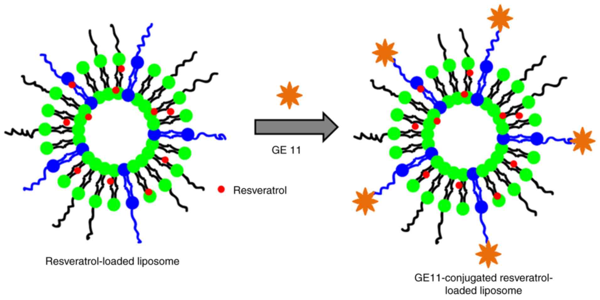
In the present study, the dodecapeptide GE11 was
conjugated onto the surface of nanoparticles (Fig. 1). RSV, which serves an important
role in the suppression of cancer cell proliferation, tumor
ablation, angiogenesis and cancer metastasis, was selected as the
nanoparticle. We hypothesized that surface conjugation of liposome
with targeting ligand would improve the anticancer effect
effectively compared with that of non-targeted nanoparticles.
Physicochemical characterizations of
RSV-GL
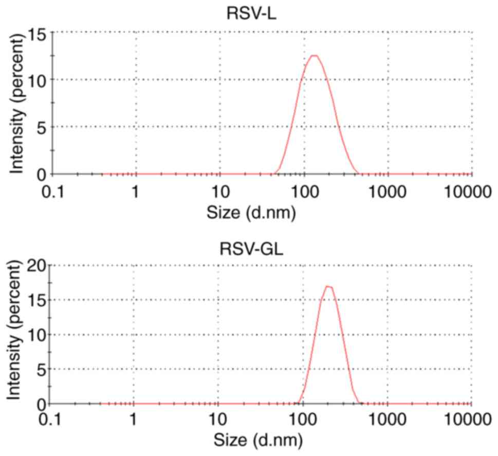
The average particle size of RSV-GL was observed to
be ~185 nm compared with ~130 nm for RSV-L (Table I) with a surface charge of
-23.4±0.98 mV. The increase in particle size in the case of RSV-GL
was primarily attributed to the conjugation of GL on its surface
(Fig. 2). The particle size of
RSV-GL in culture medium was observed to be ~208 nm. A slight
increase in the particle size in culture medium may be due to the
adsorption of proteins on the surface. It has been suggested that
nanocarriers with an average size between 100-200 nm will
extravasate into the tumor tissues preferentially via the EPR
effect (25). General features of
tumors include leaky blood vessels and poor lymphatic drainage. In
these conditions, a nanocarrier may extravasate into the tumor
tissues via the leaky vessels by the EPR effect. The dysfunctional
lymphatic drainage in tumors retains the accumulated nanocarriers
and allows them to release drugs into the vicinity of the tumor
cells. Experiments using liposomes of different mean sizes have
suggested that the threshold vesicle size for extravasation into
tumors is ~400 nm (29,30), but previous studies have indicated
that particles with diameters <200 nm are more effective
(20). In addition, the presence
of PEG on the outer surface will increase the blood circulation.
The small particle size also allows it to escape from RES-based
systemic clearances (31).
 | Table IPhysicochemical characteristics of
drug-loaded formulations. |
Table I
Physicochemical characteristics of
drug-loaded formulations.
| Nanoparticles | Size, nm Surface
charge, mV | Polydispersity
index | Entrapment
efficiency, % | Loading efficiency,
% |
|---|
| Blank L | 104.3±1.65
-21.4±1.14 | 0.089 | - | - |
| RSV-L | 129.5±1.65
-24.1±1.19 | 0.112 | 96.2±1.18 | 8.95±1.28 |
| RSV-GL | 187.5±2.13
-23.4±0.98 | 0.145 | 94.2±1.26 | 7.45±1.57 |
Entrapment efficiency and loading efficiency are two
important parameters that determine the entrapment capacity of the
liposomal carrier. The results of the present study demonstrated
that RSV-GL exhibited a high entrapment efficiency of >95% with
an active drug loading 19.5% w/w, indicating the excellent
characteristics of the present carrier (Table I). This may be attributed to the
fact that RSV has high hydrophobic characteristics that allow
integration into the lipid bilayer of the liposome.
In vitro drug release study
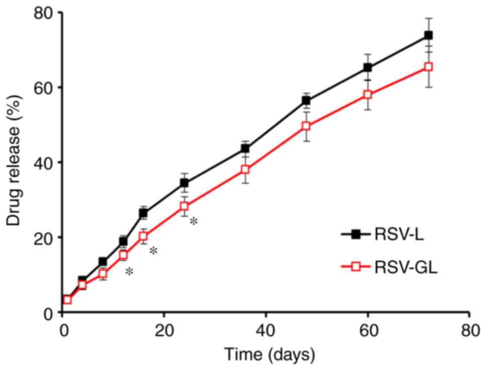
The drug release study was performed in PBS (pH 7.4)
at 37°C. It was observed that RSV was released in a slow and
sustained manner from the RSV-L and RSV-GL nanoparticulate systems
(Fig. 3). For example, ~30% of
RSV was released from the nanocarriers after 24 h, while ~70% of
the drug was released after 72 h from RSV-GL. The slightly
decreased drug release rate observed in the RSV-GL group compared
with RSV-L was primarily attributed to the presence of GL on the
outer surface. During initial time points, significant differences
between the RSV-L and RSV-GL nanoparticles were observed
(P<0.05). It should be noted that no initial high rate of
release was observed, indicating that all of the drugs were stably
loaded into the cores of the nanoparticles, and none had been
absorbed onto the surface of the carrier. This is crucial, as a
sustained release of drugs from the nanocarrier system will be a
beneficial characteristic for its proposed cancer-targeting
applications (32).
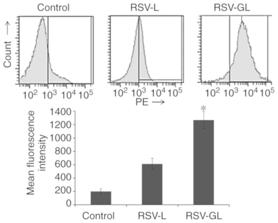
In vitro cellular uptake analysis
The cellular uptake efficiency of individual
nanoparticles was evaluated by FACS analysis. The cancer cells were
treated with respective formulations and cellular uptake was
observed after 2 h incubation (Fig.
4). As observed, rhodamine-B loaded RSV-L exhibited definite
internalization of the carrier in the cancer cells, while RSV-GL
exhibited significantly increased cellular uptake in cancer cells
compared with the RSV-L nanoparticles. The EGFR-overexpressing SCC
HN cells specifically internalized the GL11 surface conjugated
liposome in a manner that was markedly increased compared with that
of the non-targeted carrier. The liposomes were internalized via a
specific receptor-mediated active internalization triggered by the
binding of the GE11 peptides to the EGFR-overexpressing head and
neck cancer cells. This increased cellular uptake of nanocarrier is
expected to increase the therapeutic efficacy in cancer cells
(33,34).
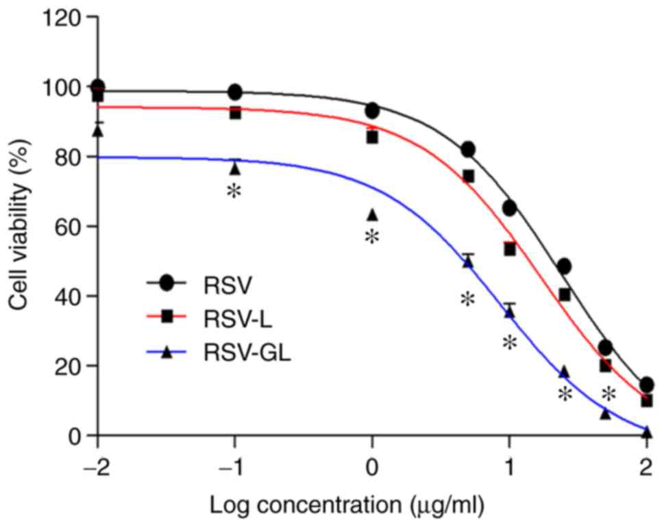
In vitro cytotoxicity assay
The in vitro cytotoxicity of individual
formulations was evaluated by MTT assay. As observed, unbound RSV
and RSV-loaded nanoparticles exhibited a typical time-dependent
cytotoxic effect in head and neck cancer cells (Fig. 5). It was observed that NP
encapsulation of RSV increased the therapeutic effect in cancer
cells. To be specific, RSV-GL exhibited a significantly increased
cytotoxic effect compared with that of the non-targeted
nanoparticles (P<0.05). The half maximal inhibitory
concentration values of unbound RSV, RSV-L and RSV-GL were 4.5,
22.5 and 34.6 μg/ml, respectively. The superior anticancer
effect of RSV-GL was attributed to the specific receptor-mediated
active internalization of RSV-GL, which triggered the binding of
the GE11 peptides to the EGFR-overexpressing head and neck cancer
cells and resulted in increased intracellular concentrations and a
cytotoxic effect.
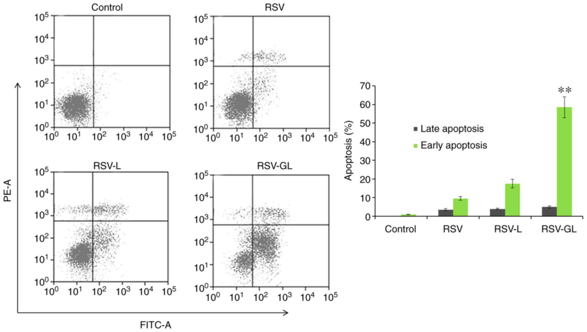
Apoptosis assay
The antitumor efficacy of individual formulations
was additionally evaluated by an apoptosis assay using a flow
cytometer (Fig. 6). For this
assay, cells were treated with respective formulations and then
stained with an Annexin V/PI staining kit. A total of ~9% of the
cells treated with RSV were identified to be in early apoptosis.
Conversely, RSV-L induced a relatively increased apoptosis effect,
with ~17.5% in early and ~4% in late apoptosis. Notably, RSV-GL
induced a significantly increased early (~60%) and late (~5%)
apoptotic effect in head and neck cancer cells (P<0.01) compared
with the RSV-L-treated cells. The increased populations of early
and late apoptotic cells in the RSV-GL-treated cancer cells
compared with control demonstrates an enhanced anticancer effect of
the ESV-GL nanoparticle system. The increased proportion of cells
in early apoptosis compared with late apoptosis may be due to the
limited incubation period of 24 h. The enhanced apoptosis effect
was due to the enhanced cellular accumulation of nanoparticles
attributed to the receptor-mediated endocytosis (35).
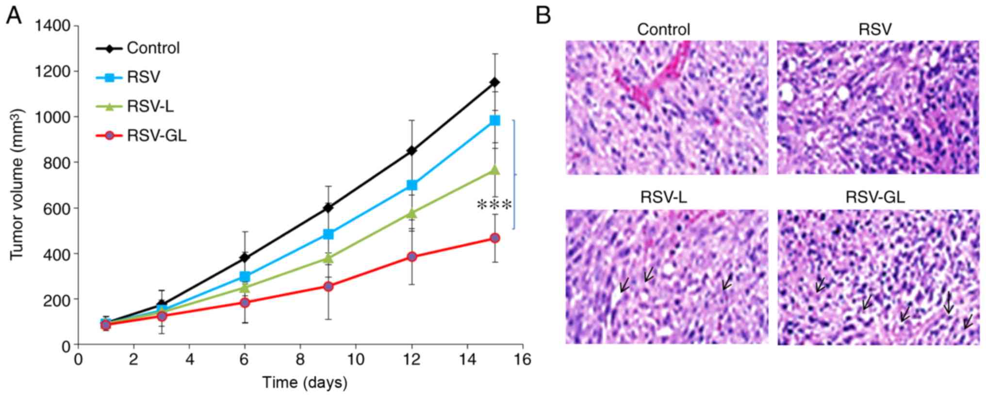
In vivo antitumor efficacy and
immunohistology analysis
The in vivo antitumor efficacy was performed
in SCC-bearing xenograft mice (Fig.
7A). As demonstrated, tumors in the untreated animal model grew
continuously at every time point. The unbound RSV exhibited a
limited effect on tumor growth, and the effect on tumor growth of
RSV-L nanoparticle was slightly improved compared with the unbound
RSV group. Notably, RSV-GL demonstrated significant antitumor
efficacy compared with any other group (P<0.0001). RSV-GL
exhibited a 2-fold decrease in tumor volume compared with the free
RSV group, and a 3-fold decrease in volume compared with the
control. The final tumor volumes were 1,152, 985, 768 and 467
mm3 for the control, unbound RSV, RSV-L and RSV-GL
groups, respectively. The improved anticancer efficacy of RSV-GL
was attributed to the specific affinity of GE11 towards the
overexpressed EGFRs in the cancer cells. The presence of PEG on the
outer surface and smaller particle size attributed to the decreased
tumor burden (36-38). The histological analysis was
performed by H&E staining analysis (Fig. 7B). The tumors in the control group
exhibited darker stained nuclei, indicating continuous tumor cell
proliferation, whereas RSV-GL demonstrated high numbers of
apoptotic nuclei and a marked decrease in the proportion of
cancerous cells.
In conclusion, the GE11-conjugated PEGylated
liposome was successfully prepared to enhance the therapeutic
effect of RSV in head and neck cancer cells. The
EGFR-overexpressing SCC HN cells specifically internalized the GE11
surface conjugated liposome in a manner that was markedly increased
compared with that of the non-targeted carrier. Consistently,
RSV-GL exhibited a significantly increased cytotoxic effect
compared with that of non-targeted NPs. Notably, RSV-GL induced
significantly increased proportions of early (~60%) and late (~10%)
apoptotic head and neck cancer cells. To the best of our knowledge,
the application and development of an EGFR-targeted
peptide-conjugated liposome system for RSV delivery has not been
studied previously in the treatment of head and neck cancer.
Overall, the nanomedicine strategy described in the present study
may potentially advance the chemotherapy-based treatment of head
and neck cancer, with promising applications in other
EGFR-overexpressing tumors.
Funding
The present study was supported by grants from the
Shenzhen Peking University-The Hong Kong University of Science and
Technology Medical Center (grant nos. 2016YFC0104707,
SZSM201512026, ZDSYS201504301045406, 2015A030313889,
JCYJ20170413100222613, JCYJ20170412171856582 and 20171228).
Availability of data and materials
All data generated and analyzed during the present
study are included in this article.
Authors’ contributions
TZ, HF, LL, JP and HX performed the experiments,
contributed to data analysis and wrote the manuscript. TY, ZZ, YL,
YZ, XB, SZ and YS analyzed the data. YC conceptualized the study
design, and contributed to data analysis and experimental
materials. All authors read and approved the final manuscript.
Ethics approval and consent to
participate
The animal study was approved by the Institutional
Animal Ethical Committee of Shenzhen Peking University-Hong Kong
University of Science and Technology Medical Center (Shenzhen,
China).
Patient consent for publication
Not applicable.
Competing interests
The authors declare that they have no competing
interests.
Acknowledgments
Not applicable.
References
|
1
|
Kamangar F, Dores GM and Anderson WF:
Patterns of cancer incidence, mortality, and prevalence across five
continents: Defining priorities to reduce cancer disparities in
different geographic regions of the world. J Clin Oncol.
24:2137–2150. 2006. View Article : Google Scholar : PubMed/NCBI
|
|
2
|
Leemans CR, Braakhuis BJ and Brakenhoff
RH: The molecular biology of head and neck cancer. Nat Rev Cancer.
11:9–22. 2011. View
Article : Google Scholar
|
|
3
|
Marur S and Forastiere AA: Head and neck
cancer: Changing epidemiology, diagnosis, and treatment. Mayo Clin
Proc. 83:489–501. 2008. View
Article : Google Scholar : PubMed/NCBI
|
|
4
|
Vermorken JB and Specenier P: Optimal
treatment for recurrent/ metastatic head and neck cancer. Ann
Oncol. 21(Suppl 7): vii252–vii261. 2010. View Article : Google Scholar
|
|
5
|
Kuczynski EA, Sargent DJ, Grothey A and
Kerbel RS: Drug rechallenge and treatment beyond
progressionimplications for drug resistance. Nat Rev Clin Oncol.
10:571–587. 2013. View Article : Google Scholar : PubMed/NCBI
|
|
6
|
Holohan C, Van Schaeybroeck S, Longley DB
and Johnston PG: Cancer drug resistance: An evolving paradigm. Nat
Rev Cancer. 13:714–726. 2013. View
Article : Google Scholar : PubMed/NCBI
|
|
7
|
Yang CS, Landau JM, Huang MT and Newmark
HL: Inhibition of carcinogenesis by dietary polyphenolic compounds.
Annu Rev Nutr. 21:381–406. 2001. View Article : Google Scholar : PubMed/NCBI
|
|
8
|
Joe AK, Liu H, Suzui M, Vural ME, Xiao D
and Weinstein IB: Resveratrol induces growth inhibition, S-phase
arrest, apoptosis and changes in biomarker expression in several
human cancer cell lines. Clin Cancer Res. 8:893–903.
2002.PubMed/NCBI
|
|
9
|
Jiang H, Zhang L, Kuo J, Kuo K, Gautam SC,
Groc L, Rodriguez AI, Koubi D, Hunter TJ, Corcoran GB, et al:
Resveratrol-induced apoptotic death in human U251 glioma cells. Mol
Cancer Ther. 4:554–561. 2005. View Article : Google Scholar : PubMed/NCBI
|
|
10
|
Buhrmann C, Shayan P, Kraehe P, Popper B,
Goel A and Shakibaei M: Resveratrol induces chemosensitization to
5-fluorouracil through up-regulation of intercellular junctions,
Epithelial-to-mesenchymal transition and apoptosis in colorectal
cancer. Biochem Pharmacol. 98:51–68. 2015. View Article : Google Scholar : PubMed/NCBI
|
|
11
|
Shen M, Wu RX, Zhao L, Li J, Guo HT, Fan
R, Cui Y, Wang YM, Yue SQ and Pei JM: Resveratrol attenuates
ischemia/reperfusion injury in neonatal cardiomyocytes and its
underlying mechanism. PLoS One. 7:e512232012. View Article : Google Scholar
|
|
12
|
Ku CR, Lee HJ, Kim SK, Lee EY, Lee MK and
Lee EJ: Resveratrol prevents streptozotocininduced diabetes by
inhibiting the apoptosis of pancreatic β-cell and the cleavage of
poly (ADP-ribose) polymerase. Endocr J. 59:103–109. 2012.
View Article : Google Scholar
|
|
13
|
Walle T, Hsieh F, DeLegge MH, Oatis JE Jr
and Walle UK: High absorption but very low bioavailability of oral
resveratrol in humans. Drug Metab Dispos. 32:1377–1382. 2004.
View Article : Google Scholar : PubMed/NCBI
|
|
14
|
Ramasamy T, Kim JH, Choi JY, Tran TH, Choi
HG, Yong CS and Kim JO: pH sensitive polyelectrolyte complex
micelles for highly effective combination chemotherapy. J Material
Chem B. 2:63242014. View Article : Google Scholar
|
|
15
|
Hare JI, Lammers T, Ashford MB, Puri S,
Storm G and Barry ST: Challenges and strategies in anti-cancer
nanomedicine development: An industry perspective. Adv Drug Deliv
Rev. 108:25–38. 2017. View Article : Google Scholar
|
|
16
|
Sundaramoorthy P, Ramasamy T, Mishra SK,
Jeong KY, Yong CS, Kim JO and Kim HM: Engineering of
caveolae-specific self-micellizing anticancer lipid nanoparticles
to enhance the chemotherapeutic efficacy of oxaliplatin in
colorectal cancer cells. Acta Biomater. 42:220–231. 2016.
View Article : Google Scholar : PubMed/NCBI
|
|
17
|
Allen TM and Cullis PR: Liposomal drug
delivery systems: From concept to clinical applications. Adv Drug
Deliv Rev. 65:36–48. 2013. View Article : Google Scholar
|
|
18
|
Kneidl B, Peller M, Winter G, Lindner LH
and Hossann M: Thermosensitive liposomal drug delivery systems:
State of the art review. Int J Nanomed. 9:4387–4398. 2014.
|
|
19
|
Mohan A, Narayanan S, Balasubramanian G,
Sethuraman S and Krishnan UM: Dual drug loaded nanoliposomal
chemotherapy: A promising strategy for treatment of head and neck
squamous cell carcinoma. Eur J Pharm Biopharm. 99:73–83. 2016.
View Article : Google Scholar
|
|
20
|
Ramasamy T, Ruttala HB, Gupta B, Poudel
BK, Choi HG, Yong CS and Kim JO: Smart chemistry-based nanosized
drug delivery systems for systemic applications: A comprehensive
review. J Control Release. 258:226–253. 2017. View Article : Google Scholar : PubMed/NCBI
|
|
21
|
Ramasamy T, Haidar ZS, Tran TH, Choi JY,
Choi HG, Yong CS and Kim JO: Layer-by-layer assembly of liposomal
nanoparticles with PEGylated polyelectrolytes enhances systemic
delivery of multiple anticancer drugs. Acta Biomaterialia.
10:5116–5127. 2014. View Article : Google Scholar : PubMed/NCBI
|
|
22
|
Rosi NL and Mirkin CA: Nanostructures in
biodiagnostics. Chem Rev. 105:1547–1562. 2005. View Article : Google Scholar : PubMed/NCBI
|
|
23
|
Sheng Q and Liu J: The therapeutic
potential of targeting the EGFR family in epithelial ovarian
cancer. Br J Cancer. 104:1241–1245. 2011. View Article : Google Scholar : PubMed/NCBI
|
|
24
|
Vidal F, de Araujo WM, Cruz AL, Tanaka MN,
Viola JP and Morgado-Díaz JA: Lithium reduces tumorigenic potential
in response to EGF signaling in human colorectal cancer cells. Int
J Oncol. 38:1365–1373. 2011.PubMed/NCBI
|
|
25
|
Acharya S, Dilnawaz F and Sahoo SK:
Targeted epidermal growth factor receptor nanoparticle
bioconjugates for breast cancer therapy. Biomaterials.
30:5737–5750. 2009. View Article : Google Scholar : PubMed/NCBI
|
|
26
|
Chen L, She X, Wang T, He L, Shigdar S,
Duan W and Kong L: Overcoming acquired drug resistance in
colorectal cancer cells by targeted delivery of 5-FU with EGF
grafted hollow meso-porous silica nanoparticles. Nanoscale.
7:14080–14092. 2015. View Article : Google Scholar : PubMed/NCBI
|
|
27
|
Kim MW, Jeong HY, Kang SJ, Choi MJ, You
YM, Im CS, Lee TS, Song IH, Lee CG, Rhee KJ, et al: Cancer-targeted
nucleic acid delivery and quantum dot imaging using EGF receptor
aptamer-conjugated lipid nanoparticles. Sci Rep. 7:94742017.
View Article : Google Scholar : PubMed/NCBI
|
|
28
|
Li Z, Zhao R, Wu X, Sun Y, Yao M, Li J, Xu
Y and Gu J: Identification and characterization of a novel peptide
ligand of epidermal growth factor receptor for targeted delivery of
therapeutics. FASEB J. 19:1978–1985. 2005. View Article : Google Scholar : PubMed/NCBI
|
|
29
|
Peer D, Karp JM, Hong S, Farokhzad OC,
Margalit R and Langer R: Nanocarriers as an emerging platform for
cancer therapy. Nat Nanotechnol. 2:751–760. 2007. View Article : Google Scholar
|
|
30
|
Liu D and Auguste DT: Cancer targeted
therapeutics: From molecules to drug delivery vehicles. J Control
Release. 219:632–643. 2015. View Article : Google Scholar : PubMed/NCBI
|
|
31
|
Huang WC, Chen SH, Chiang WH, Huang CW, Lo
CL, Chern CS and Chiu HC: Tumor microenvironment-responsive
nanoparticle delivery of chemotherapy for enhanced selective
cellular uptake and transportation within tumor. Biomacromolecules.
17:3883–3892. 2016. View Article : Google Scholar : PubMed/NCBI
|
|
32
|
Field LD, Nag OK, Sangtani A, Burns KE and
Delehanty JB: The role of nanoparticles in the improvement of
systemic anticancer drug delivery. Ther Deliv. 9:527–545. 2018.
View Article : Google Scholar : PubMed/NCBI
|
|
33
|
Ruttala HB, Chitrapriya N, Kaliraj K,
Ramasamy T, Shin WH, Jeong JH, Kim JR, Ku SK, Choi HG, Yong CS and
Kim JO: Facile construction of bioreducible crosslinked polypeptide
micelles for enhanced cancer combination therapy. Acta Biomater.
63:135–149. 2017. View Article : Google Scholar : PubMed/NCBI
|
|
34
|
Xu Y, Wang S, Chan HF, Liu Y, Li H, He C,
Li Z and Chen M: Triphenylphosphonium-modified poly(ethylene
glycol)-poly(ε-caprolactone) micelles for mitochondria- targeted
gambogic acid delivery. Int J Pharm. 522:21–33. 2017. View Article : Google Scholar : PubMed/NCBI
|
|
35
|
Sundaramoorthy P, Baskaran R, Mishra SK,
Jeong KY, Oh SH, Kyu Yoo B and Kim HM: Novel self-micellizing
anticancer lipid nanoparticles induce cell death of colorectal
cancer cells. Colloids Surf B Biointerfaces. 135:793–801. 2015.
View Article : Google Scholar : PubMed/NCBI
|
|
36
|
Ma J, Wu H, Li Y, Liu Z, Liu G, Guo Y, Hou
Z, Zhao Q, Chen D and Zhu X: Novel core-interlayer-shell DOX/ZnPc
Co-loaded MSNs@ pH-sensitive CaP@PEGylated liposome for enhanced
synergetic chemo-photodynamic therapy. Pharm Res. 35:572018.
View Article : Google Scholar : PubMed/NCBI
|
|
37
|
Gupta B, Ramasamy T, Poudel BK, Pathak S,
Regmi S, Choi JY, Son Y, Thapa RK, Jeong JH, Kim JR, et al:
Development of bioactive PEGylated nanostructured platforms for
sequential delivery of doxorubicin and imatinib to overcome drug
resistance in metastatic tumors. ACS Appl Mater Interfaces.
9:9280–9290. 2017. View Article : Google Scholar : PubMed/NCBI
|
|
38
|
Tyagi P and Subramony JA: Nanotherapeutics
in oral and parenteral drug delivery: Key learnings and future
outlooks as we think small. J Control Release. 272:159–168. 2018.
View Article : Google Scholar
|