Introduction
Pancreatic cancer is the fourth leading cause of
cancer-related deaths in the United States. As one of the most
aggressive human cancers, the 5-year survival rate of pancreatic
ductal adenocarcinoma (PDAC) is <5% (1). Recent studies suggested that
bioactive compounds from mushrooms could protect against multiple
myeloma, breast and skin cancer (2–5).
Poria cocos (also known as Wolfiporia extensa) is a
medicinal mushroom in the Polyporaceae family that grows in
pine trees and its sclerotium is widely used in traditional Asian
medicine for its sedative, diuretic, digestive and tonic effects
(6–8). Although the anticancer activity of
polysaccharides extracted from P. cocos is associated with
the stimulation of immune response and these polysaccharides
significantly enhance immunopotentiation (9), triterpenes isolated from P.
cocos have a direct inhibitory effect on cancer cells through a
variety of mechanisms including inhibition of cell proliferation,
induction of apoptosis and suppression of invasive behavior
(10–16). However the effect of triterpenes
from P. cocos against pancreatic cancer remains to be
evaluated and the mechanism determined.
In the present study, we evaluated the effects of a
triterpene mixture extracted from P. cocos (PTE) and three
purified triterpenes: pachymic acid (PA), dehydropachymic acid
(DPA) and polyporenic acid C (PPAC), on growth and invasive
behavior of human pancreatic cancer cell lines Panc-1, MiaPaca-2,
BxPc-3 and AsPc-1 and normal pancreatic duct epithelial cell line
HPDE-6. PTE as well as PA, DPA and PPAC inhibit growth of
pancreatic cancer cells and PTE and PA significantly suppress
invasive behavior of BxPc-3 cells by inhibiting expression of
MMP-7. Taken together, our results indicate that triterpenes from
P. cocos may be potentially exploited for the use in
pancreatic cancer intervention.
Materials and methods
Reagents
Dried sclerotium of P. cocos from Fujian,
P.R. China was provided by Professor Zhonglin Yang (China
Pharmaceutical University, Nanjing, P.R. China). It was
authenticated by School of Traditional Chinese Medicine at China
Pharmaceutical University. Voucher specimens were deposited at
State Key Laboratory of Natural Medicines, China Pharmaceutical
University. DMSO was purchased from Sigma (St. Louis, MO). All
other chemicals and reagents were of analytical grade. Anti-MMP-7
and anti-β-actin antibodies were obtained from Santa Cruz
Biotechnology (Santa Cruz, CA).
Extraction and purification
Pulverized sclerotium of P. cocos (2.0 kg)
was extracted three times with 95% ethanol (10 l) under reflux for
3 h at room temperature. The ethanol solution was combined and
evaporated in vacuum to give a crude extract (38 g). The crude
extract was mixed with silica gel G (size: 200–300 mesh) and
fractionated on silica column chromatography by gradient elution
using petroleum ether and ethyl acetate (100:0→75:15→1:1).
Fractions were collected, combined and subjected to further
chromatography on a silica gel→H (size: 60–120 mesh) column by step
gradients of cyclohexane-ethyl acetate (100:0→75:15). The collected
fractions were combined on the basis of their thin-layer
chromatography (TLC) characteristics to give three pooled
fractions: pooled extracts A, B and C (PEA, PEB and PEC), listed in
increasing order of polarity. Part of the PEB (PTE) was subjected
to high-performance preparative liquid chromatography (Daojing,
Japan, model: SPD-20A), from which three pure compounds, PPAC, DPA
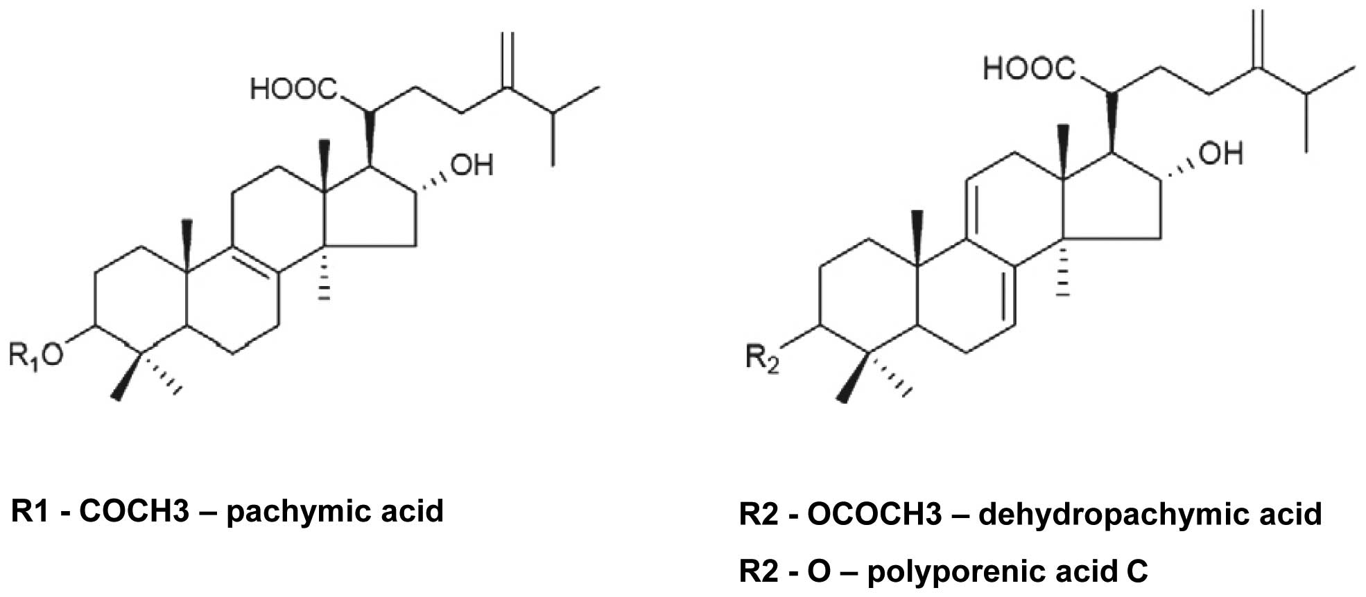
and PA (Fig. 1) were isolated with
isocratic elution of CH3OH-H2O (85:15), with
trifluoroacetic acid added at 0.05%. Quantification of HPLC
analysis demonstrated that PTE contains 55.7% PA, 31.7% DPA and
4.1% PPAC. Identification of PA, DPA and PPAC was conducted by
comparison of their physical and spectroscopic data
(1H-, 13C-NMR and MS) with the corresponding
compounds reported in the literatures. PTE, PA, DPA and PPAC were
dissolved in DMSO at a concentration of 50 mg/ml and 50 mM,
respectively then stored at −20°C.
Cell culture
The human pancreatic cancer cell lines Panc-1,
MiaPaca-2, BxPc-3 and AsPc-1 were obtained from ATCC (Manassas,
VA). Panc-1 cells were maintained in Dulbecco’s modified Eagle’s
medium containing penicillin (50 U/ml), streptomycin (50 U/ml) and
10% fetal bovine serum (FBS). MiaPaca-2 cells were maintained in
Dulbecco’s modified Eagle’s medium containing penicillin (50 U/ml),
streptomycin (50 U/ml), 10% FBS and 2.5% horse serum (HS). BxPC-3
and AsPC-1 cells were maintained in RPMI-1640 medium containing
penicillin (50 U/ml), streptomycin (50 U/ml) and 10% FBS. Media
came from ATCC. Supplements, FBS and HS were obtained from Gibco
BRL (Grand Island, NY). The normal human pancreatic duct epithelial
cell line HPDE-6 was a generous gift from Dr Ming-Sound Tsao
(University of Toronto, Toronto, Ontario, Canada). HPDE-6 cells
were routinely cultured in keratinocyte serum-free (KSF) medium
supplemented by epidermal growth factor and bovine pituitary
extract. Medium and supplements came from Gibco BRL.
Cell proliferation and cell
viability
Human pancreatic cancer cells and normal human
pancreatic duct epithelial cell line were treated with indicated
concentrations of PTE, PA, DPA or PPAC for 24–72 h and cell
proliferation determined as described (17). Cell viability was determined after
incubation with PTE, PA, DPA or PPAC for 24 h by staining with
trypan blue as described (18).
Data are the mean ± SD from three independent experiments.
Cell cycle analysis and invasive behavior
assays
Cell cycle analysis of BxPc-3 cells incubated in the
presence of PTE (0–5.0 μg/ml) for 24 h was evaluated as
described (19). Cell invasion of
BxPc-3 cells treated with PTE (0–5.0 μg/ml), PA (0–5.0
μM) or transfected with MMP-7 or control siRNA were
performed as described (20). Data
points represent the mean ± SD of three individual filters within
one representative experiment repeated at least twice.
DNA microarrays
BxPc-3 cells were treated with PTE (0, 5.0
μg/ml) for 24 h and RNA isolated with RNeasy®
Mini Kit (Qiagen, Valencia, CA). RNA quality was monitored and
quantified using the Qubit® 2.0 Fluorometer (Invitrogen,
Carlsbad, CA). Reverse transcription was performed with High
Capacity cDNA Reverse Transcription Kit (Applied Biosystems, Foster
City, CA) using 2.0 μg total RNA. PCR analysis was performed
on TaqMan® Array Human Pancreatic Adenocarcinoma and
7900HT Fast Real-Time PCR System according to the manufacturer’s
protocol (Applied Biosystems). Analysis of the relative quantity
gene expression (RQ) data was normalized by HPRT1 expression
and was performed using the 2-ΔΔCt method (21).
Western blot analysis
BxPc-3 cells were treated with PTE (0–10
μg/ml), PA (0–10 μM), DPA (0–10 μM) or PPAC
(0–10 μM) for 24 h. Whole cell extracts isolated from cells
were prepared and western blot analysis with MMP-7 antibody was
performed as previously described (17). Western blots were quantified with
HP-Scanjet 550c and analyzed by UN-SCAN-IT software (Silk
Scientific, Orem, UT).
siRNA transfection
BxPc-3 cells were transfected with human
MMP-7 siRNA or control siRNA-A using siRNA Reagent System
according to the manufacturer’s instructions at a final
concentration of 60 nM (Santa Cruz, CA). After 48 h of
transfection, the cells were harvested and MMP-7 knockdown
was verified by western blot analysis.
Statistical analysis
Data are presented as mean ± standard deviation
(SD). Statistical comparison between groups of data was carried out
using ANOVA. P<0.05 was considered to be significant.
Results
Triterpenes from P. cocos suppress
proliferation of human pancreatic cancer cell lines
As previously demonstrated P. cocos
triterpenes: PA, DPA and PPAC inhibited growth of human breast
cancer, lung cancer and prostate cancer cells (11–13,22).
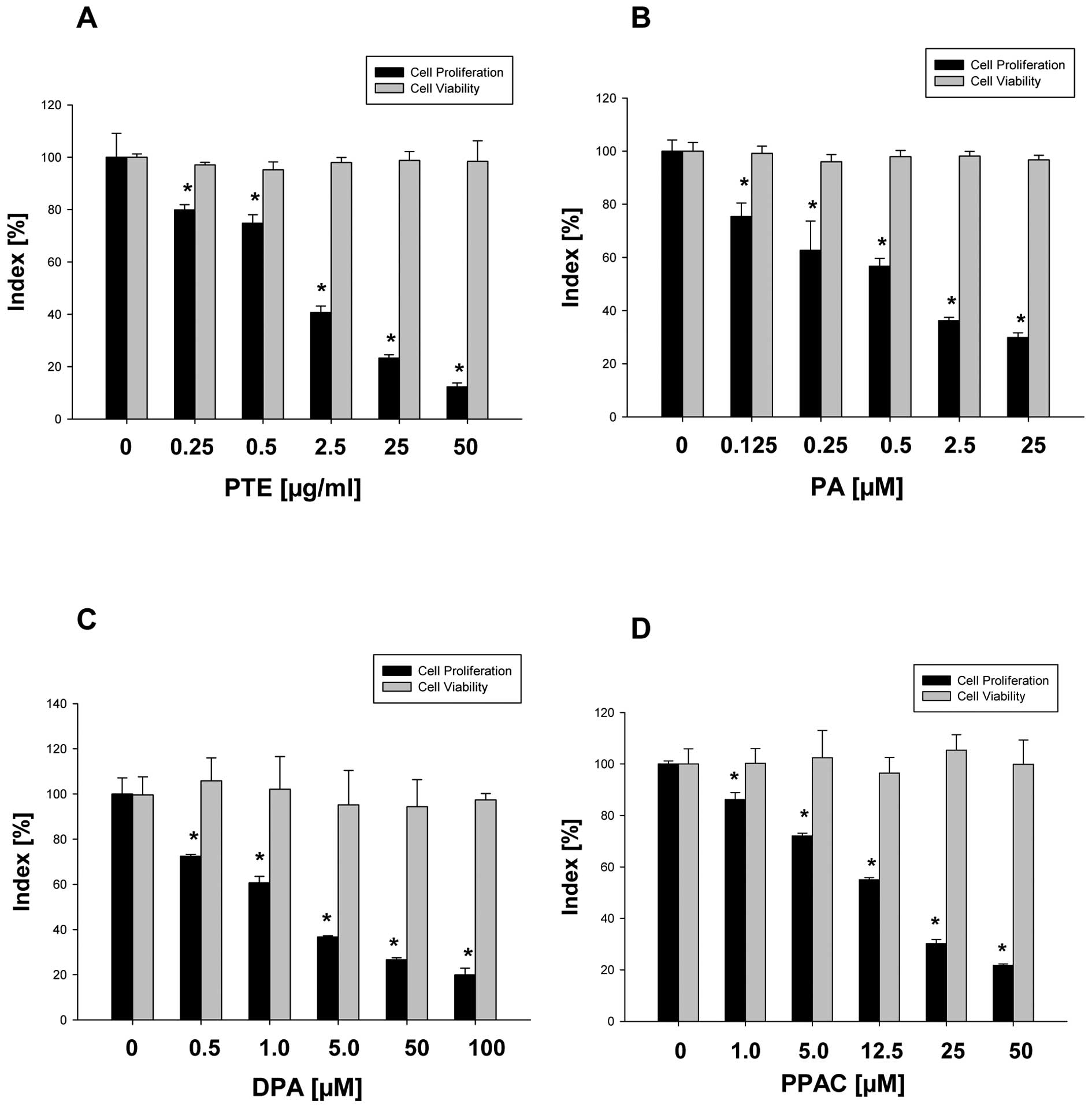
To evaluate whether these triterpenes also affect growth of
different human pancreatic cancer cell lines, Panc-1, MiaPaca-2,
AsPc-1 and BxPc-3 cells were treated with PTE (0–80 μg/ml)
for 24 and 48 h and the proliferation determined as described in
Materials and methods. PTE suppresses proliferation of Panc-1
(IC50-24 h = 28.3 μg/ml, IC50-48 h =
24.5 μg/ml), MiaPaca-2 (IC50-24 h = 29.4
μg/ml, IC50-48 h = 23.0 μg/ml), AsPc-1
(IC50-24 h = 13.7 μg/ml, IC50-48 h =
11.3 μg/ml) and BxPc-3 cells (IC50-24 h = 1.2
μg/ml, IC50-48 h = 1.0 μg/ml). Therefore,
BxPc-3 cells are most sensitive to the PTE as well as PA, DPA and
PPAC treatment (Fig. 2). PA
demonstrates the strongest activity against BxPc-3 cells with
IC50 0.26 μM (24 h), IC50 0.29
μM (48 h), IC50 0.42 μM (72 h) but only
partially affects proliferation of normal human pancreatic duct
epithelial cell line HPDE-6 with IC50 41.6 μM (24
h), IC50 34.9 μM (48 h) and IC50 76.0
μM (72 h). Moreover, PTE, PA, DPA and PPAC treatment do not
affect viability of BxPc-3 cells (Fig.
2), suggesting cytostatic effect of these P. cocos
triterpenes on BxPc-3 cells.
Effect of PTE and PA on the cell cycle
and invasive behavior of BxPc-3 cells
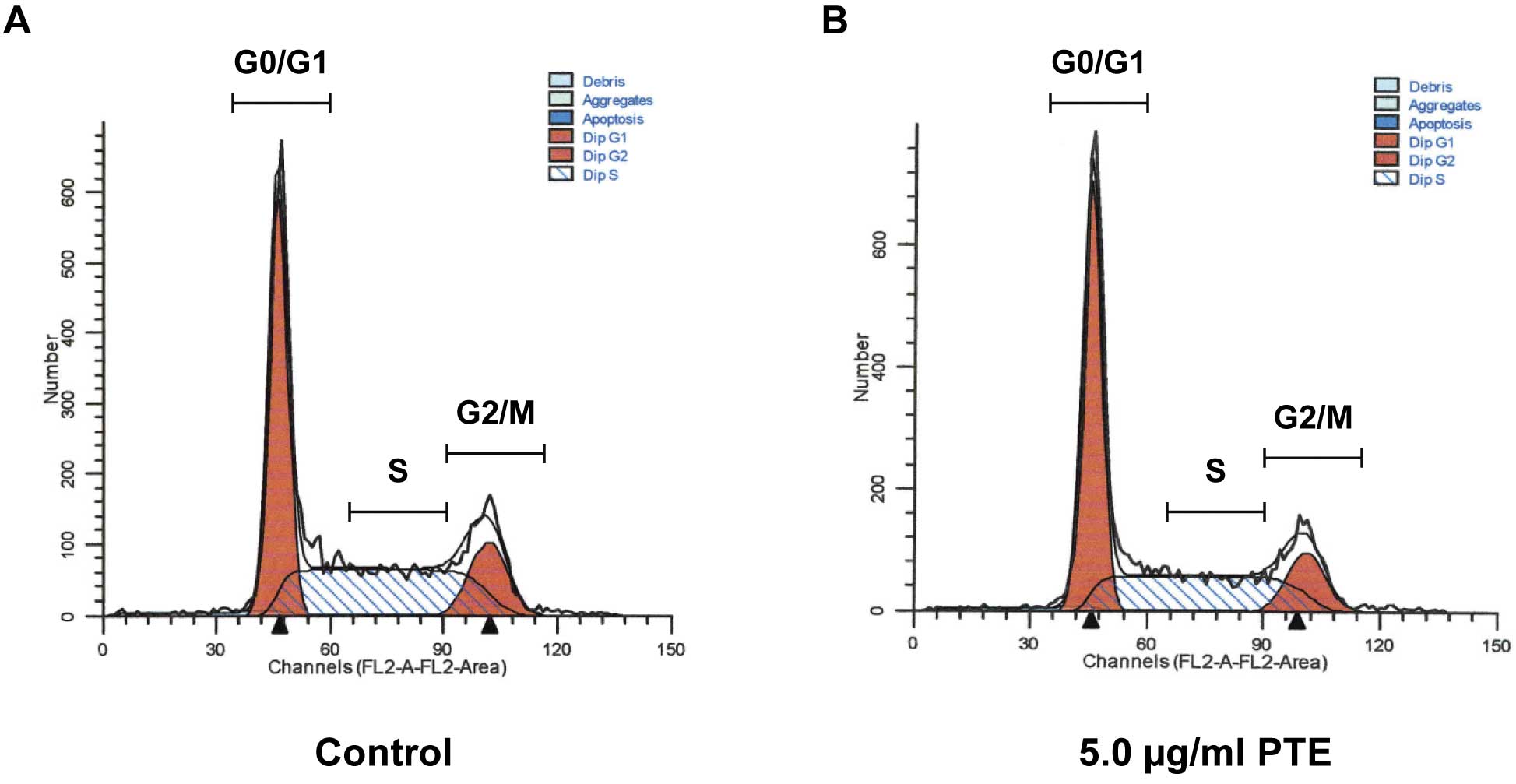
In order to evaluate whether the cytostatic effect
of PTE on pancreatic cancer cells is associated with the cell cycle
arrest, BxPc-3 cells were treated with PTE as described in
Materials and methods. Cell cycle analysis revealed that PTE
induces significant cell cycle arrest at G0/G1 phase from 44.15%
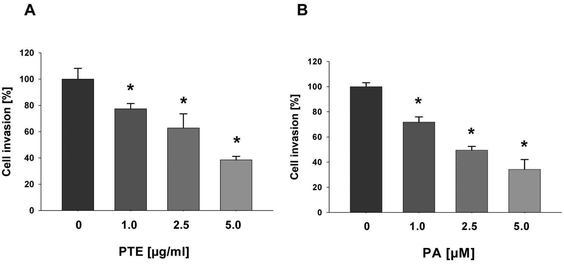
(control) to 47.66% (5.0 μg/ml) (Fig. 3 and Table I). To evaluate whether PTE and PA
suppress invasive behavior of pancreatic cancer cells, BxPc-3 cells
were treated with PTE (0–5.0 μg/ml) and PA (0–5.0 μM)
for 24 h and cell invasion was determined as described Materials
and methods. As seen in Fig. 4,
both PTE and PA markedly inhibit cell invasion through Matrigel.
Together, our data indicate that triterpenes from P. cocos
not only inhibit cell proliferation through cell cycle arrest at
G0/G1 phase but also inhibit invasive behavior of BxPc-3 cells.
 | Table IEffect of PTE on cell cycle
distribution. |
Table I
Effect of PTE on cell cycle
distribution.
| PTE
(μg/ml) | G0/G1 | S | G2/M |
|---|
| 0 | 44.15±1.06 | 40.98±1.01 | 14.87±0.10 |
| 2.5 | 44.79±0.20 | 40.54±1.18 | 14.67±0.98 |
| 5.0 | 47.66±1.57a | 38.63±1.46 | 13.70±1.04 |
Triterpenes from P. cocos downregulate
MMP-7 expression in BxPc-3 cell line
To identify possible molecular targets of
triterpenes from P. cocos, we treated BxPc-3 cells with PTE
and performed DNA-microarray analysis using Array Human Pancreatic
Adenocarcinoma genes as described in Materials and methods. In
addition to KRAS (0.74±0.02, P<0.05 vs. control), PTE
also markedly suppresses expression of MMP-7 (0.67±0.14,
P<0.05 vs control) (Table II).
Since MMP-7 is significantly overexpressed in pancreatic
cancer samples when compared to pseudotumoral chronic pancreatitis
(23), we further determined if
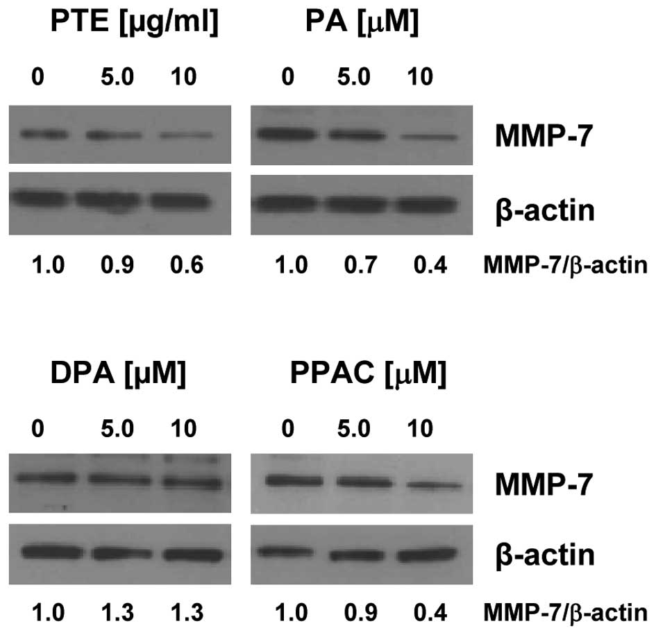
PTE, PA, DPA and PPAC inhibit MMP-7 at the translation level as
well. Western blot analysis shows that in addition to PTE and PA,
PPAC inhibits protein expression of MMP-7 in BxPc-3 cells, whereas
DPA has no effect (Fig. 5).
| Table IIEffect of PTE on the expression of
human pancreatic adenocarcinoma genes. |
Table II
Effect of PTE on the expression of
human pancreatic adenocarcinoma genes.
| Gene | Description | Fold change |
|---|
| BIRC5 | Baculoviral IAP
repeat-containing 5 (survivin) | 0.85±0.12 |
| CCNB1 | Cyclin B1 | 0.87±0.03 |
|
HSP90AA1 | Heat shock protein
90 kDa α (cytosolic), class A member 1 | 0.89±0.02 |
| IL-6 | Interleukin-6 | 0.86±0.04 |
| KRAS | V-Ki-ras2 Kirsten
rat sarcoma viral oncogene homolog | 0.86±0.04a |
| MMP-7 | Matrix
metalloproteinase-7 | 0.67±0.14a |
| MMP-9 | Matrix
metalloproteinase-9 | 0.79±0.08 |
| RELB | Transcription
factor RelB | 0.79±0.10 |
| TGFB3 | Transforming growth
factor β-3 | 0.82±0.02 |
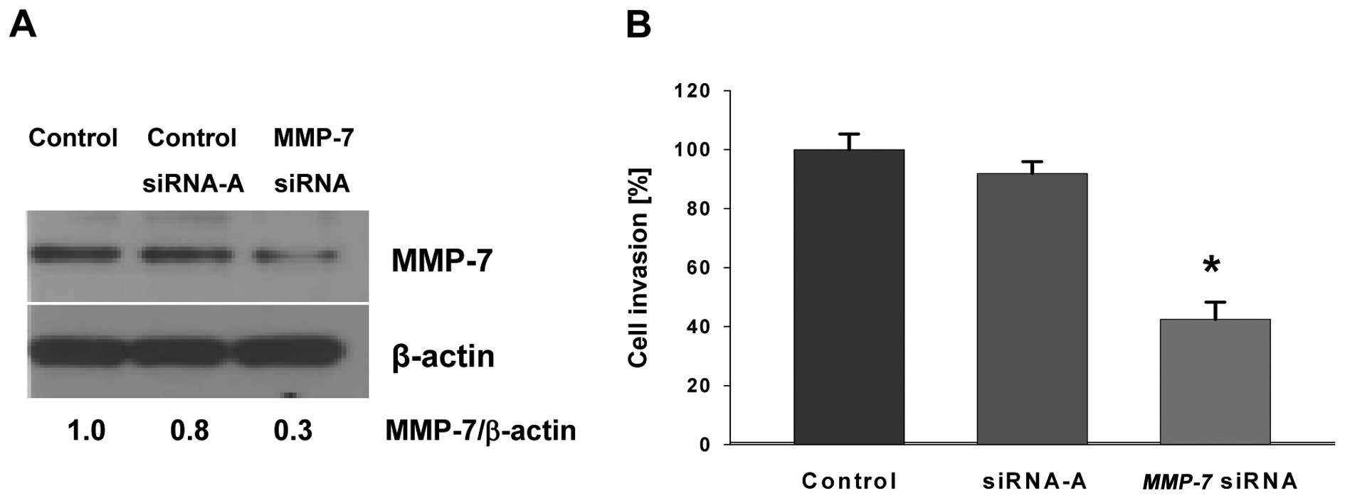
Gene silencing of MMP-7 inhibits invasive
behavior of BxPc-3 cell line
To determine whether invasive behavior of BxPc-3
cells is associated with the expression of MMP-7, we
silenced MMP-7 with siRNA as described in Materials and
methods. As shown in Fig. 6,
knockdown of MMP-7 inhibits cell invasion by >50% in
comparison with negative control cells transfected with scrambled
siRNA. These results further confirm that MMP-7 greatly contributes
to the invasive behavior and its deletion limits the invasive
capacity of BxPc-3 cells.
Discussion
The present study demonstrates that triterpenes
extracted from P. cocos suppress growth and invasive
behavior of human pancreatic cancer cells and only slightly
affecting normal pancreatic cells. Interestingly, invasive BxPc-3
cells are the most sensitive to the treatment of a characterized
triterpene mixture (PTE) as well as purified triterpenes PA, DPA
and PPAC (Fig. 1). Here we show
that PA is the most potent triterpene responsible for the
anticancer activity of the PTE mixture (containing 55.7% PA, 31.7%
DPA and 4.1% PPAC) because PA inhibits proliferation of BxPc-3
cells with the IC50 value (0.26 μM) when compared
to DPA (1.02 μM) and PPAC (21.76 μM).
To elucidate molecular mechanisms related to the
inhibition of invasiveness of pancreatic cancer cells we used
BxPc-3 cells. This cell line is the only Smad4-deficient among the
four pancreatic cancer cell lines we investigated (24,25).
Moreover, the loss of Smad4 is associated with a higher likelihood
of metastasis, poor outcome following surgical resection (26) and predict a worse prognosis in
patients with pancreatic cancer (27). Here we demonstrate, for the first
time, that both PTE and PA significantly suppress invasive behavior
of BxPc-3 cells. Evidently, this suppression is correlated with
downregulation of MMP-7 expression. MMP-7 is overexpressed in
pancreatic cancer (28),
correlates with decreased survival (29,30)
and contributes to cancer progression by supporting tumor size and
metastasis in vivo(31).
Therefore, MMP-7 is a suitable target involved in disease
progression and the downregulation of MMP-7 expression by PA may be
a useful strategy for pancreatic cancer metastasis intervention.
Interestingly, PA also suppressed invasiveness through the
inhibition of expression another matrix metalloproteinase, MMP-9 in
breast cancer cells (12).
As recently demonstrated, PA was detected in urine
and plasma of rats feed P. cocos(32), indicating that PA can be easily
absorbed into blood. Therefore, the bioavailability of PA further
promotes employment of PA or other P. cocos triterpenes for
the treatment of different cancers including pancreatic cancer.
In conclusion, our study provides new evidence that
the mixture or purified triterpenes extracted from mushroom P.
cocos inhibits growth and invasiveness of pancreatic cancer
cells. Moreover, we identified MMP-7 as a target of P. cocos
triterpenes in pancreatic cancer cells. Further studies are in
progress to investigate the exact mechanism of the inhibition of
MMP-7 expression and the evaluation of the anticancer and
anti-metastatic activity of PA in vivo.
Acknowledgements
We thank Dr Anita Thyagarajan-Sahu for
her technical assistance with the cell cycle analysis, Dr
Ming-Sound Tsao for kindly providing the normal human pancreatic
duct epithelial cell line HPDE-6, Dr Yaqiong Wang, Professor
Zhonglin Yang for kindly providing the dried sclerotium of P.
cocos, Dr Yaqiong Wang and Professor Ping Li for their helpful
suggestions on extraction, purification and identification of
triterpenes from P. cocos. This study was supported by
research grants from China Scholarship Council and EcoNugenics,
Inc., Santa Rosa, CA, USA. One of the authors, I. Eliaz,
acknowledges his interest as the formulator and owner of
EcoNugenics, Inc.
References
|
1
|
Siegel R, Naishadham D and Jemal A: Cancer
statistics, 2012. CA Cancer J Clin. 62:10–29. 2012. View Article : Google Scholar
|
|
2
|
Rhee YH, Jeong SJ, Lee HJ, et al:
Inhibition of STAT3 signaling and induction of SHP1 mediate
antiangiogenic and antitumor activities of ergosterol peroxide in
U266 multiple myeloma cells. BMC Cancer. 12:282012. View Article : Google Scholar : PubMed/NCBI
|
|
3
|
Lee CC, Yang HL, Way TD, et al: Inhibition
of cell growth and induction of apoptosis by Antrodia
camphorata in HER-2/neuoverexpressing breast cancer cells
through the induction of ROS, depletion of HER-2/neu and disruption
of the PI3K/Akt signaling pathway. Evid Based Complement Alternat
Med. 2012:7028572012.PubMed/NCBI
|
|
4
|
Kuo YC, Lai CS, Tsai CY, Nagabhushanam K,
Ho CT and Pan MH: Inotilone suppresses phorbol ester-induced
inflammation and tumor promotion in mouse skin. Mol Nutr Food Res.
56:1324–1332. 2012. View Article : Google Scholar : PubMed/NCBI
|
|
5
|
Torkelson CJ, Sweet E, Martzen MR, et al:
Phase 1 clinical trial of Trametes versicolor in women with
breast cancer. ISRN Oncol. 2012:2516322012.PubMed/NCBI
|
|
6
|
Rios JL: Chemical constituents and
pharmacological properties of Poria cocos. Planta Med.
77:681–691. 2011. View Article : Google Scholar : PubMed/NCBI
|
|
7
|
Lee SM, Lee YJ, Yoon JJ, Kang DG and Lee
HS: Effect of Poria cocos on hypertonic stress-induced water
channel expression and apoptosis in renal collecting duct cells. J
Ethnopharmacol. 141:368–376. 2012.
|
|
8
|
Zhao YY, Feng YL, Du X, Xi ZH, Cheng XL
and Wei F: Diuretic activity of the ethanol and aqueous extracts of
the surface layer of Poria cocos in rat. J Ethnopharmacol.
144:775–778. 2012. View Article : Google Scholar : PubMed/NCBI
|
|
9
|
Chen X, Zhang L and Cheung PC:
Immunopotentiation and anti-tumor activity of
carboxymethylated-sulfated beta-(1→3)-d-glucan from Poria
cocos. Int Immunopharmacol. 10:398–405. 2010.PubMed/NCBI
|
|
10
|
Kikuchi T, Uchiyama E, Ukiya M, et al:
Cytotoxic and apoptosis-inducing activities of triterpene acids
from Poria cocos. J Nat Prod. 74:137–144. 2011. View Article : Google Scholar : PubMed/NCBI
|
|
11
|
Ling H, Zhou L, Jia X, Gapter LA, Agarwal
R and Ng KY: Polyporenic acid C induces caspase-8-mediated
apoptosis in human lung cancer A549 cells. Mol Carcinog.
48:498–507. 2009. View
Article : Google Scholar : PubMed/NCBI
|
|
12
|
Ling H, Zhang Y, Ng KY and Chew EH:
Pachymic acid impairs breast cancer cell invasion by suppressing
nuclear factor-kappaB-dependent matrix metalloproteinase-9
expression. Breast Cancer Res Treat. 126:609–620. 2011. View Article : Google Scholar : PubMed/NCBI
|
|
13
|
Zhou L, Zhang Y, Gapter LA, Ling H,
Agarwal R and Ng KY: Cytotoxic and anti-oxidant activities of
lanostane-type triterpenes isolated from Poria cocos. Chem
Pharm Bull. 56:1459–1462. 2008. View Article : Google Scholar : PubMed/NCBI
|
|
14
|
Mizushina Y, Akihisa T, Ukiya M, et al: A
novel DNA topoisomerase inhibitor: dehydroebriconic acid, one of
the lanostane-type triterpene acids from Poria cocos. Cancer
Sci. 95:354–360. 2004. View Article : Google Scholar : PubMed/NCBI
|
|
15
|
Hong R, Shen MH, Xie XH and Ruan SM:
Inhibition of breast cancer metastasis via PITPNM3 by pachymic
acid. Asian Pac J Cancer Prev. 13:1877–1880. 2012. View Article : Google Scholar : PubMed/NCBI
|
|
16
|
Ling H, Jia X, Zhang Y, et al: Pachymic
acid inhibits cell growth and modulates arachidonic acid metabolism
in nonsmall cell lung cancer A549 cells. Mol Carcinog. 49:271–282.
2010.PubMed/NCBI
|
|
17
|
Jiang J, Slivova V, Harvey K,
Valachovicova T and Sliva D: Ganoderma lucidum suppresses growth of
breast cancer cells through the inhibition of Akt/NF-kappaB
signaling. Nutr Cancer. 49:209–216. 2004. View Article : Google Scholar : PubMed/NCBI
|
|
18
|
Sliva D, Jedinak A, Kawasaki J, Harvey K
and Slivova V: Phellinus linteus suppresses growth, angiogenesis
and invasive behaviour of breast cancer cells through the
inhibition of AKT signalling. Br J Cancer. 98:1348–1356. 2008.
View Article : Google Scholar : PubMed/NCBI
|
|
19
|
Sliva D, Harvey K, Mason R, Lloyd F Jr and
English D: Effect of phosphatidic acid on human breast cancer cells
exposed to doxorubicin. Cancer Invest. 19:783–790. 2001. View Article : Google Scholar : PubMed/NCBI
|
|
20
|
Lloyd FP Jr, Slivova V, Valachovicova T
and Sliva D: Aspirin inhibits highly invasive prostate cancer
cells. Int J Oncol. 23:1277–1283. 2003.PubMed/NCBI
|
|
21
|
Livak KJ and Schmittgen TD: Analysis of
relative gene expression data using real-time quantitative PCR and
the 2(-Delta Delta C(T)) method. Methods. 25:402–408. 2001.
View Article : Google Scholar : PubMed/NCBI
|
|
22
|
Gapter L, Wang Z, Glinski J and Ng KY:
Induction of apoptosis in prostate cancer cells by pachymic acid
from Poria cocos. Biochem Biophys Res Commun. 332:1153–1161.
2005. View Article : Google Scholar : PubMed/NCBI
|
|
23
|
Bournet B, Pointreau A, Souque A, et al:
Gene expression signature of advanced pancreatic ductal
adenocarcinoma using low density array on endoscopic
ultrasound-guided fine needle aspiration samples. Pancreatology.
12:27–34. 2012. View Article : Google Scholar
|
|
24
|
Nagaraj NS, Washington MK and Merchant NB:
Combined blockade of Src kinase and epidermal growth factor
receptor with gemcitabine overcomes STAT3-mediated resistance of
inhibition of pancreatic tumor growth. Clin Cancer Res. 17:483–493.
2011. View Article : Google Scholar : PubMed/NCBI
|
|
25
|
Hlavaty J, Petznek H, Holzmuller H, et al:
Evaluation of a gene-directed enzyme-product therapy (GDEPT) in
human pancreatic tumor cells and their use as in vivo models for
pancreatic cancer. PLoS One. 7:e406112012. View Article : Google Scholar : PubMed/NCBI
|
|
26
|
Tascilar M, Skinner HG, Rosty C, et al:
The SMAD4 protein and prognosis of pancreatic ductal
adenocarcinoma. Clin Cancer Res. 7:4115–4121. 2001.
|
|
27
|
Singh P, Srinivasan R and Wig JD: SMAD4
genetic alterations predict a worse prognosis in patients with
pancreatic ductal adenocarcinoma. Pancreas. 41:541–546. 2012.
View Article : Google Scholar : PubMed/NCBI
|
|
28
|
Crawford HC, Scoggins CR, Washington MK,
Matrisian LM and Leach SD: Matrix metalloproteinase-7 is expressed
by pancreatic cancer precursors and regulates acinar-to-ductal
metaplasia in exocrine pancreas. J Clin Invest. 109:1437–1444.
2002. View Article : Google Scholar : PubMed/NCBI
|
|
29
|
Fukushima H, Yamamoto H, Itoh F, et al:
Association of matrilysin mRNA expression with K-ras mutations and
progression in pancreatic ductal adenocarcinomas. Carcinogenesis.
22:1049–1052. 2001. View Article : Google Scholar : PubMed/NCBI
|
|
30
|
Jones LE: Comprehensive analysis of matrix
metalloproteinase and tissue inhibitor expression in pancreatic
cancer: increased expression of matrix metalloproteinase-7 predicts
poor survival. Clin Cancer Res. 10:2832–2845. 2004. View Article : Google Scholar
|
|
31
|
Fukuda A, Wang SC, Morris JP IV, et al:
Stat3 and MMP7 contribute to pancreatic ductal adenocarcinoma
initiation and progression. Cancer Cell. 19:441–455. 2011.
View Article : Google Scholar : PubMed/NCBI
|
|
32
|
Ling Y, Chen M, Wang K, et al: Systematic
screening and characterization of the major bioactive components of
Poria cocos and their metabolites in rats by LC-ESI-MS(n).
Biomed Chromatogr. 26:1109–1117. 2012.PubMed/NCBI
|