Introduction
Osteosarcoma (OS) is the most common and primary
bone tumor in children, adolescents and young adults (1). It happens mainly around areas with
active bone growth and repairation. Emerging evidence indicates OS
is induced by genetic and epigenetic alterations which disturb
mesenchymal stem cells to differentiate into osteoblasts (2). Over the past decade, advances in OS
therapy have improved patient outcomes (3), resulting in dramatic improvement in
the 5-year survival rate of OS patients to ~60–70%. However,
outcome is still poor and most of them will die due to local
relapse or pulmonary metastases after surgical resection and
intensive-chemotherapy (4,5). In recent years, many clinical
features, including tumor size, surgical margin and response to
chemotherapy, are useful prognostic factors for patients with
osteosarcoma. However, they lack specificity and sensitivity,
indicating that different genetic mechanisms may be operating and
altering response to chemotherapy and metastatic capability in some
tumors during the same clinical stage of the tumor (6). Consequently, it is required to
ascertain precise molecular markers for screening osteosarcoma
patients with poor prognosis, so as to provide them with more
aggressive treatment at an early stage.
Sox4, a 47-kDa protein, is a member of the
sex-determining region Y (SRY)-related high-mobility group
(HMG)-box (Sox) transcription factor family. Recently, the clinical
importance of Sox4 has gained increasing attention, and numerous
reports indicated that Sox4 may act as oncogenic gene involved in
multiple human malignancies, including hepatocellular, bladder,
lung, colon, prostate, and gastric cancers, with poor prognostic
features and advanced disease status (7–10).
There are three distinguishable domains in Sox4 gene, which contain
an HMG box, a serine-rich region, and a glycine-rich region. The
HMG box acts as a DNA-binding domain, and the serine-rich domain
acts as a transactivation domain (11). The glycine-rich region which is the
central domain located between the HMG box and serine-rich domain
acts as a novel functional region for promoting apoptotic cell
death (12). In recent years, it
has been reported that transcriptional targets of Sox4 are closely
related to tumor progression and microRNA (miRNA) processing
(11,13).
miRNAs are small (~22 nucleotides in length),
non-coding RNAs (14), miRNAs
degrade or suppress their translation and regulate a series of cell
functions such as proliferation, apoptosis, invasion and
differentiation, by binding to complementary sequences in the
3′UTRs of targeted mRNAs (15,16).
More and more evidence suggests that miRNAs are involved in various
kinds of tumors (17). Many miRNAs
have been identified to act as tumor suppressors or oncogenes in
osteosarcoma, which is dependent on the role of their target genes,
including miR-300 (18), miR-182
(19), miR-1247 (20) and miR-217 (21). These outcomes show a strong basis
for the importance of miRNAs in the pathogenesis of osteosarcoma
and emphasize the implications of miRNAs in diagnosis, therapy, and
prognosis of osteosarcoma.
At present, miR-132 has attracted much attention,
because it has been reported that it is frequently downregulated
and functions as a tumor suppressor in breast, lung, colorectal
cancer and osteosarcoma (22–25),
and is also upregulated and functions as an oncogene in glioma and
gastric cancer (26,27). It has been shown that miR-132
inhibited proliferation, invasion, migration and metastasis of
breast cancer by targeting hematopoietic- and neurologic-expressed
sequence 1 (HN1) (22). Besides,
miR-132 inhibited cell invasion in both lung and colorectal cancers
via targeting the ZEB2 (23,24).
Moreover, miR-132 suppressed cell proliferation in osteosarcoma
cells by targeting cyclin E1 (28). However, until now, the precise
mechanism of miR-132 in osteosarcoma remains unclear. In this
investigation, we also determined frequent downregulation of
miR-132 in osteosarcoma cell lines, which was consistent with a
previous study (28).
Overexpression of miR-132 inhibited cell proliferation, invasion
and epithelial-mesenchymal transition (EMT) of osteosarcoma cells.
Moreover, we found that Sox4, a novel tumor suppressor gene, was
the direct target of miR-132 in osteosarcoma. Restoration of Sox4
reversed the inhibitory effects of miR-132. Therefore, our outcomes
showed critical roles for miR-132 in the pathogenesis of
osteosarcoma and suggested its possible application in tumor
treatment.
Materials and methods
Cell culture
Osteosarcoma cell lines MG63, HOS, SaOS-2, 143B,
U2OS and the human normal osteoblastic cell line hFOB1.19 cells
were obtained from American Type Culture Collection (ATCC,
Manassas, VA, USA). The osteosarcoma cells were cultured in
RPMI-1640 (Gibco Co., New York, NY, USA) containing 10% fetal
bovine serum (FBS, Gibco), 1% penicillin and streptomycin at 37°C
in a humidified atmosphere of 5 on 0.1% gelatin-coated culture
flasks. The hFOB1.19 human osteoblasts were cultured in DMEM/Ham's
F-12 containing 10% FBS and geneticin (400 μg/ml) at 37°C in 5%
CO2 incubator.
Transient transfection
The miR-132 mimic, miR-negative control of mimics
(miR-NC), miR-132 inhibitor, miR-negative control of inhibitor
(anti-miR-NC), siRNA for Sox4 (si-Sox4) and siRNA-negative control
(NC) were synthesized and purified by Gene-Pharma (Shanghai,
China). miR-132 mimic, miR-132 inhibitor, and si-Sox4 were
transfected at a final concentration of 50 nM using Lipofectamine
2000 reagent (Invitrogen) following the manufacturer's protocols.
Total RNA and protein were collected 24 h after transfection.
Reverse transcription polymerase chain
reaction
Total RNA of SaOS-2 and 143B cells was isolated by
TRIzol reagent (Invitrogen) following the manufacturer's protocol.
RNA (2 μg) was used for gene-specific reverse transcription
polymerase chain reaction (RT-PCR) using one-step RT-PCR kit
(Qiagen, Venlo, The Netherlands) following the manufacturer's
protocols. Denaturation was performed at 94°C for 3 min, annealing
at 95°C for 1 min, and elongation at 94°C for 30 sec for 40 cycles,
followed by 72°C for 5 min. The following primers were used:
miR-132 forward, 5′-TGGATCCCCCCCA GTCCCCGTCCCTCAG-3′; reverse,
5′-TGAATTCGGATA CCTTGGCCGGGAGGAC-3′. U6 forward, 5′-CTCGCTTC
GGCAGCACA-3′; reverse, 5′-AACGCTTCACGAATTTG CGT-3′. U6 was used for
normalization. Each sample was assessed in triplicate.
Cell proliferation assay
To explore the effect of miR-132 on proliferation of
SaOS-2 and 143B cells, 5×10³ cells were seeded in
96-well plate and allowed to grow overnight in complete RPMI-1640
medium. The medium was then removed, and cells were transfected
with miR-132 mimic or inhibitor for 24 h at 37°C. Cell
Proliferation ELISA-BrdU (colorimetric) kit (Roche Diagnostics,
USA) was used to detect the cell proliferation according to the
manufacturer's protocols.
Cell cycle analysis
To detect cell cycle distribution, the SaOS-2 and
143B cells were transfected with miR-132 mimic or inhibitor for 24
h. After transfection, SaOS-2 and 143B cells were collected by
trypsinization, washed with ice-cold PBS, and fixed in ice-cold 70%
methanol overnight. Then, cells were centrifuged, resuspended in
ice-cold phosphate-buffered saline (PBS), and incubated with RNase
(Sigma Chemical Co., USA) for 30 min at 37°C, and then were
incubated with propidium iodide (PI; Sigma) at room temperature for
30 min. The analyses of cell cycle distribution were performed by
FACScan flow cytometer (BD Biosciences, San Jose, CA, USA).
Annexin V-FITC/PI analysis
SaOS-2 and 143B cells were transfected with miR-132
mimic or inhibitor for 24 h. After transfection, cells were
harvested and washed twice in PBS and double-stained with Annexin
V-FITC and PI by using the Annexin V-FITC Apoptosis Detection kit
(BD Biosciences) following the manufacturer's protocols. Then, each
sample was quantitatively analyzed at 488 nm emission and 570 nm
excitation by FACSCalibur flow cytometer (BD Biosciences).
Transwell invasion assay
To determine cell invasion, Transwell matrigel
invasion assay using Transwell chambers (8-mm pore size; Millipore)
precoated with Matrigel (BD Biosciences, Franklin Lakes, NJ, USA)
that contained extracellular matrix proteins was used following the
manufacturer's protocol. In brief, 1×10⁵ SaOS-2 and 143B
cells transfected with miR-132 mimic or inhibitor were suspended in
200 μl RPMI-1640 containing 1% FBS and seeded on the upper chamber.
RPMI-1640 (600 ml) containing 10% FBS was added to the lower
chamber. After 24-h incubation at 37°C in a 5% CO2
atmosphere, cells that remained in the upper chamber were removed
by cotton swabs and penetrating cells were fixed in methanol, and
then stained with 0.1% crystal violet. Cell invasion was quantified
by counting cells on the lower surface using phase contrast
microscopy (Olympus IX83, Japan).
Western blot analysis
To extract the proteins, SaOS-2 and 143B cells were
washed twice in cold PBS, and then lysed in RIPA lysis buffer
(Beyotime Institute of Biotechnology Jiangsu, China) with protease
inhibitor cocktail (Merk, Germany). The protein concentration of
cell lysates was quantified by BCA kit (Beyotime Institute of
Biotechnology), and 50 μg of each of proteins were separated by
SDS-PAGE on 8% gels, and then transferred to a polyvinylidene
fluoride (PVDF) membrane (Millipore, USA). The membranes were
blocked in 5% shimmed milk diluted with Tri-buffered saline
Tween-20 (TBST) (in mmol/l: Tris-HCl 20, NaCl 150, pH 7.5, 0.1%
Tween-20) at room temperature for 1 h and incubated overnight at
4°C with primary antibody, respectively: anti-Sox4 (no. ab80261;
1:500; Abcam, USA); anti-cyclin D1 (no. 2978), anti-CDK4 (no.
12790), anti-Bcl-2 (no. 2870), anti-E-cadherin (no. 5296),
anti-N-cadherin (no. 3195), anti-vimentin (no. 3390) (1:1,000; Cell
Signaling Technology Inc., MA, USA). The membranes were then
incubated with a goat anti-rabbit (no. 14708) or anti-mouse (no.
14709) IgG conjugated to horseradish peroxidase secondary antibody
(1:1,000; Cell Signaling Technology) for 2 h. The proteins were
visualized using ECL-plus reagents (Amersham Biosciences Corp.,
USA). The density of the bands was measured using ImageJ software
(USA), and values were normalized to the densitometric values of
α-tubulin (T5168; 1:1,000; Sigma) in each sample.
Luciferase reporter assay
SaOS-2 and 143B cells (1×10⁵/well) were
seeded in 24-well plates and incubated for one day before
transfection. Cells were cotransfected with 0.5 μg pGL3-Sox4-3′UTR
wild-type or mutant reporter plasmid, 50 nM miR-132 mimic or
miR-NC, and 20 ng pRL-SV40 Renilla plasmid (Promega, USA)
using Lipofectamine 2000. At 24 h after transfection, both firefly
and Renilla luciferase activities were quantified using the
Dual-Luciferase reporter system (Promega) according to the
manufacturer's instructions. All experiments were performed in
triplicate.
Statistical analysis
All statistical analyses were performed using
GraphPad Prism 5.0 (GraphPad software, Inc., USA). Data from each
group were expressed as mean ± standard error of the mean (SEM) and
statistically analyzed by Student's t-test. Differences were
considered statistically significant at a p-value of <0.05.
Results
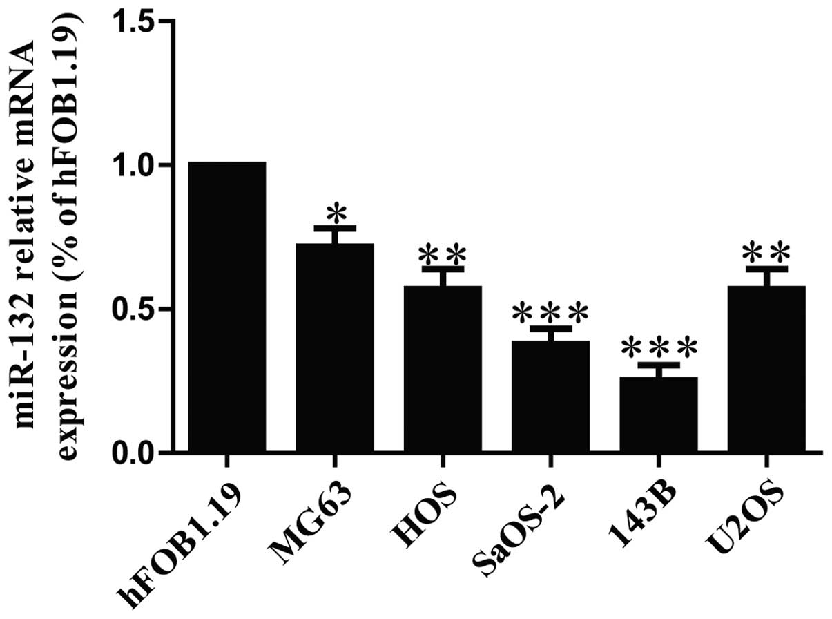
The expression of miR-132 is
downregulated in osteosarcoma cell lines
To determine the levels of miR-132 in OS cells, five
osteosarcoma cell lines (MG63, HOS, SaOS-2, 143B and U2OS) and a
human normal osteoblastic cell line (hFOB1.19) were used to detect
the level of miR-132 by real time-PCR. Our results demonstrated
that the level of miR-132 was significantly decreased in all five
OS cell lines compared to that in human normal osteoblastic cell
line hFOB1.19, as shown in Fig. 1.
Among these OS cell lines, SaOS-2 and 143B cells were used for
further study.
miR-132 inhibites cell proliferation,
induces G1-phase arrest and cell apoptosis in both SaOS-2 and 143B
cells
Based on the downregulation of miR-132, we believed
that miR-132 could act as a suppressor of cell growth. After
transfection with miR-132 mimic, the RT-PCR analysis showed that
mRNA level of miR-132 was significantly upregulated in miR-132
mimic group compared to miR-NC group (Fig. 2A). These data demonstrated that we
efficiently enhanced or reduced miR-132 expression in SaOS-2 and
143B cells. To determine the role of miR-132 in proliferation of
osteosarcoma cells, the results from Brdu-ELISA assay demonstrated
that overexpression of miR-132 dramatically inhibited the
proliferation of SaOS-2 and 143B cells (Fig. 2B). Because miR-132 significantly
inhibited proliferation of SaOS-2 and 143B cells, we speculated
that miR-132 could induce cell cycle arrest in osteosarcoma cells,
and proved this tentatively by flow cytometry. Our finding showed
that upregulation of miR-132 induced a dramatic G1-phase arrest and
decreased the percentage of cells in the S-phase in both SaOS-2 and
143B cells compared with cells transfected with miR-NC (Fig. 2C). Therefore, miR-132 might inhibit
the proliferation of osteosarcoma cells by impeding the G1/S cell
cycle transition. In order to explore whether pro-apoptosis
participated in miR-132 mimic-induced anti-proliferative effect,
the total apoptosis rates of SaOS-2 and 143B cells were detected by
flow cytometry analysis. As shown in Fig. 2D, flow cytometry analysis showed
that the number of apoptotic SaOS-2 and 143B cells was evidently
higher in miR-132 mimic than that in miR-NC group. However, the
cell proliferation and cell cycle were increased and cell apoptosis
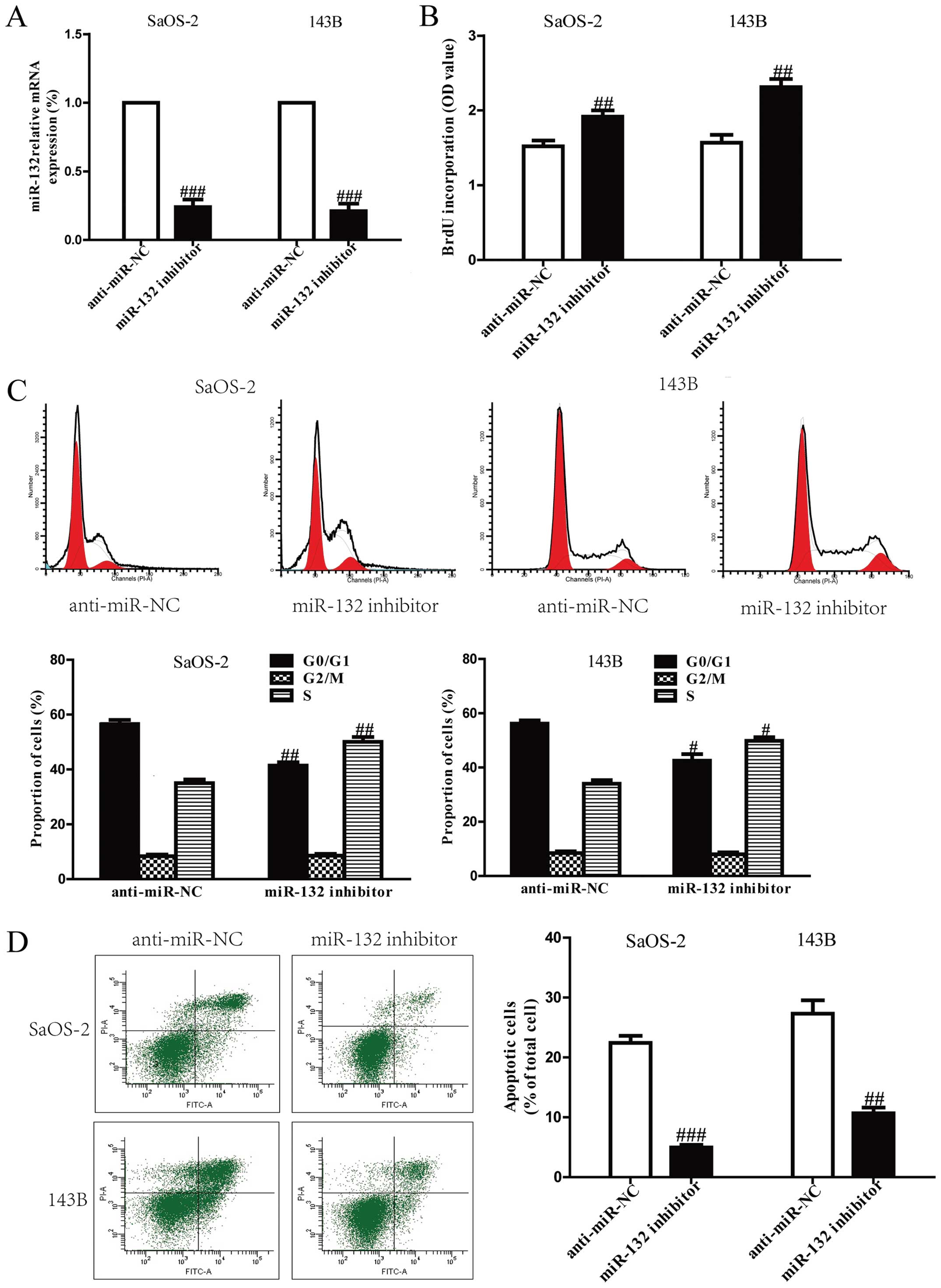
was inhibited in both SaOS-2 and 143B cells transfected with
miR-132 inhibitor compared with anti-miR-NC group (Fig. 3).
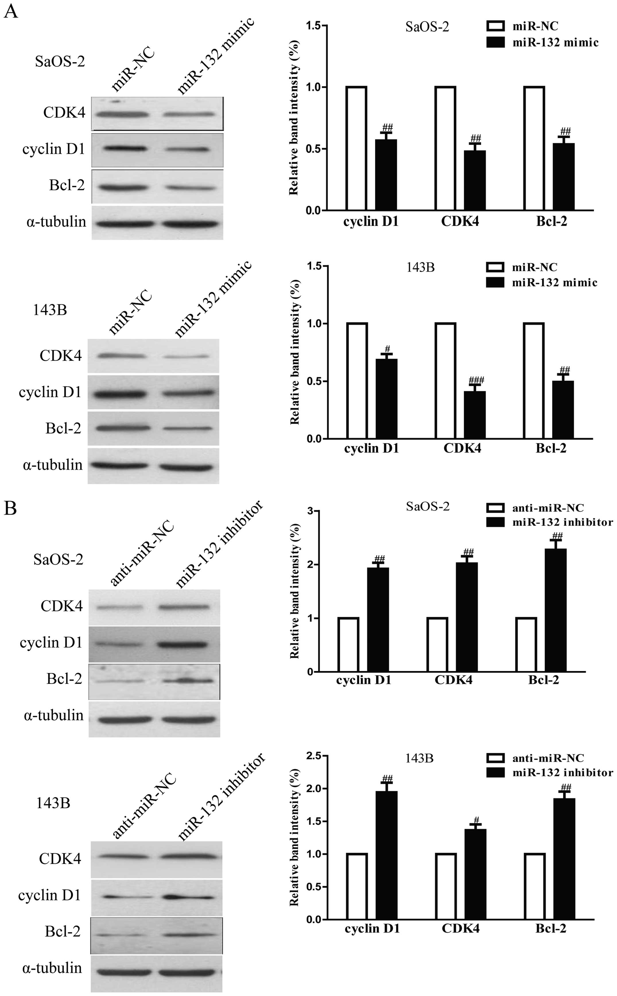
The effects of miR-132 on the expressions
of cell cycle and apoptosis-related proteins in osteosarcoma
cells
To investigate the possible mechanism of miR-132 on
cell proliferation, cell cycle and apoptosis, we tested the effects
of miR-132 on several cell cycle and apoptosis-related molecules.
As shown in Fig. 4A, upregulation
of miR-132 decreased the protein levels of cyclin D1, CDK4 and
Bcl-2 in SaOS-2 and 143B cells, which suggested that miR-132
inhibited cell proliferation, cell cycle and induced apoptosis by
downregulation of cyclin D1, CDK4 and Bcl-2. In addition,
downregulation of miR-132 increased expression of these proteins
(Fig. 4B).
The effects of miR-132 on the invasion
and EMT in osteosarcoma cells
To evaluate the effects of miR-132 on invasion and
EMT in osteosarcoma cells, we further transfected miR-132 mimic or
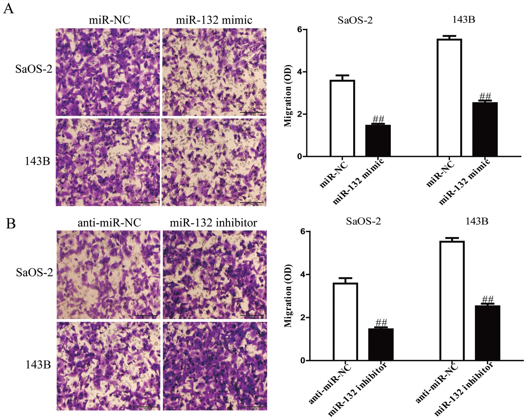
inhibitor into SaOS-2 and 143B cells, and the invasive capacities
of SaOS-2 and 143B cells were evaluated by Transwell invasion
chamber experiments. The results from Transwell assays showed that
the number of invading SaOS-2 and 143B cells was significantly
reduced in miR-132 mimic group compared to miR-NC group (Fig. 5A). However, miR-132 inhibitor could
evidently increase the number of invading SaOS-2 and 143B cells
(Fig. 5B). Furthermore, we
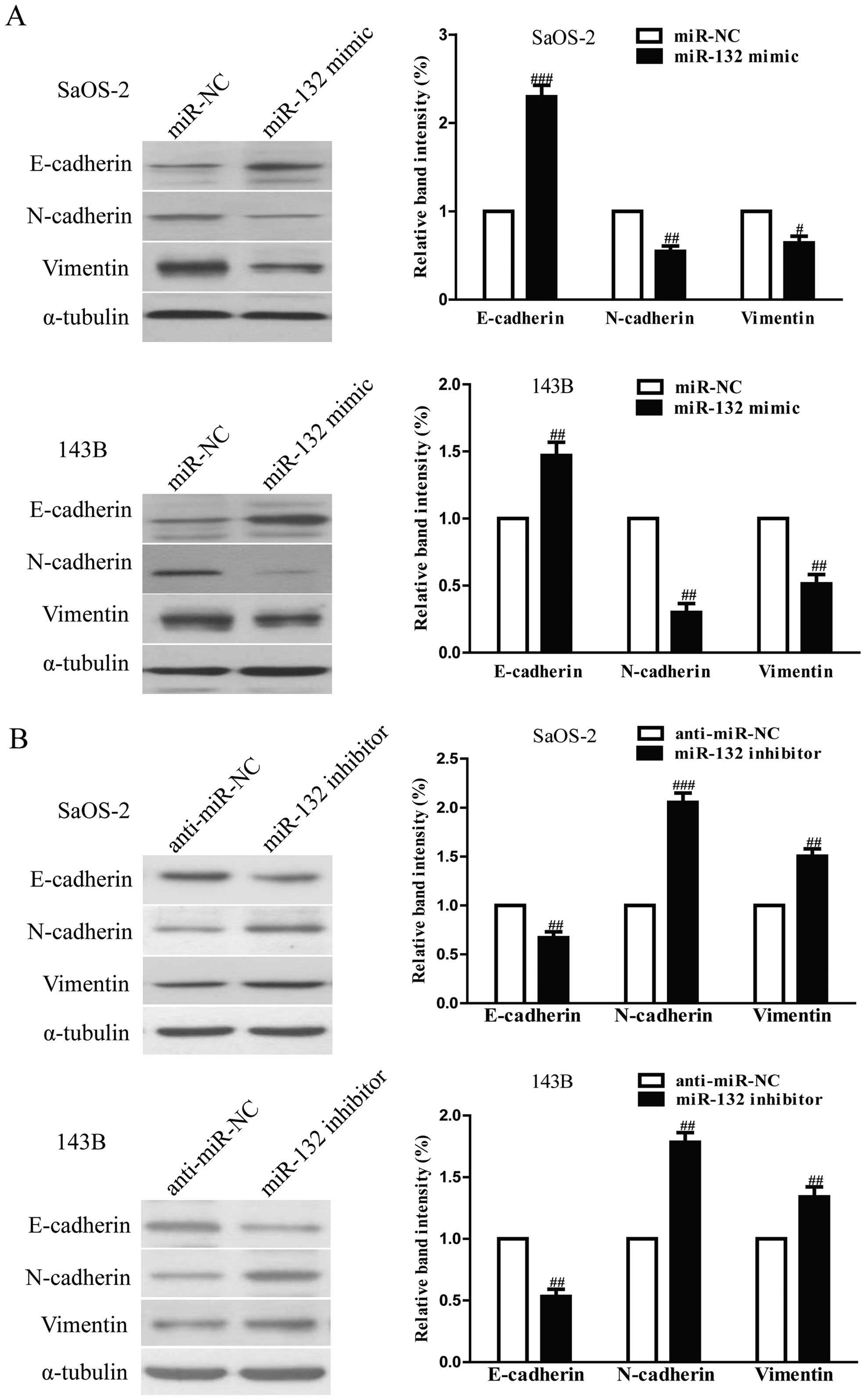
examined the effect of miR-132 mimic or inhibitor on the
expressions of EMT markers in SaOS-2 and 143B cells using western
blotting. Overexpression of miR-132 was able to upregulate the
expression of epithelial marker E-cadherin, and downregulate the
expression of mesenchymal markers N-cadherin and vimentin in SaOS-2
and 143B cells (Fig. 6A), but the
miR-132 inhibitor had the opposing effects on these EMT expression
markers (Fig. 6B). Taken together,
our results indicated that miR-132 was able to inhibit the invasion
and EMT in osteosarcoma cells.
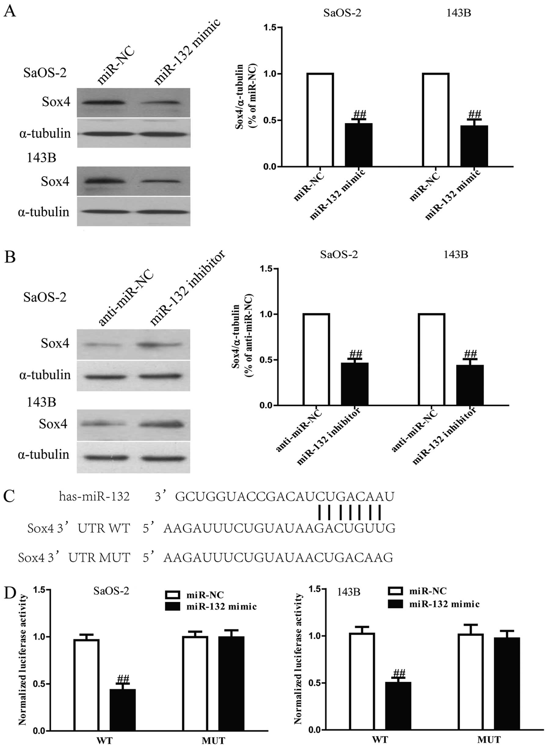
Sox4 is a direct target of miR-132 in
osteosarcoma cells
Since Sox4 was a binding target of miR-132 predicted
by the online database, TargetScan 6.2, we performed western
blotting and RT-PCR to observe the expression of Sox4 on protein
level in SaOS-2 and 143B cells transfected with miR-132 mimic or
inhibitor. Our results showed that protein level of Sox4 was
remarkably decreased after upregulation of miR-132 (Fig. 7A), but was evidently increased
after downregulation of miR-132 (Fig.
7B). To further demonstrate whether Sox4 was a direct target of
miR-132, Sox4 3′-UTR was cloned into a luciferase reporter vector
and the putative miR-132 binding site in the Sox4 3′-UTR was
mutated (Fig. 7C). The effect of
miR-132 was determined using luciferase reporter assay. The results
showed that overexpression of miR-132 significantly inhibited the
luciferase activity of pGL3-Sox4 3′-UTR WT (Fig. 7D). Mutation of the miR-132-binding
site in the Sox4 3′-UTR abolished the effect of miR-132, which
suggested that Sox4 was directly and negatively regulated by
miR-132.
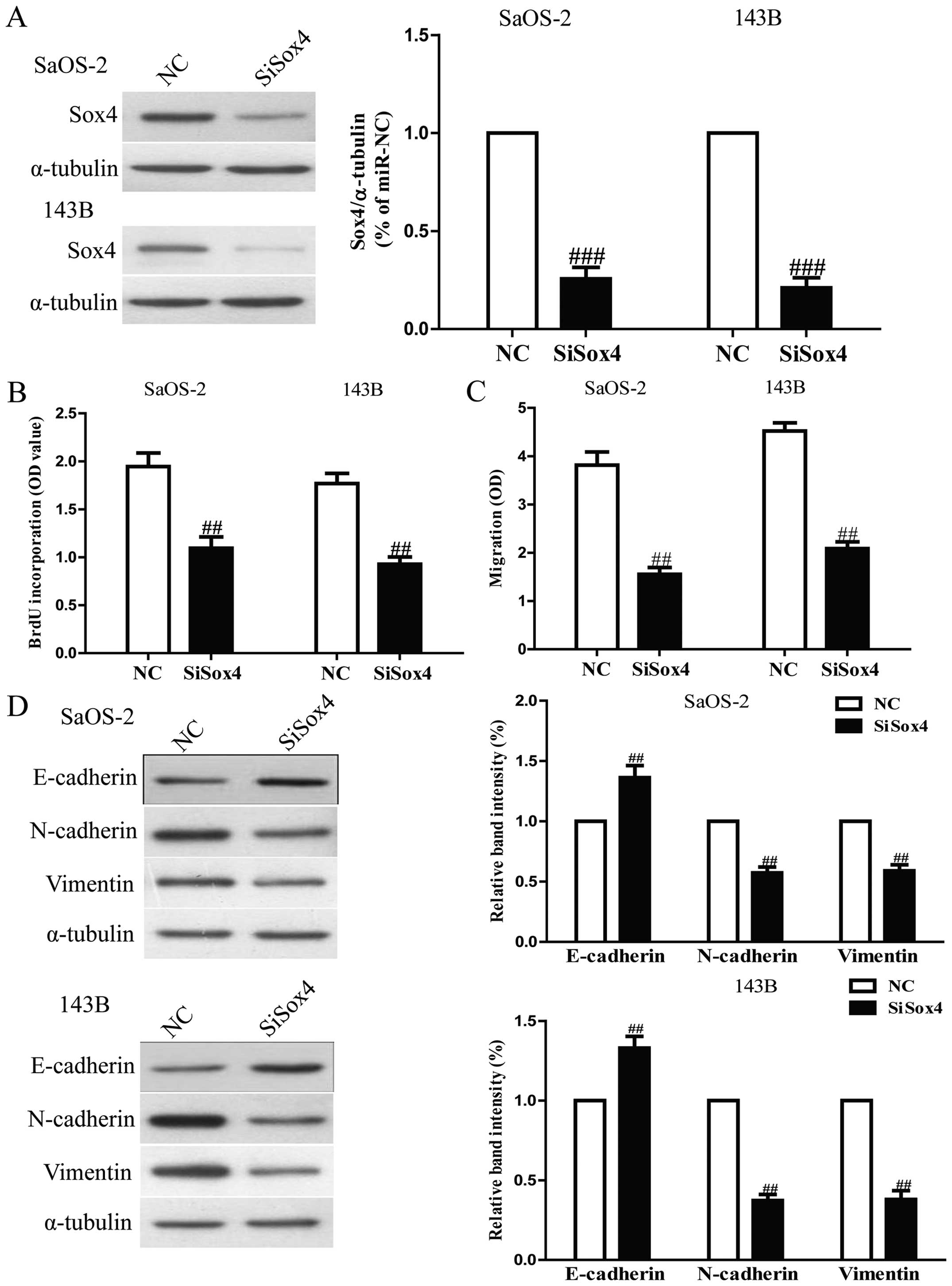
Downregulation of Sox4 by siRNA had
similar effects with miR-132 overexpression
To explore the function of Sox4 in osteosarcoma
cells, SaOS-2 and 143B cells were transfected with si-Sox4. Western
blot analysis indicated that protein expression of Sox4 was
significantly decreased after 24 h in both SaOS-2 and 143B cells
transfected with si-Sox4 (Fig.
8A). The Brdu-ELISA assay revealed that downregulation of Sox4
also inhibited osteosarcoma cell proliferation (Fig. 8B). Furthermore, Transwell assay
suggested that down-regulation of Sox4 expression inhibited
invasion capability of osteosarcoma cells (Fig. 8C). Downregulation of Sox4 resulted
in upregulation of the epithelial marker E-cadherin, and
downregulation of the mesenchymal markers N-cadherin and vimentin
(Fig. 8D). Sox4 silencing induced
a very similar phenotype to miR-132 expression in osteosarcoma
cells. These results indicated that miR-132 downregulated Sox4,
thus inhibiting osteosarcoma cell growth and metastasis.
Suppression of Sox4 is essential for
miR-132-inhibited cell proliferation, invasion and EMT in
osteosarcoma cells
To determine whether miR-132 reduced the
proliferation, invasion and EMT of osteosarcoma cells in a
Sox4-dependent manner, we cotransfected SaOS-2 and 143B cells with
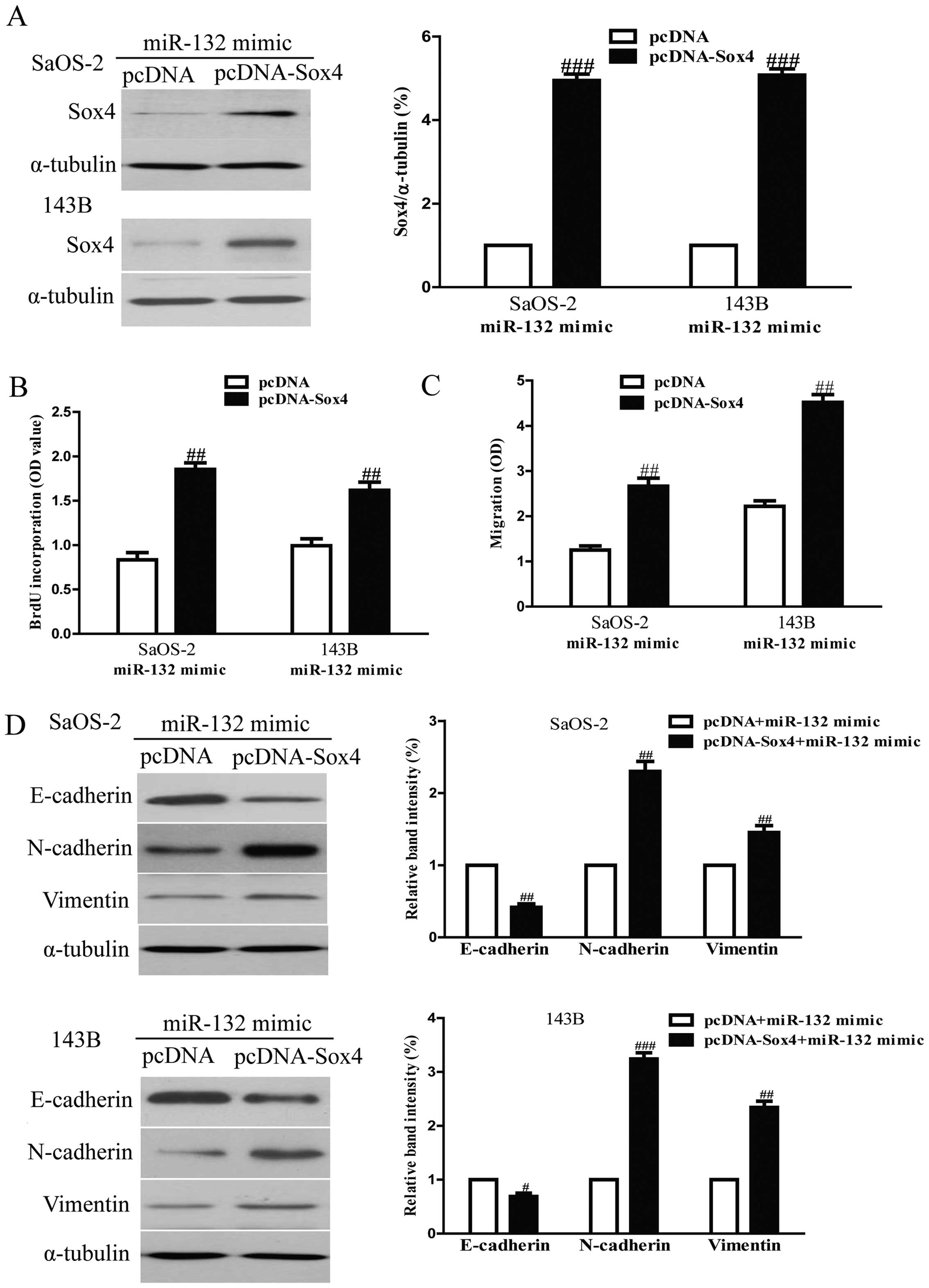
miR-132 mimic and pcDNA3.1-Sox4 vector. We found that the
expression of Sox4 was significantly increased after transfection
with miR-132 and pcDNA-Sox4 compared with miR-132 and pcDNA vector
in both SaOS-2 and 143B cells (Fig.
9A). Analysis by Brdu-ELISA assay indicated that overexpression
of Sox4 in cells transfected with the miR-132 mimic enhanced the
proliferation of osteosarcoma cells (Fig. 9B). The Transwell assay showed that
upregulating Sox4 expression could reverse the inhibitory effect of
the miR-132 mimic on invasion of osteosarcoma cells (Fig. 9C). Moreover, increased Sox4
expression downregulated the expression of epithelial marker
E-cadherin, and upregulated the expression of mesenchymal marker
N-cadherin and vimentin in SaOS-2 and 143B cells transfected with
miR-132 mimic (Fig. 9D).
Therefore, the inhibitory effects of miR-132 were reversed by Sox4
overexpression. Our results clearly demonstrated that miR-132
inhibited cell proliferation, invasion and EMT in osteosarcoma
cells by downregulation of Sox4, and that knockdown of Sox4 was
essential for the miR-132-inhibited cell proliferation, invasion
and EMT in osteosarcoma cells.
Discussion
The miRNAs have been reported as important
regulators involved in different biological processes such as cell
proliferation, metastasis, differentiation, transcriptional
regulation and tumorigenesis (29). Globally miRNA dysregulation of
tumors have provided major insights into the molecular mechanisms
of neoplasia (30). As one of the
most prominent miRNAs implicated in tumorigenesis, miR-132 has been
presented with a controversial role during tumor progression
(31). miR-132 was found to be
decreased in many human cancers, including breast, lung, colorectal
cancers and osteosarcoma (22–25),
but increased in glioma and gastric cancer (26,27).
The precise mechanism of miR-132 in osteosarcoma remained unclear.
Therefore, in this study, we aimed to elucidate the biological
functions and its mechanism of miR-132 in osteosarcoma. Our results
demonstrated that miR-132 was frequently downregulated in
osteosarcoma cell lines compared to human normal osteoblastic
cells. According to these findings, we speculated that miR-132
might be a potential anti-oncogene in osteosarcoma, which was
consistent with a previous study (28). As expected, upregulation of miR-132
inhibited proliferation, invasion, EMT and induced apoptosis of
SaOS-2 and 143B cells. Our current findings indicate that miR-132
played important roles in regulation of proliferation, apoptosis,
invasion and metastasis in osteosarcoma and may be potential
diagnostic and predictive biomarkers.
We also explored the exact molecular mechanism of
miR-132 in suppressing proliferation, invasion, EMT and inducing
apoptosis in osteosarcoma cells. As a result, the real-time PCR,
western blotting and luciferase reporter assay demonstrated that
Sox4 is a direct target of miR-132. Importantly, we also showed
that the proliferation-, invasion- and EMT-inhibiting effects of
miR-132 overexpression were partly reversed by upregulating Sox4
expression. Thus, we confirmed that miR-132 played critical roles
in the inhibition of proliferation, invasion and metastasis in
osteosarcoma cells, partially by downregulating the protein
expression of Sox4.
In this study, Brdu-ELISA assays showed that
overexpression of miR-132 was able to significantly inhibit the
proliferation of SaOS-2 and 143B cells. Cell cycle analyses also
showed that the percentage of cells in the G1-phase was increased
and the percentage of cells in the S-phase was decreased in cells
transfected with miR-132 mimic compared to cells transfected with
miR-NC. Moreover, flow cytometry analysis demonstrated that miR-132
mimic could evidently induce apoptosis of SaOS-2 and 143B cells
compared with miR-NC group. However, the cell proliferation and
cell cycle were increased and cell apoptosis was inhibited in both
SaOS-2 and 143B cells transfected with miR-132 inhibitor compared
with anti-miR-NC group. It is known that cell cycle progression and
apoptosis are regulated by numerous proteins. To confirm the
possible mechanism of miR-132 on regulation of cell cycle and
apoptosis, we investigated the effects of miR-132 mimic or
inhibitor on cell cycle- and apoptosis-related proteins. We
detected the expression of cyclin D1, CDK4 and Bcl-2. From our
data, we found that upregulation or downregulation of miR-132
decreased or increased the protein levels of cyclin D1, CDK4 and
Bcl-2, respectively. Cyclin D1 interacts with CDK4 to form the
cyclin D-CDK4 complex, and then phosphorylates Rb, which plays a
critical role in carcinogenesis. The cyclin-D1/CDK4/p-Rb pathway
has been proved to be changed in most of human cancers (32,33).
It is a pivotal regulator of the G1 to S phase transition of the
cell cycle. Bcl-2, an anti-apoptotic protein, is considered to be
resistant to conventional treatment of cancer (34,35).
In this report, our finding showed that miR-132 mimic reduced Bcl-2
protein and miR-132 inhibitor increased Bcl-2 protein, which
indicated that miR-132 regulated cell apoptosis via Bcl-2
modulation. Altogether, these outcomes indicated that miR-132
affected the cell cycle and apoptosis by regulating cyclin D1, CDK4
and Bcl-2. In addition, Transwell assay showed that miR-132 mimic
or inhibitor dramatically inhibited or enhanced the invasion of
SaOS-2 and 143B cells compared with miR-NC or anti-miR-NC group,
respectively. Furthermore, we determined the change of EMT markers
in SaOS-2 and 143B cells transfected with miR-132 mimic or
inhibitor. Our results showed that upregulation of miR-132 could
markedly suppress invasive ability of osteosarcoma cells by
dramatically upregulating the epithelial marker E-cadherin and
downregulating the mesenchymal marker N-cadherin and vimentin, and
miR-132 inhibitor had the opposing effect on expression of EMT
markers, which supported that miR-132 might suppress the EMT
process to restrain cell invasion and metastasis.
At the molecular level, our results demonstrated
that Sox4 was a direct target of miR-132 in osteosarcoma cells.
Some reports have indicated that overexpression of Sox4 exists in
multiple human cancers, including endometrial (36), esophageal (37), ovarian (38), gastric cancers (39) and osteosarcoma (28), suggesting that Sox4 might be a
vital oncogene affecting progression and metastasis of tumors. In
the present study, Sox4 was also found to be upregulated in
osteosarcom cells. Furthermore, we found that knockdown of Sox4
using siRNA oligos inhibited the proliferation, invasion and EMT of
osteosarcoma cells, which had similar effects with miR-132
overexpression. Besides, restoration of Sox4 reversed the
inhibitory effects of miR-132, suggesting that Sox4 may play a
critical role in osteosarcoma progression and metastasis.
In conclusion, our results indicate that miR-132 was
dramatically downregulated in osteosarcoma cells. Overexpression of
miR-132 inhibited proliferation, invasion, EMT and induced
apoptosis of osteosarcoma cells through directly targeting Sox4.
This novel miR-132/Sox4 axis might provide new insights into the
molecular mechanisms underlying progression and metastasis of
tumors, and upregulation of miR-132 expression might be a possible
therapeutic strategy for the therapy of osteosarcoma in the
future.
References
|
1
|
Mirabello L, Troisi RJ and Savage SA:
Osteosarcoma incidence and survival rates from 1973 to 2004: Data
from the Surveillance, Epidemiology, and End Results Program.
Cancer. 115:1531–1543. 2009. View Article : Google Scholar : PubMed/NCBI
|
|
2
|
Yan K, Gao J, Yang T, Ma Q, Qiu X, Fan Q
and Ma B: MicroRNA-34a inhibits the proliferation and metastasis of
osteosarcoma cells both in vitro and in vivo. PLoS One.
7:e337782012. View Article : Google Scholar : PubMed/NCBI
|
|
3
|
Amankwah EK, Conley AP and Reed DR:
Epidemiology and therapies for metastatic sarcoma. Clin Epidemiol.
5:147–162. 2013.PubMed/NCBI
|
|
4
|
Bacci G, Briccoli A, Rocca M, Ferrari S,
Donati D, Longhi A, Bertoni F, Bacchini P, Giacomini S, Forni C, et
al: Neoadjuvant chemotherapy for osteosarcoma of the extremities
with metastases at presentation: Recent experience at the Rizzoli
Institute in 57 patients treated with cisplatin, doxorubicin, and a
high dose of methotrexate and ifosfamide. Ann Oncol. 14:1126–1134.
2003. View Article : Google Scholar : PubMed/NCBI
|
|
5
|
Rainusso N, Wang LL and Yustein JT: The
adolescent and young adult with cancer: State of the art - bone
tumors. Curr Oncol Rep. 15:296–307. 2013. View Article : Google Scholar : PubMed/NCBI
|
|
6
|
Szuhai K, Cleton-Jansen AM, Hogendoorn PC
and Bovée JV: Molecular pathology and its diagnostic use in bone
tumors. Cancer Genet. 205:193–204. 2012. View Article : Google Scholar : PubMed/NCBI
|
|
7
|
Pramoonjago P, Baras AS and Moskaluk CA:
Knockdown of Sox4 expression by RNAi induces apoptosis in ACC3
cells. Oncogene. 25:5626–5639. 2006. View Article : Google Scholar : PubMed/NCBI
|
|
8
|
Medina PP, Castillo SD, Blanco S,
Sanz-Garcia M, Largo C, Alvarez S, Yokota J, Gonzalez-Neira A,
Benitez J, Clevers HC, et al: The SRY-HMG box gene, SOX4, is a
target of gene amplification at chromosome 6p in lung cancer. Hum
Mol Genet. 18:1343–1352. 2009. View Article : Google Scholar : PubMed/NCBI
|
|
9
|
Liao YL, Sun YM, Chau GY, Chau YP, Lai TC,
Wang JL, Horng JT, Hsiao M and Tsou AP: Identification of SOX4
target genes using phylogenetic footprinting-based prediction from
expression microarrays suggests that overexpression of SOX4
potentiates metastasis in hepatocellular carcinoma. Oncogene.
27:5578–5589. 2008. View Article : Google Scholar : PubMed/NCBI
|
|
10
|
Liu P, Ramachandran S, Ali Seyed M,
Scharer CD, Laycock N, Dalton WB, Williams H, Karanam S, Datta MW,
Jaye DL, et al: Sex-determining region Y box 4 is a transforming
oncogene in human prostate cancer cells. Cancer Res. 66:4011–4019.
2006. View Article : Google Scholar : PubMed/NCBI
|
|
11
|
Scharer CD, McCabe CD, Ali-Seyed M, Berger
MF, Bulyk ML and Moreno CS: Genome-wide promoter analysis of the
SOX4 transcriptional network in prostate cancer cells. Cancer Res.
69:709–717. 2009. View Article : Google Scholar : PubMed/NCBI
|
|
12
|
Dy P, Penzo-Méndez A, Wang H, Pedraza CE,
Macklin WB and Lefebvre V: The three SoxC proteins - Sox4, Sox11
and Sox12 - exhibit overlapping expression patterns and molecular
properties. Nucleic Acids Res. 36:3101–3117. 2008. View Article : Google Scholar : PubMed/NCBI
|
|
13
|
Tavazoie SF, Alarcón C, Oskarsson T, Padua
D, Wang Q, Bos PD, Gerald WL and Massagué J: Endogenous human
microRNAs that suppress breast cancer metastasis. Nature.
451:147–152. 2008. View Article : Google Scholar : PubMed/NCBI
|
|
14
|
Bartel DP: MicroRNAs: Genomics,
biogenesis, mechanism, and function. Cell. 116:281–297. 2004.
View Article : Google Scholar : PubMed/NCBI
|
|
15
|
Kim VN, Han J and Siomi MC: Biogenesis of
small RNAs in animals. Nat Rev Mol Cell Biol. 10:126–139. 2009.
View Article : Google Scholar : PubMed/NCBI
|
|
16
|
Thomson DW, Bracken CP and Goodall GJ:
Experimental strategies for microRNA target identification. Nucleic
Acids Res. 39:6845–6853. 2011. View Article : Google Scholar : PubMed/NCBI
|
|
17
|
Wiemer EA: The role of microRNAs in
cancer: No small matter. Eur J Cancer. 43:1529–1544. 2007.
View Article : Google Scholar : PubMed/NCBI
|
|
18
|
Xue Z, Zhao J, Niu L, An G, Guo Y and Ni
L: Up-regulation of miR-300 promotes proliferation and invasion of
osteosarcoma by targeting BRD7. PLoS One. 10:e01276822015.
View Article : Google Scholar : PubMed/NCBI
|
|
19
|
Hu J, Lv G, Zhou S, Zhou Y, Nie B, Duan H,
Zhang Y and Yuan X: The downregulation of MiR-182 is associated
with the growth and invasion of osteosarcoma cells through the
regulation of TIAM1 expression. PLoS One. 10:e01211752015.
View Article : Google Scholar : PubMed/NCBI
|
|
20
|
Zhao F, Lv J, Gan H, Li Y, Wang R, Zhang
H, Wu Q and Chen Y: MiRNA profile of osteosarcoma with CD117 and
stro-1 expression: miR-1247 functions as an onco-miRNA by targeting
MAP3K9. Int J Clin Exp Pathol. 8:1451–1458. 2015.PubMed/NCBI
|
|
21
|
Sun B, Yang M, Li M and Wang F: The
microRNA-217 functions as a tumor suppressor and is frequently
downregulated in human osteosarcoma. Biomed Pharmacother. 71:58–63.
2015. View Article : Google Scholar : PubMed/NCBI
|
|
22
|
Zhang ZG, Chen WX, Wu YH, Liang HF and
Zhang BX: MiR-132 prohibits proliferation, invasion, migration, and
metastasis in breast cancer by targeting HN1. Biochem Biophys Res
Commun. 454:109–114. 2014. View Article : Google Scholar : PubMed/NCBI
|
|
23
|
You J, Li Y, Fang N, Liu B, Zu L, Chang R,
Li X and Zhou Q: MiR-132 suppresses the migration and invasion of
lung cancer cells via targeting the EMT regulator ZEB2. PLoS One.
9:e918272014. View Article : Google Scholar : PubMed/NCBI
|
|
24
|
Zheng YB, Luo HP, Shi Q, Hao ZN, Ding Y,
Wang QS, Li SB, Xiao GC and Tong SL: miR-132 inhibits colorectal
cancer invasion and metastasis via directly targeting ZEB2. World J
Gastroenterol. 20:6515–6522. 2014. View Article : Google Scholar : PubMed/NCBI
|
|
25
|
Yang J, Gao T, Tang J, Cai H, Lin L and Fu
S: Loss of microRNA-132 predicts poor prognosis in patients with
primary osteosarcoma. Mol Cell Biochem. 381:9–15. 2013. View Article : Google Scholar : PubMed/NCBI
|
|
26
|
Liu Q, Liao F, Wu H, Cai T, Yang L, Wang
ZF and Zou R: Upregulation of miR-132 expression in glioma and its
clinical significance. Tumour Biol. 35:12299–12304. 2014.
View Article : Google Scholar : PubMed/NCBI
|
|
27
|
Liu X, Yu H, Cai H and Wang Y: The
expression and clinical significance of miR-132 in gastric cancer
patients. Diagn Pathol. 9:572014. View Article : Google Scholar : PubMed/NCBI
|
|
28
|
Wang J, Xu G, Shen F and Kang Y: miR-132
targeting cyclin E1 suppresses cell proliferation in osteosarcoma
cells. Tumour Biol. 35:4859–4865. 2014. View Article : Google Scholar : PubMed/NCBI
|
|
29
|
Wu WK, Lee CW, Cho CH, Fan D, Wu K, Yu J
and Sung JJ: MicroRNA dysregulation in gastric cancer: A new player
enters the game. Oncogene. 29:5761–5771. 2010. View Article : Google Scholar : PubMed/NCBI
|
|
30
|
Miao J, Wu S, Peng Z, Tania M and Zhang C:
MicroRNAs in osteosarcoma: Diagnostic and therapeutic aspects.
Tumour Biol. 34:2093–2098. 2013. View Article : Google Scholar : PubMed/NCBI
|
|
31
|
Yin J, Lin J, Luo X, Chen Y, Li Z, Ma G
and Li K: miR-137: A new player in schizophrenia. Int J Mol Sci.
15:3262–3271. 2014. View Article : Google Scholar : PubMed/NCBI
|
|
32
|
Vogelstein B and Kinzler KW: Cancer genes
and the pathways they control. Nat Med. 10:789–799. 2004.
View Article : Google Scholar : PubMed/NCBI
|
|
33
|
Nevins JR: The Rb/E2F pathway and cancer.
Hum Mol Genet. 10:699–703. 2001. View Article : Google Scholar : PubMed/NCBI
|
|
34
|
Yao Q, Chen J, Lv Y, Wang T, Zhang J, Fan
J and Wang L: The significance of expression of autophagy-related
gene Beclin, Bcl-2, and Bax in breast cancer tissues. Tumour Biol.
32:1163–1171. 2011. View Article : Google Scholar : PubMed/NCBI
|
|
35
|
Korbakis D and Scorilas A: Quantitative
expression analysis of the apoptosis-related genes BCL2, BAX and
BCL2L12 in gastric adenocarcinoma cells following treatment with
the anticancer drugs cisplatin, etoposide and taxol. Tumour Biol.
33:865–875. 2012. View Article : Google Scholar : PubMed/NCBI
|
|
36
|
Huang YW, Liu JC, Deatherage DE, Luo J,
Mutch DG, Goodfellow PJ, Miller DS and Huang TH: Epigenetic
repression of microRNA-129-2 leads to overexpression of SOX4
oncogene in endometrial cancer. Cancer Res. 69:9038–9046. 2009.
View Article : Google Scholar : PubMed/NCBI
|
|
37
|
Kang M, Li Y, Liu W, Wang R, Tang A, Hao
H, Liu Z and Ou H: miR-129-2 suppresses proliferation and migration
of esophageal carcinoma cells through downregulation of SOX4
expression. Int J Mol Med. 32:51–58. 2013.PubMed/NCBI
|
|
38
|
Yeh YM, Chuang CM, Chao KC and Wang LH:
MicroRNA-138 suppresses ovarian cancer cell invasion and metastasis
by targeting SOX4 and HIF-1α. Int J Cancer. 133:867–878. 2013.
View Article : Google Scholar : PubMed/NCBI
|
|
39
|
Zhou X, Li L, Su J and Zhang G: Decreased
miR-204 in H pylori-associated gastric cancer promotes cancer cell
proliferation and invasion by targeting SOX4. PLoS One.
9:e1014572014. View Article : Google Scholar
|