Introduction
Gastric cancer is one of the most commonly occurring
malignancies worldwide (1,2). The detection of early-stage gastric
cancer is associated with improved survival and the potential for
curative resection (3,4). Conventional endoscopy with
white-light imaging is an important method for the screening and
detection of early-stage gastric cancer. Furthermore, recent
advanced endoscopic technologies, including chromoendoscopy,
high-resolution endoscopy and magnification endoscopy (ME), have
greatly improved endoscopic image quality. However, formal
histological diagnosis is difficult, even with these endoscopic
techniques and a biopsy of the targeted lesion is necessary
(5).
The narrow band imaging (NBI) system is a recently
developed advanced endoscopic imaging technique based on the light
of two specific wavelengths (blue: 390–445 nm and green: 530–550
nm). Magnifying endoscopy combined with narrow band imaging
(ME-NBI) enables a clear visualization of the microvascular
architecture and the microsurface structure of the superficial
section of the mucosa. Certain studies have demonstrated that
ME-NBI findings are closely associated with the histological type
of gastric mucosal lesion (2,6–9). In
particular, irregularities of microstructures have been revealed to
be crucial for the endoscopic diagnosis of early gastric cancer
(10–14). However, the evaluation of combined
irregular microvascular and microstructural patterns is complicated
and may lead to a large variation in diagnostic results among
observers. Therefore, it is necessary to construct simple
evaluation criteria for ME-NBI in the diagnosis of depressed early
gastric cancer. Yao et al reported that ME findings based on
microvascular architecture showed high diagnostic accuracy for flat
early gastric cancer (15).
However, no report evaluating the diagnostic accuracy of ME-NBI
findings based on microvascular architecture for early gastric
cancer is available.
The aim of this study was to devise a novel
diagnostic algorithm based on ME-NBI findings relating to
microvascular architecture and to evaluate the potential usefulness
of this algorithm in the differential diagnosis of depressed
gastric lesions.
Patients and methods
Patients
This retrospective study was performed at the Kurume
University School of Medicine. Between August 2007 and May 2011,
110 lesions from 90 patients [62 males and 28 females with a median
age of 70 years (range, 28–89)] were detected using conventional
white light endoscopy as depressed gastric lesions and were
included in the study. Of the 110 lesions, 68 were diagnosed as
differentiated adenocarcinoma (well- and/or moderately
differentiated adenocarcinoma), 32 as undifferentiated
adenocarcinoma (poorly differentiated adenocarcinoma and/or signet
ring cell carcinoma), and 10 as non-cancerous by the
histopathological evaluation of the biopsy specimens. The
histological findings were classified according to the Japanese
classification (16). The invasion
depth of the 100 cancerous lesions was examined by conventional
white light endoscopy, endoscopic ultrasonography, and upper
gastrointestinal X-ray. Following the evaluation of invasion depth,
the lesions diagnosed as early gastric cancer were endoscopically
resected with the endoscopic submucosal dissection (ESD) technique
according to the Japanese gastric cancer treatment guidelines
(17,18), and pathologic evaluation of the
resected specimens confirmed complete resection and curative
treatment. The study protocol was approved by the ethics committee
of Kurume University (Kurume, Japan).
Endoscopy procedures
The patients submitted their written informed
consent prior to examination and treatment. A high resolution
magnifying upper gastrointestinal endoscope (GIF-Q240Z, GIF-H260Z;
Olympus, Tokyo, Japan) and an electronic endoscopy system (EVIS
Lucera Spectrum; Olympus) were used in the screening endoscopic
examinations. Screening endoscopy procedures were as follows: i)
examination of a target lesion with conventional white light
endoscopy, ii) examination with chromoendoscopy with an indigo
carmine dye solution, and iii) examination with ME-NBI. The
endoscopic images of the entire procedure were recorded and stored
in a digital filing system for subsequent evaluation.
Assessment of ME-NBI
The recorded ME-NBI images were evaluated
independently by five experienced endoscopists (H.S., Y.W., K.M.,
M.M and T.S.) blinded to patient information and histological
features. In order to evaluate the depressed gastric lesions
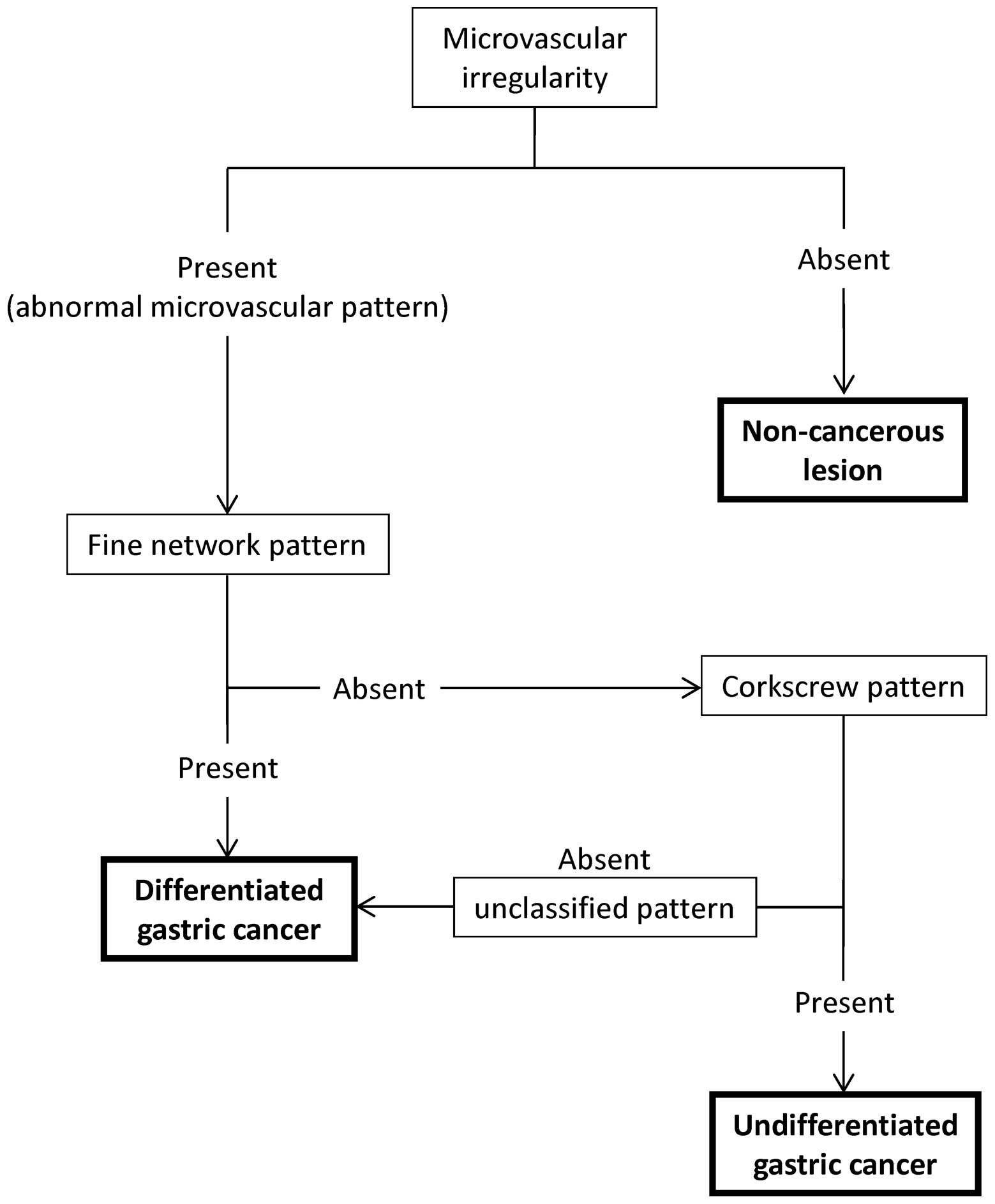
included in this study, a diagnostic algorithm based on the
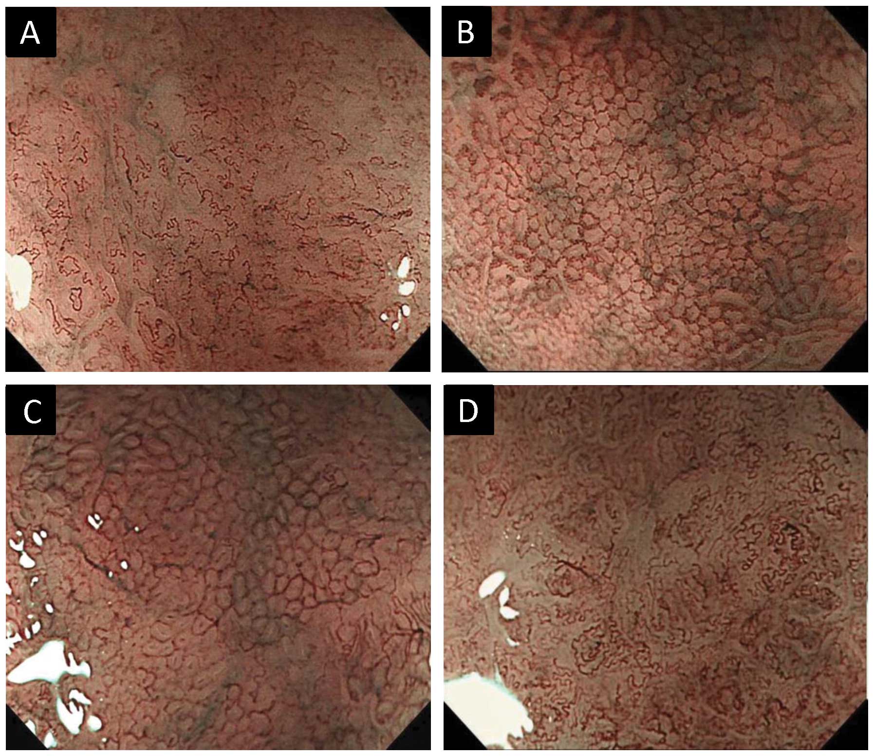
microvascular findings of ME-NBI was devised (Fig. 1). The diagnostic algorithm was
composed of ME-NBI microvascular findings of the superficial mucosa
as follows: i) microvascular irregularity (Fig. 2A and B) and ii) abnormal
microvascular patterns (10) [fine
network pattern (Fig. 2C),
corkscrew pattern (Fig. 2D), or
unclassified pattern)]. The definitions of micro-vascular
irregularity were denoted in two previous reports [Yao et al
(11) and Kaise et al
(12)]. A lesion was considered to
have microvascular irregularity if the ME-NBI findings were as
follows: ‘heterogeneity of shape’ and ‘asymmetries and
irregularities of arrangement and distribution’ (11) as well as ‘dilation’, ‘tortuousness’
and ‘abrupt caliber alteration’ as the abnormalities of individual
microvessels (12). If the lesion
had no microvascular irregularity, it was considered a
non-cancerous lesion. If the lesion had microvascular irregularity,
we assessed its abnormal microvascular pattern of ME-NBI to predict
the histological grade of gastric cancer. If it had a fine network
pattern or was regarded as an unclassified pattern, we identified
it as a differentiated adenocarcinoma. If it had a corkscrew
pattern, we identified it as an undifferentiated
adenocarcinoma.
Statistical analysis
Continuous variables were expressed as the median
(range). Sensitivity, specificity, positive predictive value (PPV),
negative predictive value (NPV) and accuracy were calculated in
order to evaluate the diagnostic efficiency of the algorithm for
gastric cancer and histological grade based on the microvascular
findings of ME-NBI. Cohen’s κ statistics were calculated for intra-
and inter-observer agreement. Inter-observer variation was
calculated from the results of the first reading, with 10 pairs in
total. Intra-observer variation was determined by comparing the
first and the second assessments for each endoscopist, with 5 pairs
in all. A κ value of ≤0.20 indicated poor agreement; 0.21–0.40,
fair agreement; 0.41–0.60, moderate agreement; 0.61–0.80, good
agreement; and 0.81–1.00, excellent agreement. Statistical analysis
was performed with SPSS software version 20 (SPSS Inc., Chicago,
IL, USA).
Results
The patients analyzed included 62 males (69%) and 28
females (31%), with a median age of 70 years (range, 28–89). The
median lesion size was 14 mm (range, 3–30). The location was the
gastric lower third (L) for 60 lesions (55%), gastric middle third
(M) for 33 lesions (30%), and gastric upper third (U) for 17
lesions (15%) (16).
In the predictions of gastric cancer by the five
endoscopists, the ME-NBI diagnostic algorithm had a mean
sensitivity, specificity, PPV, NPV, and accuracy of 86.7, 48.0,
94.4, 26.7 and 83.2%, respectively (Table I). Furthermore, in the prediction
of the histological grade of gastric cancer by the five
endoscopists, the ME-NBI diagnostic algorithm had a mean
sensitivity, specificity, PPV, NPV, and accuracy of 61.6, 86.3,
69.0, 84.8 and 79.1%, respectively (Table II).
 | Table I.Performance factors of the ME-NBI
diagnostic algorithm in predicting gastric cancer. |
Table I.
Performance factors of the ME-NBI
diagnostic algorithm in predicting gastric cancer.
| Factors | Observer A (%) | Observer B (%) | Observer C (%) | Observer D (%) | Observer E (%) | Mean (%) |
|---|
| Sensitivity | 88.0 | 88.5 | 85.0 | 81.0 | 91.0 | 86.7 |
| Specificity | 70.0 | 25.0 | 50.0 | 55.0 | 40.0 | 48.0 |
| PPV | 96.7 | 92.2 | 94.5 | 94.8 | 93.9 | 94.4 |
| NPV | 37.0 | 17.7 | 25.3 | 22.7 | 31.0 | 26.7 |
| Accuracy | 86.4 | 82.7 | 81.8 | 78.7 | 86.4 | 83.2 |
| Table II.Performance factors of the ME-NBI
diagnostic algorithm in predicting histological grade of gastric
cancer. |
Table II.
Performance factors of the ME-NBI
diagnostic algorithm in predicting histological grade of gastric
cancer.
| Factors | Observer A (%) | Observer B (%) | Observer C (%) | Observer D (%) | Observer E (%) | Mean (%) |
|---|
| Sensitivity | 57.8 | 73.5 | 64.1 | 65.6 | 46.9 | 61.6 |
| Specificity | 91.1 | 68.6 | 87.8 | 87.8 | 96.2 | 86.3 |
| PPV | 74.9 | 49.0 | 68.6 | 69.0 | 83.6 | 69.0 |
| NPV | 84.2 | 86.3 | 85.6 | 86.2 | 81.5 | 84.8 |
| Accuracy | 81.4 | 70.0 | 80.9 | 81.4 | 81.8 | 79.1 |
For evaluations of the ME-NBI features and the final
diagnosis, the mean κ values for inter-observer agreement were as
follows: microvascular irregularity, 0.49 (range, 0.32–0.74); fine
network pattern, 0.55 (range, 0.38–0.65); corkscrew pattern, 0.60
(range, 0.35–0.83); and the ME-NBI final diagnosis, 0.50 (range,
0.32–0.67). The mean κ values for intra-observer agreement were as
follows: microvascular irregularity, 0.61 (range, 0.39–0.84); fine
network pattern, 0.75 (range, 0.64–0.84); corkscrew pattern, 0.78
(range, 0.64–0.86); and ME-NBI final diagnosis, 0.77 (range,
0.66–0.84) (Table III).
| Table III.Inter- and intra-observer agreements
for evaluations of ME-NBI features and ME-NBI final diagnosis. |
Table III.
Inter- and intra-observer agreements
for evaluations of ME-NBI features and ME-NBI final diagnosis.
| Variables | Inter-observer κ
value mean (range) | Intra-observer κ
value mean (range) |
|---|
| ME-NBI features | | |
| Microvascular
irregularity | 0.49 (0.32–0.74) | 0.61 (0.39–0.84) |
| Fine network
pattern | 0.55 (0.38–0.65) | 0.75 (0.64–0.84) |
| Corkscrew
pattern | 0.60 (0.35–0.83) | 0.78 (0.64–0.86) |
| ME-NBI final
diagnosis | 0.50 (0.32–0.67) | 0.77 (0.66–0.84) |
Discussion
Early gastric cancers can be classified
macroscopically into elevated and depressed types. It is reported
that the frequency of the depressed type (∼60–70%) was higher than
that of the elevated type in early gastric cancer (19–21).
Therefore, it is important to enhance the diagnostic accuracy of
early depressed gastric cancers. ME-NBI yields clear visualization
of the microvasculature and microsurface structure of the
gastrointestinal mucosa. In a previous study it was demonstrated
that the diagnostic accuracy was significantly higher with ME-NBI
than with conventional endoscopy with white light imaging for the
differential diagnosis of depressed gastric lesions (13). Thus, we devised a diagnostic
algorithm based on ME-NBI findings to evaluate depressed gastric
lesions. ME-NBI findings, such as irregular microvascular and
microstructural patterns, are closely associated with gastric
cancer. As the microsurface structure of depressed gastric lesions
is frequently unclear, objective evaluation is difficult by ME-NBI.
Kaise et al reported that disappearance of the microsurface
structure was observed frequently in depressed gastric cancers
(12). As a consequence, this
algorithm was constructed based on ME-NBI findings relating to
microvasculature alone.
In this study, ME-NBI diagnosis with microvascular
irregularities demonstrated a high sensitivity (86.7%) and accuracy
(83.2%) for predicting gastric cancer. Previous studies revealed
that ME-NBI exhibited high sensitivity (69–93%), specificity
(85–95%) and accuracy (79–90%) in the differential diagnosis of
depressed gastric cancers from non-cancerous lesions (12–14).
Those studies evaluated the ME-NBI features of combined irregular
microvascular and microstructural patterns. By contrast, we
evaluated only ME-NBI microvascular findings and obtained good
sensitivity and accuracy equivalent to previous studies. However,
the specificity of ME-NBI diagnosis based on microvascular
irregularity was unsatisfactory. This result suggested that our
definition of microvascular irregularity included the non-specific
findings, thus, the highly specific findings for predicting gastric
cancer should be investigated. The ME-NBI diagnosis based on
abnormal microvascular patterns (fine network pattern, corkscrew
pattern, or unclassified pattern) showed high specificity (86.3%)
and accuracy (79.1%) in distinguishing undifferentiated from
differentiated adenocarcinoma. Nakayoshi et al reported that
differentiated adenocarcinoma frequently showed a fine network
pattern, and differentiated adenocarcinoma frequently showed a
corkscrew pattern on ME-NBI (10).
In depressed early gastric cancers, this study showed that 105 of
109 differentiated lesions (96.3%) had a fine network pattern or
unclassified pattern, and 48 of 56 undifferentiated lesions (85.7%)
had a corkscrew pattern. Therefore, the ME-NBI diagnosis based on
abnormal microvascular patterns was useful for predicting the
histological grade of depressed gastric cancers.
We also investigated inter- and intra-observer
agreements in the ME-NBI diagnosis with microvascular findings. To
the best of our knowledge, no complete study on the reliability and
the reproducibility of an ME-NBI diagnosis with micro-vascular
findings for depressed gastric lesions is currently available. In
the evaluation of depressed gastric lesions by the diagnostic
algorithm, inter-observer reproducibility of the ME-NBI
microvascular findings (κ value 0.49–0.60) and the ME-NBI final
diagnosis (κ value 0.50) showed fair agreement. Furthermore, the
intra-observer reproducibility of the ME-NBI microvascular findings
(κ value 0.61–0.78) and the ME-NBI final diagnosis (κ value 0.77)
exhibited a moderate agreement. Kaise et al reported that
the κ value of inter-observer concordance for an ME-NBI diagnosis
was 0.47 in the evaluation of depressed gastric lesions based on
the ME-NBI features of combined irregular microvascular and
microstructural patterns (12).
Consequently, our results are satisfactory and representative of
the observer agreement to be expected in clinical practice when
ME-NBI findings are assessed with this diagnostic algorithm.
In conclusion, the diagnostic algorithm utilized in
this study, based on ME-NBI microvascular findings was convenient
and had high diagnostic accuracy, reliability and reproducibility
in the differential diagnosis of depressed gastric lesions. Based
on these results, a prospective study is essential to evaluate the
benefit of this diagnostic algorithm.
References
|
1.
|
Parkin DM, Bray F, Ferlay J and Pisani P:
Global cancer statistics, 2002. CA Cancer J Clin. 55:74–108. 2005.
View Article : Google Scholar
|
|
2.
|
Catalano V, Labianca R, Beretta GD, Gatta
G, de Braud F and Van Cutsem E: Gastric cancer. Crit Rev Oncol
Hematol. 71:127–164. 2009. View Article : Google Scholar
|
|
3.
|
Oliveira FJ, Ferrao H, Furtado E, Batista
H and Conceicao L: Early gastric cancer: report of 58 cases.
Gastric Cancer. 1:51–56. 1998. View Article : Google Scholar : PubMed/NCBI
|
|
4.
|
Isomoto H, Shikuwa S, Yamaguchi N, et al:
Endoscopic submucosal dissection for early gastric cancer: a
large-scale feasibility study. Gut. 58:331–336. 2009. View Article : Google Scholar : PubMed/NCBI
|
|
5.
|
Tajiri H, Ohtsu A, Boku N, et al: Routine
endoscopy using electronic endoscopes for gastric cancer diagnosis:
retrospective study of inconsistencies between endoscopic and
biopsy diagnoses. Cancer Detect Prev. 25:166–173. 2001.
|
|
6.
|
Capelle LG, Haringsma J, de Vries AC, et
al: Narrow band imaging for the detection of gastric intestinal
metaplasia and dysplasia during surveillance endoscopy. Dig Dis
Sci. 55:3442–3448. 2010. View Article : Google Scholar : PubMed/NCBI
|
|
7.
|
Tahara T, Shibata T, Nakamura M, et al:
Gastric mucosal pattern by using magnifying narrow-band imaging
endoscopy clearly distinguishes histological and serological
severity of chronic gastritis. Gastrointest Endosc. 70:246–253.
2009. View Article : Google Scholar
|
|
8.
|
Tsuji Y, Ohata K, Sekiguchi M, et al:
Magnifying endoscopy with narrow-band imaging helps determine the
management of gastric adenomas. Gastric Cancer. 15:414–418. 2012.
View Article : Google Scholar : PubMed/NCBI
|
|
9.
|
Hirata I, Nakagawa Y, Ohkubo M, Yahagi N
and Yao K: Usefulness of magnifying narrow-band imaging endoscopy
for the diagnosis of gastric and colorectal lesions. Digestion.
85:74–79. 2012. View Article : Google Scholar : PubMed/NCBI
|
|
10.
|
Nakayoshi T, Tajiri H, Matsuda K, Kaise M,
Ikegami M and Sasaki H: Magnifying endoscopy combined with narrow
band imaging system for early gastric cancer: correlation of
vascular pattern with histopathology (including video). Endoscopy.
36:1080–1084. 2004. View Article : Google Scholar
|
|
11.
|
Yao K, Anagnostopoulos GK and Ragunath K:
Magnifying endoscopy for diagnosing and delineating early gastric
cancer. Endoscopy. 41:462–467. 2009. View Article : Google Scholar : PubMed/NCBI
|
|
12.
|
Kaise M, Kato M, Urashima M, et al:
Magnifying endoscopy combined with narrow-band imaging for
differential diagnosis of superficial depressed gastric lesions.
Endoscopy. 41:310–315. 2009. View Article : Google Scholar : PubMed/NCBI
|
|
13.
|
Ezoe Y, Muto M, Horimatsu T, et al:
Magnifying narrow-band imaging versus magnifying white-light
imaging for the differential diagnosis of gastric small depressive
lesions: a prospective study. Gastrointest Endosc. 71:477–484.
2010. View Article : Google Scholar
|
|
14.
|
Kato M, Kaise M, Yonezawa J, et al:
Magnifying endoscopy with narrow-band imaging achieves superior
accuracy in the differential diagnosis of superficial gastric
lesions identified with white-light endoscopy: a prospective study.
Gastrointest Endosc. 72:523–529. 2010. View Article : Google Scholar
|
|
15.
|
Yao K, Iwashita A, Tanabe H, et al: Novel
zoom endoscopy technique for diagnosis of small flat gastric
cancer: a prospective, blind study. Clin Gastroenterol Hepatol.
5:869–878. 2007. View Article : Google Scholar : PubMed/NCBI
|
|
16.
|
Japanese Gastric Cancer Association:
Japanese classification of gastric carcinoma: 3rd English edition.
Gastric Cancer. 14:101–112. 2011. View Article : Google Scholar : PubMed/NCBI
|
|
17.
|
Ono H, Kondo H, Gotoda T, et al:
Endoscopic mucosal resection for treatment of early gastric cancer.
Gut. 48:225–229. 2001. View Article : Google Scholar : PubMed/NCBI
|
|
18.
|
Japanese Gastric Cancer Association:
Japanese gastric cancer treatment guidelines 2010 (ver. 3). Gastric
Cancer. 14:113–123. 2011. View Article : Google Scholar : PubMed/NCBI
|
|
19.
|
Xuan ZX, Ueyama T, Yao T and Tsuneyoshi M:
Time trends of early gastric carcinoma. A clinicopathologic
analysis of 2846 cases. Cancer. 72:2889–2894. 1993. View Article : Google Scholar : PubMed/NCBI
|
|
20.
|
Hirota T, Itabashi M, Daibo M, et al:
Chronological changes in the morphological features of early
gastric cancer, especially recent changes in macroscopic findings.
Jpn J Clin Oncol. 14:181–199. 1984.
|
|
21.
|
Hyung WJ, Cheong JH, Kim J, Chen J, Choi
SH and Noh SH: Application of minimally invasive treatment for
early gastric cancer. J Surg Oncol. 85:181–185. 2004. View Article : Google Scholar : PubMed/NCBI
|