Introduction
Adipose-derived stem cells (ADSCs) are multipotent
mesenchymal cells, which present similar cellular characteristics
to bone marrow-derived stem cells (BMSCs) (1). ADSCs can be acquired from adipose
tissue in large quantities during surgical procedures. The yield of
mesenchymal stem cells from adipose tissue is markedly higher than
that from bone marrow tissue. Similar to BMSCs, ADSCs secrete
various dynamic cytokines. These cytokines are extensively involved
in regenerative medicine, such as wound healing, tissue repair and
anti-aging therapies (2–5).
Human dermal fibroblasts (HDFs) are imperative in
human skin aging. They function in repairing dermal tissue and
maintaining the integrity and youth of skin. During the aging
process of skin, dermal fibroblasts exhibit a decline in cell
number and cell function (6). The
altered function of HDFs is a vital target for skin rejuvenation
therapy. Traditional anti-aging therapy involves activation of
dermal fibroblasts through laser or topical agents (5). However, stem cell therapy for aging
has recently started to evolve. The therapeutic effects of
menchymal stem cells have been widely studied. Previous studies
have shown that ADSCs can promote fibroblast function in wounded or
normal skin through paracrine effects (2). Since these growth factors secreted by
ADSCs can be produced in large quantities and applied with few
ethical issues, there is a promising future for the use of ADSCs in
anti-aging therapy (7). In
addition, a recent study has demonstrated that cytokines from ADSCs
have protective properties against photo-aging (8).
There is limited information regarding the exact
mechanism of the paracrine effects of ADSCs on fibroblasts.
Previous research has failed to consider whether the cytokines
produced by ADSCs genetically rejuvenate fibroblasts, or improve
fibroblast function to compensate for accelerated cell aging. In
addition, few reports have shown the change in the senescence state
of fibroblasts modulated by ADSCs.
In order to clarify whether ADSCs could rejuvenate
naturally aged human dermal fibroblasts through paracrine
mechanisms, the present study investigated the paracrine effects of
ADSC-secreted cytokines on HDFs, and particularly analyzed the
change in the senescence state of fibroblasts.
Materials and methods
Subjects
Adipose tissues were harvested during cosmetic
abdominal liposuction surgery from three healthy female donors
(mean age, 24.6 years). Full skin samples were obtained from the
abdomen in selective plastic surgery, including abdominoplasty,
skin grafts and scar plasty, from three young donors (mean age,
22.3 years) and three aged donors (mean age, 64.5 years). The study
protocol was approved by the Ethics Committee of the First
Affiliated Hospital of Chongqing Medical University (Chongqing,
China). Informed consent was obtained from all donors.
Isolation and culture of ADSCs and
HDFs
Human ADSCs were prepared as previously described,
with slight modifications (9).
Briefly, the adipose aspirate tissue was washed extensively with
phosphate-buffered saline (PBS) and digested with 0.01% type I
collagenase (Sigma-Aldrich, St. Louis, MO, USA) with gentle
agitation for 60 min at 37°C. The collagenase activity was ceased
by adding Dulbecco’s modified Eagle’s media (DMEM). Then
centrifugation was performed at 160 g for 10 min. The pellet was
filtered with a 100-mesh nylon mesh filter. The retrieved cell
fraction was incubated in T225 flasks overnight at 37°C in a
humidified atmosphere with DMEM (F12; Gibco-BRL, Carlsbad, CA, USA)
with 10% fetal bovine serum (FBS) and 1% penicillin-streptomycin
solution. The cells were passaged by 0.05% trypsin digestion and
plated at a density of 5×10³ cells/cm2 until
they reached 75–90% confluence (~6 days later). ADSCs were used in
experiments at passage 4–6.
The harvested skin samples were de-epithelialized
and minced. The dermal tissue was incubated in serum free DMEM
(F12) with 0.25% trypsin for 3 h in a 37°C, 5% CO2
atmosphere. Centrifugation was performed (120 g for 5 min) and the
pellets were seeded in DMEM (F12) containing 10% FBS and 1%
penicillin-streptomycin solution. Fibroblasts of passage 4–6 were
used for the subsequent experiments.
Identification of immunophenotype of
ADSCs by flow-cytometry
Cultured ADSCs at passage 3 were analyzed by
fluorescence-activated cell sorting (FACS) for the expression of
cluster of differentiation (CD)34, CD90 and CD105. The cells were
re-suspended in PBS and incubated with fluorescein
isothiocyanate-conjugated mouse antibodies against CD90 (Becton
Dickinson, Franklin Lakes, NJ, USA), CD34 and CD44 (Millipore,
Billerica, MA, USA) for 30 min at 4°C. Cells were analyzed using a
FACScan flow cytometer (BD Facs Vantage SE; BD Biosciences,
Franklin Lakes, NJ, USA). Data were processed by CellQuest Pro
software (BD Biosciences).
Co-culture of ADSCs and HDFs
Four groups of cell culture were established,
including the control groups. For the control groups, HDFs from the
young or the aged patients were cultured respectively with DMEM
(F12) containing 10% FBS. For the experimental co-culture groups,
ADSCs from passage 4–6 were seeded in the upper chamber of 0.45-μm
pore, collagen-coated Transwell culture plates (Corning Costar,
Cambridge, MA, USA). In the lower chambers, 1×10⁵/ml
HDFs from young or aged donors at passage 3 were seeded in DMEM
(F12) containing 10% FBS, accordingly.
Proliferation of HDFs with the MTT
assay
Cell proliferation was assessed by a colorimetric
MTT (tetrazolium salts) assay. After removing the medium from each
well, 500 μl of 0.5 mg/ml MTT solution (KeyGen Biotech, Nanjing,
China) was added and incubated for 4 h at 37°C in the dark. The
supernatant was removed, and formazan crystals were dissolved in
200 μl dimethyl sulfoxide (DMSO) for 10 min. The DMSO solution was
transferred into 24-well plates and the optical density (OD) was
measured using a spectrophotometer (Hach Company, Loveland, CO,
USA) at 490 nm. Each test was repeated three times. Data were
measured every 24 h for 3 days from 0 h in all groups.
Quantitative polymerase chain reaction
(qPCR) assay of type I collagen, MMP-1 and senescence associated
β-galactosidase (SA-β-GAL) in HDFs
The mRNA expression of type I collagen, MMP-1 and
SA-β-GAL in dermal fibroblasts were assessed by qPCR. Cultured HDFs
at 0, 24, 48 and 72 h were used for analysis. Total RNA was
isolated from HDFs using the TRIzol (Tiangen, Beijing, China)
according to the manufacturer’s instructions and quantified using a
spectrophotometer. Total RNA (1 μg) was reverse transcribed using
the ReverTra Ace qPCR RT kit (Toyobo Biotech, Shanghai, China). The
PCR reaction was then conducted according to the manufacturer’s
instructions (Applied Biosystems, Foster City, CA, USA). Briefly,
50 μl reaction mixture, including 2.5 units Taq polymerase
(Tiangen, Beijing, China), 5 μl 10X buffer, 1.5 mm
MgCl2, 200 μm dNTPs, 1 μl of first-strand cDNA, and 25
pm of each primer, were subjected to 28 cycles (denaturation at
94°C for 1.5 min, annealing at 58°C for 1 min and polymerization at
72°C for 1 min). The PCR products were analyzed on a 1.5% agarose
gel. The primer sequences and product sizes used for this study
were as follows: Forward: 5′-ACCCCGTGCTGCTGACCGAG-3′ and reverse:
5′-TCCCGGCCAGCCAGGTCCA-3′ for β-actin-1, 249 base pairs (bp);
forward: 5′-CCCCAA AAGCGTGTGACAGTAAG-3′ and reverse: 5′-GAA
GGGATTTGTGCGCATGTAGA-3′ for MMP-1, 200 bp; forward:
5′-GCACGAAACACACTGGGAATG-3′ and reverse:
5′-GGCCAACGTCCACACCAAATTC-3′ for type I collagen, 262 bp; and
forward: 5′-GTGCATTGGCCATACCCTTAGG-3′ and reverse:
5′-CACACGGTCAGCATGCATAAATA-3′ for SA-β-Gal, 259 bp. GAPDH (forward
5′-CGGAGTCAACGGATTTGGTCGTAT-3′ and reverse,
5′-AGCCTTCTCCATGGTGGTGAAGAC-3′) was included in each run as a
template control. PCR bands were visualized by UV illumination
following electrophoresis on 1.5% agarose gel.
Statistical analysis
All results are presented as the mean ± standard
deviation. Data were analyzed using SPSS package 19.0 (SPSS Inc.,
Chicago, IL, USA). Comparisons among the groups were analyzed with
one way analysis of variance and P<0.05 was considered to
indicate a statistically significant difference.
Results
Characteristics of ADSCs
For the purpose of investigating the paracrine
effects of ADSCs on aged human dermal fibroblasts, human ADSCs were
co-cultured with human dermal fibroblasts in Transwell plates,
which allow for paracrine interactions without direct cell contact.
Young and aged HDFs alone were set as control groups. The ADSCs
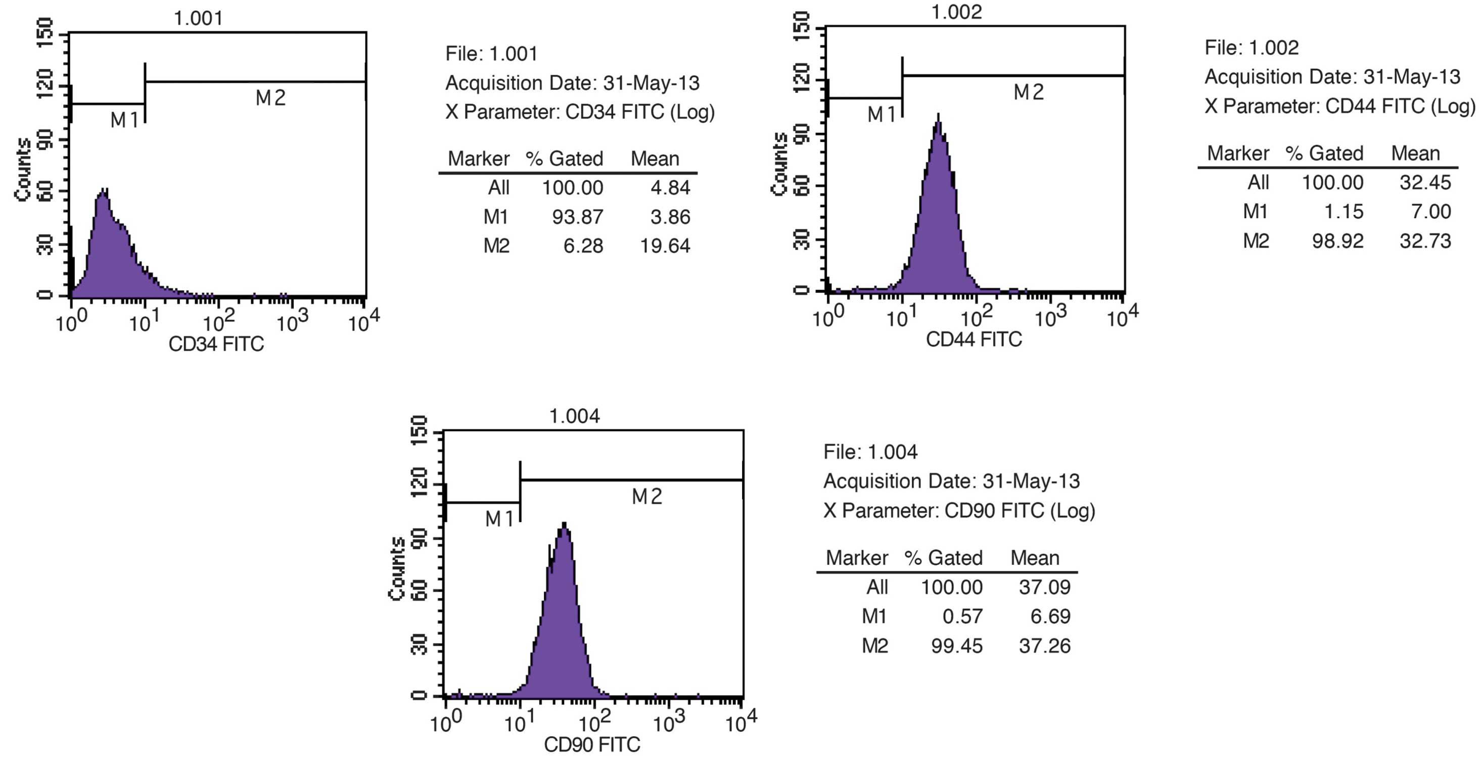
were assayed at passage 3. ADSCs were characterized with the
profile of surface CD markers. The results obtained from
flow-cytometry showed that ADSCs exhibited positive expression of
CD90, CD44 and negative expression of CD34 (Fig. 1).
ADSCs increase the proliferation of human
dermal fibroblasts through paracrine effects
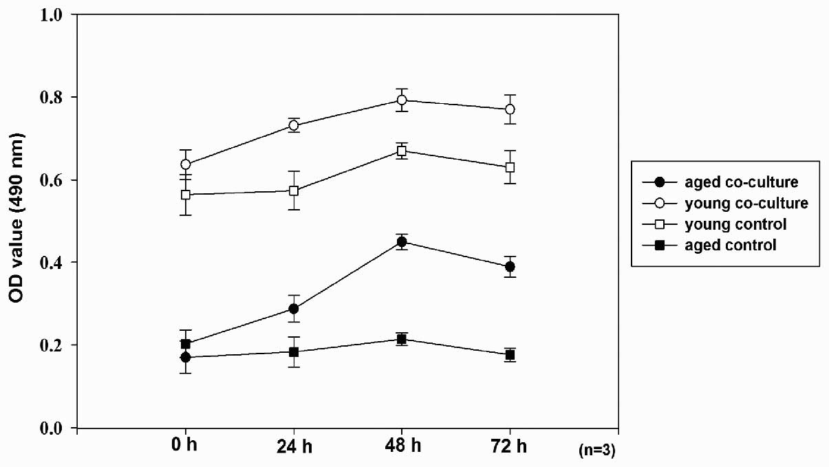
The proliferation status of HDFs in each group was
investigated by an MTT assay (Fig.
2). The MTT assays showed that the co-culture of young and aged
HDFs had exhibited significant higher optical densities (OD, 490
nm) values as compared with the control groups (P<0.01). The
time-course effects showed that proliferation of co-cultured young
HDFs gradually increased during the first 48 h (P<0.05), and
then decreased at 72 h (P<0.05). The control group of young HDFs
had higher OD values as compared with the aged control group at
each time point (P<0.05). The young and aged fibroblasts group
shared a similar OD trend with co-cultured aged HDFs (Fig. 2).
ASDCs induce increased cellular function
of human dermal fibroblasts through paracrine effects
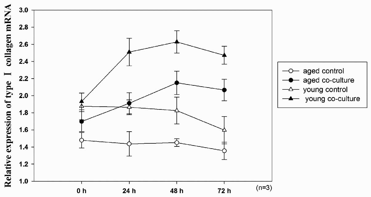
In order to investigate the paracrine effects of
ADSCs on HDFs, the mRNA expression of type I collagen and MMP-1 in
HDFs was analyzed using qPCR (Fig.
3). During the first 48 h, the type I collagen mRNA expression
of co-cultured aged HDFs exhibited a marked increase as compared
with the control groups (P<0.01). Following this, the type I
collagen expression decreased significantly at 72 h (P<0.05).
The aged control group showed no significant difference in the type
I collagen mRNA expression over 72 h (P>0.05), however, the
young and aged control groups showed a marginal decrease in mRNA
expression at 72 h (P<0.05). The co-cultured young HDF group
exhibited a steady increase in type I collagen mRNA expression
during the first 48 h, of which a higher level of expression was
reached overall as compared with the aged co-culture groups
(P<0.05). These cells expressed a similar decreased expression
at 72 h as the aged co-culture group (Fig. 3).
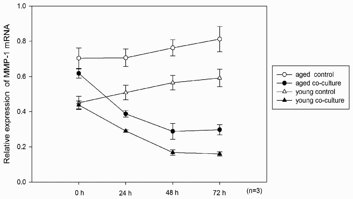
The mRNA expression of MMP-1 in the co-culture aged
groups decreased significantly as compared with the control groups
(P<0.01). The evaluation of the time-course effects showed that
MMP-1 expression increased gradually in the control groups
(P<0.05). There was no significant difference in MMP-1 mRNA
expression between the 48 and 72 h time points of the two
co-culture groups (P>0.05), indicating that the descending trend
ceased after 48 h in the two groups (Fig. 4).
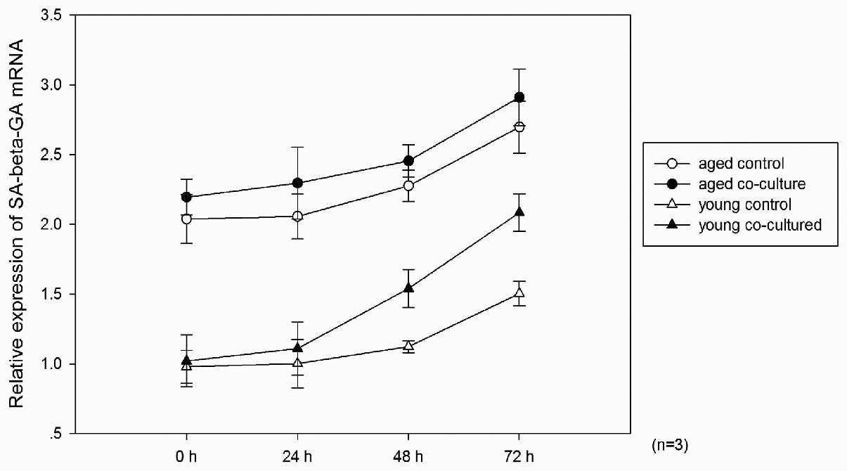
ADSCs increase the SA-β-Gal levels in
fibroblasts through a paracrine mechanism
To evaluate the change in senescence stage of
cultured HDFs, the SA-β-Gal mRNA expression was measured. The two
control groups showed no significant differences in SA-β-Gal mRNA
expression during the first 24 h (P>0.05) and then exhibited a
gradual increase in the level of SA-β-Gal mRNA expression after 24
h (P<0.05). The aged groups exhibited a higher expression level
of SA-β-Gal at each time point (P<0.01). The SA-β-Gal mRNA
expression increased markedly in the two co-cultured groups
(P<0.05). The SA-β-Gal mRNA expression in every group
significantly increased at 72 h (P<0.01; Fig. 5).
Discussion
Adipose derived stem cells (ADSCs) are mesenchymal
stem cells residing in adipose tissue. They were first identified
in 2003 by Zuk et al (10).
ADSCs are readily accessible, and are therefore a valuable source
of a large number of somatic stem cells. ADSCs exhibit a
multi-lineage differentiation capacity, with the differential
potential to become osteocytes, chondrocytes and adipocytes
(11). Similar to BMSCs, ADSCs are
reported to secrete a profile of cytokines (3). They are capable of secreting various
cytokines, including platelet-derived growth factor, insulin-like
growth factor, keratinocyte growth factor, fibroblast growth
factor, transforming growth factor-β, vascular endothelial growth
factor, interleukin (IL)-6, IL-8, IL-11, IL-17 and monocyte
chemo-attractant protein (12,13).
These dynamic growth factors are regarded to communicate with
surrounding tissues and are reported to show positive effects on
wound healing, tissue repair and anti-aging (14). Since these growth factors can be
administered with few ethical implications, the paracrine effects
of ADSCs have proposed a novel strategy for numerous clinical
treatments. These are more likely organizers than participants as,
considering their relatively small cell quantity, ADSCs exhibit
regenerative bioactivities mainly through paracrine factors, rather
than directly differentiating to surrounding tissue. ADSCs modify
the microenvironment, which sequentially modifies the peripheral
cells. Numerous data have indicated that stem cells implement their
therapeutic effects through a paracrine mechanism (4,5,8,14,15).
Dermal fibroblasts serve to maintain dermis
homeostasis and participate in skin tissue repair. Alternations of
their function, such as reduced production of collagen and
hyaluronic acid, increase of MMP activity, slowed proliferation and
a decrease in cell number, are vital manifestations of human skin
aging (16). Several studies have
demonstrated the paracrine effects of ADSCs on fibroblasts. Kim
et al (17) reported that
injection of ADSCs in moderately aged mice resulted in increased
dermal thickness and collagen density. It was considered that ADSCs
had anti-aging properties by stimulating collagen synthesis and
increasing dermal angiogenesis through paracrine effects. Park
et al (2) showed in an
animal model that ADSCs and their conditioned media could stimulate
collagen synthesis and promote the migration of fibroblasts to
wounds. It was proposed that this was associated with the collagen
synthesis of ADSCs or the secretion of various growth factors. A
study by Kim et al (5)
isolated and cultured ADSCs and HDFs separately, then added the
conditioned medium of ADSCs to the fibroblasts. It was shown that
the migration, proliferation and collagen production of fibroblasts
increased accordingly, and the wound healing ratio was markedly
increased. The paracrine mechanism of ADSCs was proposed; however,
the influence of cell senescence and age difference as well as the
time-course effect, was not discussed.
In the present study, it was confirmed that ADSCs
promote proliferation in young and aged fibroblasts through a
paracrine mechanism. ADSCs showed mild promoting effects on the
proliferation of co-cultured aged fibroblasts in the early phase,
and the proliferation peaked at 48 h, then declined at 72 h. There
was a decreasing trend after 48 h. It was assumed that the effects
of ADSCs in promoting proliferation had certain limitations as
determined by the potential restoration of fibroblasts. Further
investigation and a longer time period are required to verify these
assumptions.
The results of the present study have shown a
decreased expression of MMP-1 and increased expression of type I
collagen in co-culture groups. MMP-1 is a tissue inhibitor that is
found to increase in the degradation process and decrease in the
regeneration process, whereas type I collagen is positively
correlated with dermal tissue repair. MMP-1 is known to initiate
photo-aging and tissue degeneration. A decrease in the level of
MMP-1 in fibroblasts suggests improved tissue regeneration. The
results of the MMP-1 and type I collagen mRNA assays were
consistent with a previous study by Song et al (15). The aged fibroblast groups showed a
higher expression of MMP-1 mRNA. In the co-culture groups, the
MMP-1 mRNA expression decreased significantly as compared with the
control groups. However, the decreasing trend ceased after 48 h in
the co-culture groups. This may suggest that the promoting effect
of ADSCs may reach the maximum at 48 h. Although the expression of
type I collagen mRNA of co-cultured aged fibroblasts showed no
significant difference at 24 and 48 h, an increasing trend over
time was observed.
A previous study by Song et al (15) showed that ADSCs induced decreased
expression of p16 in photo-aged fibroblasts. P16 is a
tumor-suppressor and a senescence marker. It was proposed that the
decrease of p16 in photo-aged fibroblasts indicated a reverse of
the aging process at the genetic level. Although various biomarkers
have been reported to identify senescent cells, none of them are
regarded to be specific. SA-β-GAL has long been considered as a
reliable biomarker for cell senescence (18). In the present study; however, the
results of SA-β-GAL mRNA expression assay showed a significant
increase in co-cultured young and aged fibroblasts. The increased
expression of SA-β-GAL may indicate acceleration of fibroblast
senescence. It may be implied from these results that ADSC
co-cultured fibroblasts present improved cellular function with
accelerated senescence. The exact mechanism underlying the effects
of the secreted factors from ADSCs on fibroblasts remains unknown.
It is assumed that these cytokines may improve the function of
fibroblasts to a certain extent; however, they could not rejuvenate
fibroblasts, or accelerate senescence at the genetic level. As
cells in vitro have distinct biological characteristics,
in vivo investigations are required to confirm these
assumptions. Additional genetic senescence markers should be tested
and a longer culture time is also required for determining the
long-term effects of ADSC-secreted cytokines on fibroblasts.
Although ADSCs are generally regarded as a promising
source of cells for anti-aging therapies, the exact mechanism of
action remains unclear. Favorable effects have been widely
reported, however, the side effects require additional attention.
Meticulous study of long term side effects is imperative prior to
any clinic applications.
Acknowledgements
This study was financed by a CMA-L’OREAL China
Skin/Hair grant (grant no. S2012091001).
References
|
1
|
Wagner W, Wein F, Seckinger A, et al:
Comparative characteristics of mesenchymal stem cells from human
bone marrow, adipose tissue, and umbilical cord blood. Exp Hematol.
33:1402–1416. 2005. View Article : Google Scholar : PubMed/NCBI
|
|
2
|
Park BS, Jang KA, Sung JH, et al:
Adipose-derived stem cells and their secretory factors as a
promising therapy for skin aging. Dermatol Surg. 34:1323–1326.
2008.PubMed/NCBI
|
|
3
|
Kilroy GE, Foster SJ, Wu X, et al:
Cytokine profile of human adipose-derived stem cells: expression of
angiogenic, hematopoietic and pro-inflammatory factors. J Cell
Physiol. 212:702–709. 2007. View Article : Google Scholar : PubMed/NCBI
|
|
4
|
Lee SH, Jin SY, Song JS, Seo KK and Cho
KH: Paracrine effects of adipose-derived stem cells on
keratinocytes and dermal fibroblasts. Ann Dermatol. 24:136–143.
2012. View Article : Google Scholar : PubMed/NCBI
|
|
5
|
Kim WS, Park BS, Sung JH, et al: Wound
healing effect of adipose-derived stem cells: A critical role of
secretory factors on human dermal fibroblasts. J Dermatol Sci.
48:15–24. 2007. View Article : Google Scholar : PubMed/NCBI
|
|
6
|
Zouboulis CC and Makrantonaki E: Clinical
aspects and molecular diagnostics of skin aging. Clin Dermatol.
29:3–14. 2011. View Article : Google Scholar : PubMed/NCBI
|
|
7
|
Watson RE and Griffiths CE: Pathologic
aspects of cutaneous photoaging. J Cosmet Dermatol. 4:230–236.
2005. View Article : Google Scholar
|
|
8
|
Kim W, Park B and Sung J: Protective role
of adipose-derived stem cells and their soluble factors in
photoaging. Arch Dermatol Res. 301:329–336. 2009. View Article : Google Scholar : PubMed/NCBI
|
|
9
|
Gimble J and Guilak F: Adipose-derived
adult stem cells: isolation, characterization, and differentiation
potential. Cytotherapy. 5:362–369. 2003. View Article : Google Scholar : PubMed/NCBI
|
|
10
|
Zuk PA, Zhu M, Ashjian P, et al: Human
adipose tissue is a source of multipotent stem cells. Mol Biol
Cell. 13:4279–4295. 2002.PubMed/NCBI
|
|
11
|
Strem BM, Hicok KC, Zhu M, et al:
Multipotential differentiation of adipose tissue-derived stem
cells. Keio J Med. 54:132–141. 2005. View Article : Google Scholar : PubMed/NCBI
|
|
12
|
Rehman J, Traktuev D, Li J, et al:
Secretion of angiogenic and antiapoptotic factors by human adipose
stromal cells. Circulation. 109:1292–1298. 2004. View Article : Google Scholar : PubMed/NCBI
|
|
13
|
Kondo K, Shintani S, Shibata R, et al:
Implantation of adipose-derived regenerative cells enhances
ischemia-induced angiogenesis. Arterioscler Thromb Vasc Biol.
29:61–66. 2009. View Article : Google Scholar : PubMed/NCBI
|
|
14
|
Hong SJ, Traktuev DO and March KL:
Therapeutic potential of adipose-derived stem cells in vascular
growth and tissue repair. Curr Opin Organ Transplant. 15:86–91.
2010. View Article : Google Scholar : PubMed/NCBI
|
|
15
|
Song SY, Jung JE, Jeon YR, Tark KC and Lew
DH: Determination of adipose-derived stem cell application on
photo-aged fibroblasts, based on paracrine function. Cytotherapy.
13:378–384. 2011. View Article : Google Scholar : PubMed/NCBI
|
|
16
|
Varani J, Dame MK, Rittie L, et al:
Decreased collagen production in chronologically aged skin: roles
of age-dependent alteration in fibroblast function and defective
mechanical stimulation. Am J Pathol. 168:1861–1868. 2006.
View Article : Google Scholar
|
|
17
|
Kim JH, Jung M, Kim HS, Kim YM and Choi
EH: Adipose-derived stem cells as a new therapeutic modality for
ageing skin. Exp Dermatol. 20:383–387. 2011. View Article : Google Scholar : PubMed/NCBI
|
|
18
|
Maier AB, Westendorp RG and van Heemst D:
Beta-galactosidase activity as a biomarker of replicative
senescence during the course of human fibroblast cultures. Ann NY
Acad Sci. 1100:323–332. 2007. View Article : Google Scholar : PubMed/NCBI
|