Introduction
Natriuretic peptides comprise a family of three
structurally related peptides: Atrial natriuretic peptide (ANP),
brain natriuretic peptide (BNP) and C-type natriuretic peptide
(CNP). ANP and BNP are the primary cardiac hormones in the
myocardium, which regulate cardiac remodeling through
paracrine/autocrine actions. However, CNP, which was originally
isolated from porcine brain extracts (1), not only has a regulatory effect in
the central nervous system, but is also involved in the
cardiovascular system (2). Besides
the central nervous system and vascular endothelial cells, CNP is
also synthesized by cardiac ventricular cells, mainly cardiac
fibroblasts (CFs) in neonatal rats (3). Multifaceted cadioprotective effects
of CNP have been previously reported (4–6), and
it has been suggested that CNP may have more potent antifibrotic
effects compared to ANP and BNP in cultured CFs (3).
Cardiac fibrosis is a significant aspect of cardiac
remodeling, in which CFs are essential (7). Evidence reveals that CNP has a
suppressive effect on cardiac fibrosis; however, the underlying
cellular and molecular mechanisms have not been fully investigated.
Whether and how CNP affects the function of CFs remains to be
elucidated. Therefore, experiments were conducted in isolated and
purified CFs in order to investigate whether CNP suppresses the
differentiation of CFs into myofibroblasts and whether the
secretion of profibrotic factors in CFs is affected by CNP. The
present study demonstrated that CNP inhibits the differentiation of
CFs into myofibroblasts in vitro, inhibits the migrational
ability of CFs, and reduces the expression and secretion of
monocyte chemoattractant protein-1 (MCP-1) and plasminogen
activator inhibitor-1 (PAI-1) from CFs, which was mediated by the
inhibition of the activity of extracellular signal-regulated kinase
1/2 (ERK1/2).
Materials and methods
Reagents
Dulbecco’s modified Eagle’s medium (DMEM), fetal
bovine serum (FBS) and TRIzol reagent were purchased from
Invitrogen Life Technologies (Carlsbad, CA, USA). CNP was purchased
from Bachem (Torrance, CA, USA). Mouse monoclonal antibody against
α-smooth muscle actin (α-SMA) was purchased from Sigma-Aldrich (St.
Louis, MO, USA). Mouse monoclonal antibodies against glyceraldehyde
3-phosphate dehydrogenase (GAPDH), extra domain-A (ED-A)
fibronectin, collagen I and III, and vimentin were purchased from
Abcam (Cambridge, MA, USA). Mouse monoclonal antibodies against von
Willebrand factor and troponin I were purchased from Santa Cruz
Biotechnology, Inc. (Santa Cruz, CA, USA). Horseradish peroxidase
(HRP)-conjugated, rhodamine-conjugated and fluorescein
isothiocyanate (FITC)-conjugated secondary antibodies were obtained
from BD Biosciences (San Jose, CA, USA). 4,
6-diamidino-2-phenylindole (DAPI) was purchased from Beyotime
(Jiangsu, China). Antibodies against ERK1/2 and phospho (p)-ERK1/2
were purchased from Cell Signaling Technology, Inc. (Beverly, MA,
USA). U0126 was purchased from Calbiochem (San Diego, CA, USA). An
MCP-1 ELISA kit was obtained from R&D Systems (Minneapolis, MN,
USA) and the PAI-1 ELISA kit was from American Diagnostica
(Greenwich, CT, USA).
Cell cultures
Primary cultures of neonatal rat CFs were prepared
as described previously (8).
Briefly, the hearts were excised from three-day-old Sprague-Dawley
rats (n=46; Vital River Laboratory Animal Technology Co., Ltd.,
Beijing, China) and rinsed several times with phosphate-buffered
saline. The ventricles were minced and trypsinized. Following
centrifugation at 180 × g for 10 min, the cell pellets were
resuspended. Floating cardiomyocytes and attached fibroblasts were
separated. The purity of the cultured CFs was >95% on the basis
of positive staining for vimentin and negative staining of troponin
I and von Willebrand factor. Third-passage CFs were used in all the
experiments. For subsequent experiments, cells at 80% confluence
were growth-arrested by serum starvation for 24 h For myofibroblast
differentiation and the cytokine secretion assay, CFs were cultured
in DMEM with 10% FBS, with or without different concentrations of
CNP (10−9, 10−8, 10−7 mol/l) for
24 h. In order to investigate the potential involvement of the ERK
signaling pathway in the effects of CNP, CFs were pretreated with
the pharmacological kinase inhibitor U0126 (10−4 mol/l)
for 30 min, followed by treatment with CNP (10−7 mol/l).
The cells and supernatants were harvested and stored at −80°C. All
the procedures using animals were reviewed and approved by the
Experimental Laboratory Animal Ethics Committee of Capital Medical
University (Beijing, China) and were performed according to the
criteria outlined by the National Ministry of Health.
Western blot analysis
Western blot analysis was performed as previously
described (9). Anti-α-SMA
(1:1,000), anti-ED-A fibronectin (1:400), anti-collagen I
(1:1,000), anti-collagen III (1:500), anti-ERK1/2 (1:2,000),
anti-p-ERK1/2 (1:2,000) and anti-GAPDH (1:2,000) mouse monoclonal
antibodies were used as the primary antibodies. The secondary
antibody was HRP-conjugated anti-mouse IgG (1:4,000). The
immunoreactive protein bands were detected using an enhanced
chemiluminescence detection method (Applygen Technologies Inc.,
Beijing, China). Equal loading of the samples was further verified
by staining of the blots with monoclonal antibodies against GAPDH.
All the western blots were quantified using densitometry (Universal
Hood; Bio-Rad Laboratories, Inc., Hercules, CA, USA).
Immunofluorescence
CFs at passage 3 were seeded on coverslips and
allowed to attach overnight. The cells were serum-starved for 24 h
and subsequently treated with CNP for 24 h prior to staining with
anti-α-SMA antibody (1:400) or anti-ED-A fibronectin antibody
(1:200). Then cultures were incubated with Rhodamine- or
FITC-conjugated secondary antibodies. Mounting medium containing
DAPI was used to visualize the cell nuclei.
Transwell migration assay
The ability of CFs to migrate across a matrix
barrier towards chemotactic stimuli (in the present study 2% FBS
was used) was investigated using the colorimetric transwell system
QCM™ (Millipore Corporation, Billerica, MA, USA), which allows
cells to migrate through a polycarbonate membrane with 8-μm pores.
The CFs were serum starved for 24 h prior to the experiment. The
cells were harvested and resuspended with serum-free DMEM, diluted
to 5×10⁵ cells/ml. Subsequently, 300 μl cell suspensions
were added to the upper chamber of each insert, 500 μl serum-free
DMEM with 2% FBS was added to the lower chamber. CNP at the
appropriate concentration was added to the upper and lower chambers
of the experimental wells and incubation was at room temperature
for 6 h. The migrated cells on the underside of the membrane were
fixed by methanol treatment and stained with 0.1% crystal violet.
Inserts were then dried and added to the extraction buffer. The
optical density (OD) of the dye extract was measured at 560 nm
using a microplate reader (Bio-Rad Laboratories, Inc.).
Reverse transcription-quantitative
polymerase chain reaction (RT-qPCR)
The total RNA was isolated using TRIzol reagent
according to the manufacturer’s instructions. cDNA was generated
with oligo (dT)15 primers (Promega Corporation, Madison,
WI, USA). RT-qPCR was performed with Applied Biosystems 7500 real
time PCR system using QuantiTect SYBR Green PCR Master mix (Applied
Biosystems, Carlsbad, CA, USA). For normalization the housekeeping
gene GAPDH was applied as a reference gene. The primer sequences
are as follows: Forward: 5′-TGGCTCAGCCAGATGCAGT-3′ and reverse:
5′-ATTGGGATCATCTTGCTGGTG-3′ for MCP-1; PAI-1, forward:
5′-AACCCAGGCCGACTTCA-3′ and reverse: 5′-CATGCGGGCTGAGACTAGAAT-3′
for PAI-1; and forward: 5′-GGCAAATTCAACGGCACAGT-3′ and reverse:
5′-AGATGGTGATGGGCTTCCC-3′ for GAPDH. The comparative Ct (cycle
threshold) method, also referred to as the 2−ΔΔCT
method, was used to quantify the gene expression. The relative gene
expression was expressed as the ratio of CNP-treated to
non-CNP-treated samples.
ELISA assay
MCP-1 and PAI-1 concentrations in the supernatants
were determined by ELISA kits, according to the manufacturer’s
instructions. The OD of each well at 450 nm was measured using a
microplate reader (Bio-Rad Laboratories, Inc.). The relative
protein secretion was expressed as the ratio of CNP-treated to
non-CNP-treated samples.
Statistical analysis
The values are shown as the mean ± standard error of
the mean. Analysis of variance and Student’s t-test were used for
statistical analysis of the data. P<0.05 was considered to
indicate a statistically significant difference.
Results
CNP inhibits myofibroblast
differentiation of CFs
To investigate the effect of CNP on myofibroblast
differentiation of CFs, CFs isolated from neonatal rat ventricles
were cultured in complete medium (DMEM with 10% FBS), and only
third-passage CFs were analyzed. Prominent expression of α-SMA and
ED-A fibronectin is the most well-characterized feature of
myofibroblasts (10–14). In CFs treated with CNP, the protein
expression of α-SMA and ED-A fibronectin was suppressed in a
dose-dependent manner (Fig. 1A).
Previous studies have shown that CFs cultured in vitro with
DMEM containing FBS acquired a myofibroblast phenotype (13,15).
Consistently, in the present study, it was revealed that CFs
differentiated into myofibroblats with prominent stress fibers
after 24 h in culture, demonstrated by the immunofluorescence
staining of α-SMA, and the immunofluorescence signal was markedly
reduced in the CNP-treated CFs. The inhibitory effect of CNP on the
protein expression of ED-A fibronectin was also validated by
immunofluorescence staining (Fig.
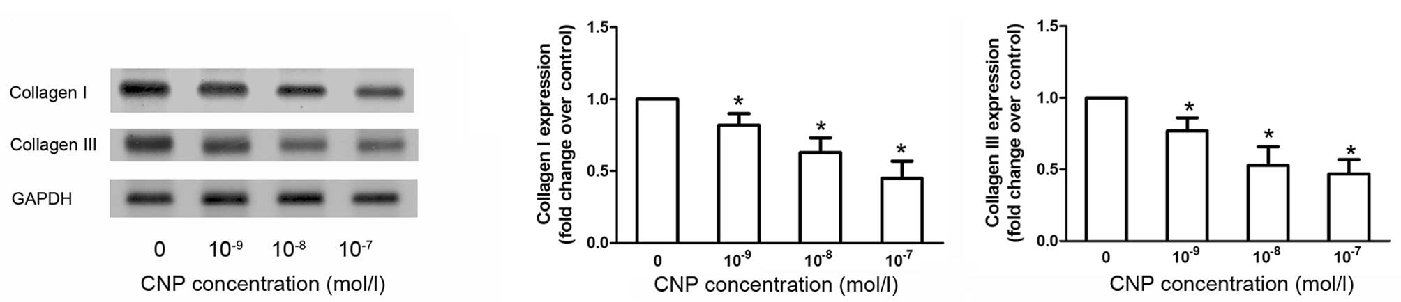
1B). In addition, western blot analysis results also revealed
that CNP treatment decreased the expression of collagen I and III
in CFs (Fig. 2).
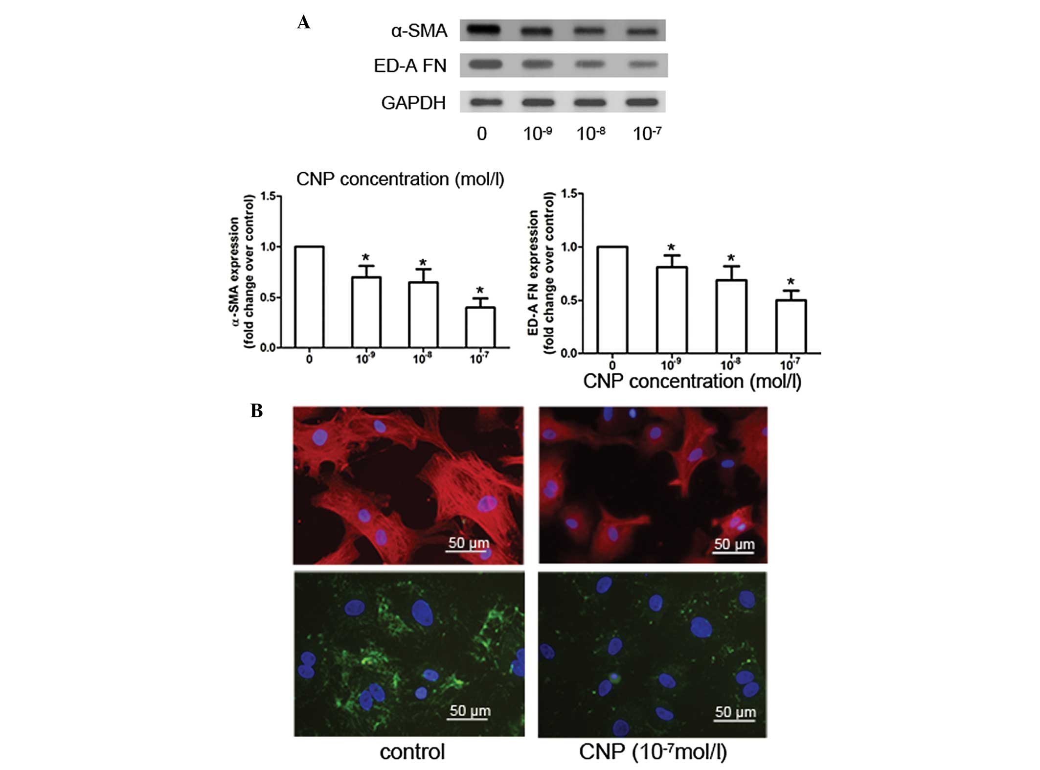 | Figure 1CNP inhibits α-SMA and ED-A
fibronectin expression in CFs. (A) Western blot analysis and the
corresponding densitometric quantification of α-SMA, ED-A
fibronectin and GAPDH for culture after 24 h incubation with CNP
concentrations of 10−9 to 10−7 mol/l. (B)
α-SMA and ED-A FN immunofluorescent staining in CFs following
treatment with 10−7 mol/l CNP. α-SMA and ED-A
fibronectin were labeled with rhodamine (red) and FITC (green), and
the nuclei were stained with DAPI (blue). Scale bar, 50 μm.
*P<0.05 compared with untreated CFs. CNP, C-type
natriuretic peptide; α-SMA, α-smooth muscle actin; ED-A FN, extra
domain-A fibronectin; CFs, cardiac fibroblasts; GAPDH,
glyceraldehyde 3-phosphate dehydrogenase; DAPI, 4,
6-diamidino-2-phenylindole. |
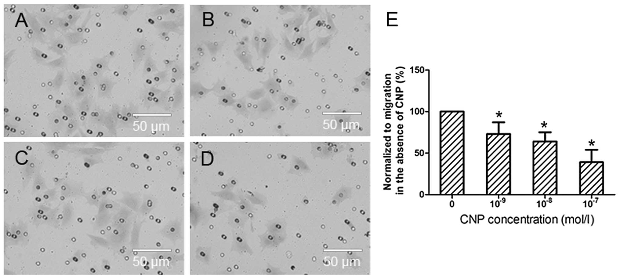
CNP inhibits cardiac fibroblast
migration
The effects of CNP on cardiac fibroblast migration
were examined by a transwell assay using 2% FBS as a chemotactic
stimulus. The results demonstrated that treatment with CNP
significantly reduced the number of migrating cells compared with
the control group, as indicated by the cellular staining and the OD
analysis of the cellular extraction (Fig. 3).
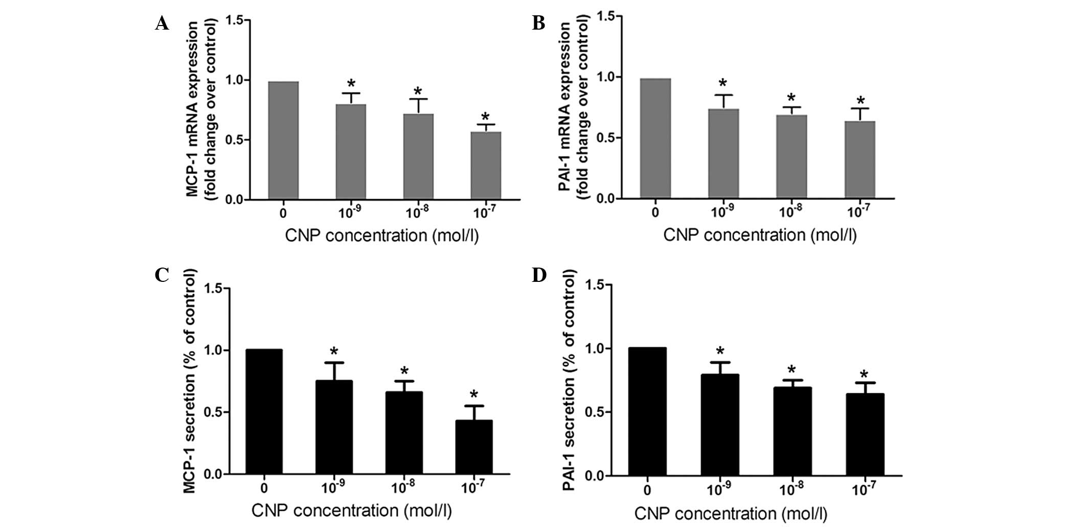
CNP inhibits the expression and secretion
of MCP-1 and PAI-1
RT-qPCR and ELISA assays were used to determine the
effect of CNP on the RNA expression and protein secretion of MCP-1
and PAI-1 in CFs. RT-qPCR analyses demonstrated that treatment of
CFs with CNP for 24 h resulted in a significant decrease in the
mRNA expression of MCP-1 and PAI-1 (Fig. 4A and B). MCP-1 and PAI-1 protein
concentrations in the culture medium of CNP-treated CFs were also
significantly decreased compared with the control group (Fig. 4C and D).
CNP inhibits activation of ERK1/2
In order to examine the influence of CNP on the
downstream signaling pathways in CFs, experiments were conducted to
analyze the effect of CNP on the activation of ERK1/2. The present
study demonstrated that CNP effectively inhibited the protein
expression of p-ERK1/2 (Fig. 5A).
Additionally, the ELISA experiments revealed that the protein
secretion of MCP-1 and PAI-1 was significantly decreased in the
presence of the ERK1/2-specific inhibitor U0126 alone, and U0126 in
combination with CNP (Fig.
5B).
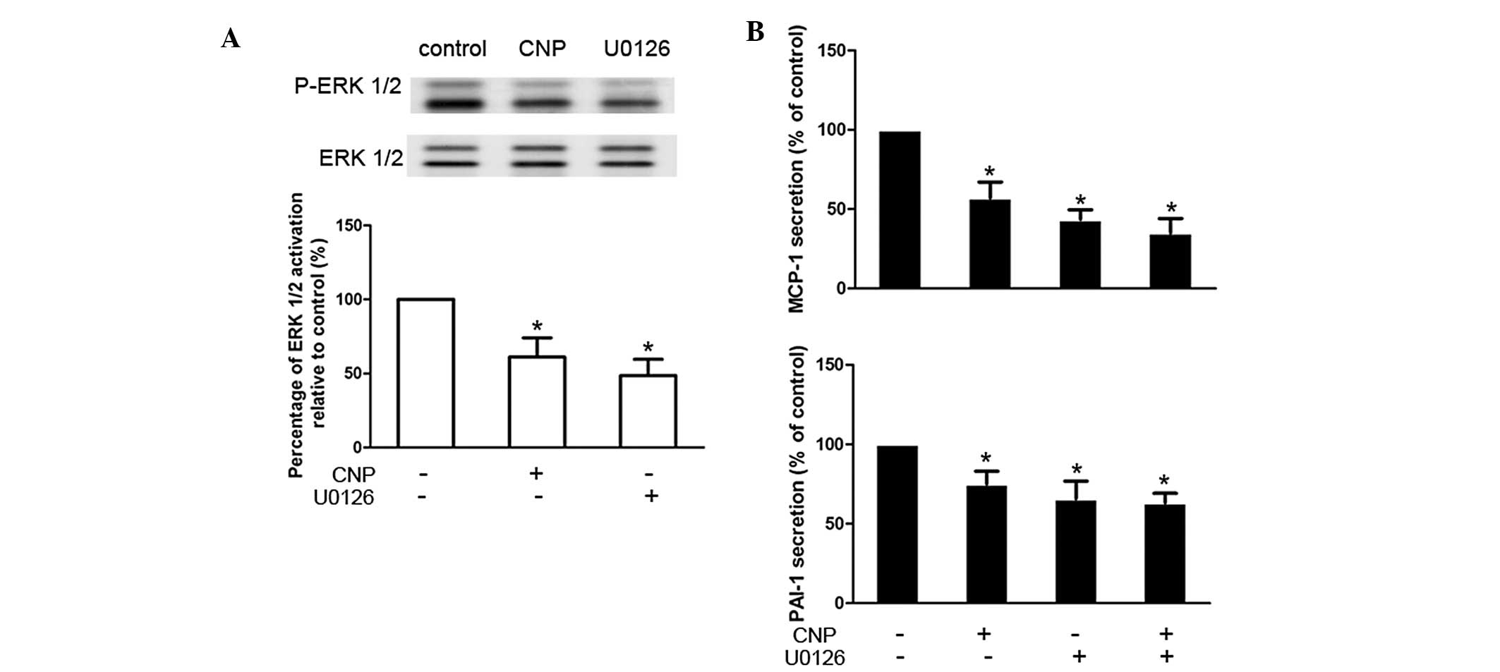 | Figure 5CNP inhibits the activation of
ERK-MAPK. (A) Serum-starved cells were treated with CNP
(10−7 mol/l) or pretreated with the ERK1/2 inhibitor
U0126 (10−4 mol/l) for 30 min prior to culture with DMEM
with 10% FBS for 30 min and then ERK1/2 phosphorylation was
analyzed by western blot analysis. The signal was quantified by
densitometry. (B) Serum-starved cells were treated with CNP
(10−7 mol/l), or pretreated with the ERK1/2 inhibitor
U0126 (10−4 mol/l) for 30 min prior to culture with DMEM
with 10% FBS for 24 h, or pretreated with U0126 (10−4
mol/l) for 30 min prior to treatment with CNP (10−7
mol/l) for 24 h. MCP-1 and PAI-1 protein secretion in supernatants
was determined by ELISA. *P<0.05 compared with
untreated CFs. CNP, C-type natriuretic peptide; ERK, extracellular
signal-regulated kinase; MAP, mitogen activated protein; MCP,
monocyte chemoattractant protein; PAI-1, plasminogen activator
inhibitor-1; FBS, fetal bovine serum; DMEM, Dulbecco’s modified
Eagle’s medium; CFs, cardiac fibroblasts. |
Discussion
Out of the three natriuretic peptides, CNP was the
last to be identified (1). It has
slowly emerged that CNP has a significant role in regulating
cardiac function since its identification approximately two decades
ago (16). The ability of CNP to
protect against myocardial ischemia-reperfusion injury, and to
inhibit cardiac fibrosis and hypertrophy has been reported
(4–6). Additionally, elevated plasma CNP
levels are identified in patients with heart failure (17,18).
CFs are fundamental to the normal structure of the
heart and are essential during cardiac fibrosis, a pathological
process that can ultimately result in heart failure. CFs are
responsible for the deposition of the extracellular matrix and also
secrete a number of inflammatory cytokines. Although previous
studies have identified that CNP is a potent antifibrotic agent
following myocardial infarction (5,19),
the cellular mechanisms underlying this in vivo antifibrotic
action of CNP are not fully understood. It has been reported that
CNP inhibits proliferation and collagen synthesis of cultured CFs
(3,5). However, the effects of CNP on other
significant cardiac fibroblast functions, including
differentiation, migration and cytokine secretion have not been
investigated.
Cardiac myofibroblast differentiation is pivotal in
the process of cardiac fibrosis. Following myocardial infarction,
CFs migrate into the infarct border zone and differentiate into
myofibroblasts promoting contraction of the scar (7,20).
In the present study, it was revealed that CNP can inhibit the
conversion of CFs to cardiac myofibroblasts, which was demonstrated
by attenuated protein expression of α-SMA and fibronectin, which
are key marker proteins of myofibroblast differentiation (10–14).
This effect may confer beneficial antifibrotic effects. In
addition, the expression of collagen I and III, which has also been
demonstrated to be corerelated with the differentation of CFs into
myofibroblasts (21–23), was investigated and results
revealed that collagen I and III expression in CFs was inhibited
following treatment with CNP. Cardiac fibroblast migration is
another key process in cardiac fibrosis that usually accompanies
differentiation (24). The in
vivo migration was mimicked by a transwell assay, which enables
CFs to traverse the matrigel-coated filters. The results
demonstrated that CNP effectively inhibited the migration of
CFs.
The inflammatory response, and cytokine secretion
and production are particularly active following myocardial
infarction (MI) and contribute to cardiac fibrosis and eventual
cardiac dysfunction (25–27). MCP-1 is one of the most
well-investigated cytokines in cardiac fibrosis (28). An elevated MCP-1 level was observed
in the myocardium following MI and it has been demonstrated that
MCP-1 has significant effects on macrophage recruitment and
activation, cytokine synthesis and myofibroblast accumulation in
healing infarcts (27,29–31).
Disruption of the MCP-1 axis reduces fibrosis and attenuates
dilation of the infarcted ventricle (32). With regard to PAI-1, a potent
inhibitor of urokinase and tissue-type plasminogen activator, it is
also critical in tissue fibrosis, including cardiac fibrosis
(33). Evidence obtained in
gene-deficient mice reveal that PAI-1 contributes to cardiac
fibrosis (34,35), and it was reported that inhibition
of PAI-1 is protective against the development of cardiac fibrosis
(36). Therefore, as two
profibrotic factors, MCP-1 and PAI-1 are key in the pathogenesis of
cardiac fibrosis. In the present study, it was demonstrated that
the mRNA expression and protein secretion of MCP-1 and PAI-1 in CFs
were attenuated by CNP treatment, which may serve as another
mechanism that underlies the anticardiac fibrotic properties of
CNP.
The bioactivity of CNP is mainly mediated by its
specific receptor NPR-B (2);
however, the downstream signaling pathways have not been fully
examined. In the present study, it was identified that CNP
significantly decreased the activity of ERK1/2, a member of the
mitogen-activated protein kinase (MAPK) superfamily, which was
demonstrated to be involved in the regulation of various cytokines
amongst numerous cell types (37).
At the same time, the level of MCP-1 and PAI-1 was significantly
decreased in the presence of U0126, an ERK1/2 inhibitor, which
indicates that CNP inhibits MCP-1 and PAI-1 secretion via an
ERK1/2-dependent mechanism.
In conclusion, CNP inhibited cardiac fibroblast
differentiation and migration, and reduced MCP-1 and PAI-1
secretion in the CFs via the ERK1/2-MAPK signaling pathway, which
implies novel mechanisms to explain the antifibrotic effect of CNP
in the pathological process of cardiac remodeling.
Abbreviations:
|
CFs
|
cardiac fibroblasts
|
|
α-SMA
|
α-smooth muscle actin
|
|
ED-A FN
|
extra domain-A fibronectin
|
|
MCP-1
|
monocyte chemotattractant
protein-1
|
|
PAI-1
|
plasminogen activator inhibitor-1
|
References
|
1
|
Sudoh T, Minamino N, Kangawa K and Matsuo
H: C-type natriuretic peptide (CNP): a new member of natriuretic
peptide family identified in porcine brain. Biochem Biophys Res
Commun. 168:863–870. 1990. View Article : Google Scholar : PubMed/NCBI
|
|
2
|
Lumsden NG, Khambata RS and Hobbs AJ:
C-type natriuretic peptide (CNP): cardiovascular roles and
potential as a therapeutic target. Curr Pharm Des. 16:4080–4088.
2010. View Article : Google Scholar : PubMed/NCBI
|
|
3
|
Horio T, Tokudome T, Maki T, et al: Gene
expression, secretion, and autocrine action of C-type natriuretic
peptide in cultured adult rat cardiac fibroblasts. Endocrinology.
144:2279–2284. 2003. View Article : Google Scholar : PubMed/NCBI
|
|
4
|
Hobbs A, Foster P, Prescott C, Scotland R
and Ahluwalia A: Natriuretic peptide receptor-C regulates coronary
blood flow and prevents myocardial ischemia/reperfusion injury:
novel cardioprotective role for endothelium-derived C-type
natriuretic peptide. Circulation. 110:1231–1235. 2004. View Article : Google Scholar
|
|
5
|
Soeki T, Kishimoto I, Okumura H, et al:
C-type natriuretic peptide, a novel antifibrotic and
antihypertrophic agent, prevents cardiac remodeling after
myocardial infarction. J Am Coll Cardiol. 45:608–616. 2005.
View Article : Google Scholar : PubMed/NCBI
|
|
6
|
Wang Y, de Waard MC, Sterner-Kock A, et
al: Cardiomyocyte-restricted over-expression of C-type natriuretic
peptide prevents cardiac hypertrophy induced by myocardial
infarction in mice. Eur J Heart Fail. 9:548–557. 2007. View Article : Google Scholar : PubMed/NCBI
|
|
7
|
Porter KE and Turner NA: Cardiac
fibroblasts: at the heart of myocardial remodeling. Pharmacol Ther.
123:255–278. 2009. View Article : Google Scholar : PubMed/NCBI
|
|
8
|
Kim NN, Villarreal FJ, Printz MP, Lee A
and Dillmann WH: Trophic effects of angiotensin II on neonatal rat
cardiac myocytes are mediated by cardiac fibroblasts. Am J Physiol.
269:E426–E437. 1995.PubMed/NCBI
|
|
9
|
Swaney JS, Roth DM, Olson ER, et al:
Inhibition of cardiac myofibroblast formation and collagen
synthesis by activation and overexpression of adenylyl cyclase.
Proc Natl Acad Sci USA. 102:437–442. 2005. View Article : Google Scholar : PubMed/NCBI
|
|
10
|
Desmoulière A, Chaponnier C and Gabbiani
G: Tissue repair, contraction, and the myofibroblast. Wound Repair
Regen. 13:7–12. 2005.PubMed/NCBI
|
|
11
|
Petrov VV, Fagard RH and Lijnen PJ:
Transforming growth factor-beta(1) induces angiotensin-converting
enzyme synthesis in rat cardiac fibroblasts during their
differentiation to myofibroblasts. J Renin Angiotensin Aldosterone
Syst. 1:342–352. 2000. View Article : Google Scholar
|
|
12
|
Olson ER, Naugle JE, Zhang X, Bomser JA
and Meszaros JG: Inhibition of cardiac fibroblast proliferation and
myofibroblast differentiation by resveratrol. Am J Physiol Heart
Circ Physiol. 288:H1131–H1138. 2005. View Article : Google Scholar : PubMed/NCBI
|
|
13
|
Wang J, Chen H, Seth A and McCulloch CA:
Mechanical force regulation of myofibroblast differentiation in
cardiac fibroblasts. Am J Physiol Heart Circ Physiol.
285:H1871–H1881. 2003.PubMed/NCBI
|
|
14
|
Santiago JJ, Dangerfield AL, Rattan SG, et
al: Cardiac fibroblast to myofibroblast differentiation in
vivo and in vitro: expression of focal adhesion
components in neonatal and adult rat ventricular myofibroblasts.
Dev Dyn. 239:1573–1584. 2010.PubMed/NCBI
|
|
15
|
Teunissen BE, Jansen AT, van Amersfoorth
SC, O’Brien TX, Jognsma HJ and Bierhuizen MF: Analysis of the rat
connexin 43 proximal promoter in neonatal cardiomyocytes. Gene.
322:123–136. 2003. View Article : Google Scholar : PubMed/NCBI
|
|
16
|
Potter LR, Yoder AR, Flora DR, Antos LK
and Dickey DM: Natriuretic peptides: their structures, receptors,
physiologic functions and therapeutic applications. Handb Exp
Pharmacol. 341–366. 2009. View Article : Google Scholar : PubMed/NCBI
|
|
17
|
Kalra PR, Clague JR, Bolger AP, et al:
Myocardial production of C-type natriuretic peptide in chronic
heart failure. Circulation. 107:571–573. 2003. View Article : Google Scholar : PubMed/NCBI
|
|
18
|
Del Ry S, Maltinti M, Piacenti M, et al:
Cardiac production of C-type natriuretic peptide in heart failure.
J Cardiovasc Med (Hagerstown). 7:397–399. 2006.PubMed/NCBI
|
|
19
|
Izumiya Y, Araki S, Usuku H, Rokutanda T,
Hanatani S and Ogawa H: Chronic C-type natriuretic peptide infusion
attenuates angiotensin II-induced myocardial superoxide production
and cardiac remodeling. Int J Vasc Med. 2012:2460582012.PubMed/NCBI
|
|
20
|
van den Borne SW, Diez J, Blankesteijn WM,
et al: Myocardial remodeling after infarction: the role of
myofibroblasts. Nat Rev Cardiol. 7:30–37. 2010.PubMed/NCBI
|
|
21
|
Lijnen PJ, Petrov VV and Fagard RH:
Induction of cardiac fibrosis by transforming growth
factor-beta(1). Mol Genet Metab. 71:418–435. 2000. View Article : Google Scholar : PubMed/NCBI
|
|
22
|
Petrov VV, Fagard RH and Lijnen PJ:
Stimulation of collagen production by transforming growth
factor-beta1 during differentiation of cardiac fibroblasts to
myofibroblasts. Hypertension. 39:258–263. 2002. View Article : Google Scholar : PubMed/NCBI
|
|
23
|
Herum KM, Lunde IG, Skrbic B, et al:
Syndecan-4 signaling via NFAT regulates extracellular matrix
production and cardiac myofibroblast differentiation in response to
mechanical stress. J Mol Cell Cardiol. 54:73–81. 2013. View Article : Google Scholar : PubMed/NCBI
|
|
24
|
Dobaczewski M, Bujak M, Li N, et al: Smad3
signaling critically regulates fibroblast phenotype and function in
healing myocardial infarction. Circ Res. 107:418–428. 2010.
View Article : Google Scholar : PubMed/NCBI
|
|
25
|
Nian M, Lee P, Khaper N and Liu P:
Inflammatory cytokines and postmyocardial infarction remodeling.
Circ Res. 94:1543–1553. 2004. View Article : Google Scholar : PubMed/NCBI
|
|
26
|
Ono K, Matsumori A, Shioi T, Furukawa Y
and Sasayama S: Cytokine gene expression after myocardial
infarction in rat hearts: possible implication in left ventricular
remodeling. Circulation. 98:149–156. 1998. View Article : Google Scholar : PubMed/NCBI
|
|
27
|
Frangogiannis NG, Smith CW and Entman ML:
The inflammatory response in myocardial infarction. Cardiovasc Res.
53:31–47. 2002. View Article : Google Scholar : PubMed/NCBI
|
|
28
|
Dobaczewski M and Frangogiannis NG:
Chemokines and cardiac fibrosis. Front Biosci (Schol Ed).
1:391–405. 2009. View
Article : Google Scholar
|
|
29
|
Koyanagi M, Egashira K, Kitamoto S, et al:
Role of monocyte chemoattractant protein-1 in cardiovascular
remodeling induced by chronic blockade of nitric oxide synthesis.
Circulation. 102:2243–2248. 2000. View Article : Google Scholar : PubMed/NCBI
|
|
30
|
Dewald O, Zymek P, Winkelmann K, et al:
CCL2/monocyte chemoattractant protein-1 regulates inflammatory
responses critical to healing myocardial infarcts. Circ Res.
96:881–889. 2005. View Article : Google Scholar : PubMed/NCBI
|
|
31
|
Frangogiannis NG, Dewald O, Xia Y, et al:
Critical role of monocyte chemoattractant protein-1/CC chemokine
ligand 2 in the pathogenesis of ischemic cardiomyopathy.
Circulation. 115:584–592. 2007. View Article : Google Scholar : PubMed/NCBI
|
|
32
|
Kaikita K, Hayasaki T, Okuma T, et al:
Targeted deletion of CC chemokine receptor 2 attenuates left
ventricular remodeling after experimental myocardial infarction. Am
J Pathol. 165:439–447. 2004. View Article : Google Scholar : PubMed/NCBI
|
|
33
|
Ghosh AK and Vaughan DE: PAI-1 in tissue
fibrosis. J Cell Physiol. 227:493–507. 2012. View Article : Google Scholar
|
|
34
|
Kaikita K, Fogo AB, Ma L, et al:
Plasminogen activator inhibitor-1 deficiency prevents hypertension
and vascular fibrosis in response to long-term nitric oxide
synthase inhibition. Circulation. 104:839–844. 2001. View Article : Google Scholar : PubMed/NCBI
|
|
35
|
Takeshita K, Hayashi M, Iino S, et al:
Increased expression of plasminogen activator inhibitor-1 in
cardiomyocytes contributes to cardiac fibrosis after myocardial
infarction. Am J Pathol. 164:449–456. 2004. View Article : Google Scholar : PubMed/NCBI
|
|
36
|
Boe AE, Eren M, Murphy SB, et al:
Plasminogen activator inhibitor-1 antagonist TM5441 attenuates
Nω-nitro-L-arginine methyl ester-induced hypertension and vascular
senescence. Circulation. 128:2318–2324. 2013.PubMed/NCBI
|
|
37
|
Rozengurt E: Mitogenic signaling pathways
induced by G protein-coupled receptors. J Cell Physiol.
213:589–602. 2007. View Article : Google Scholar : PubMed/NCBI
|