Introduction
Oral cancer is the sixth most common type of cancer
in humans and poses a significant global threat to public health.
The five-year survival rate of oral cancer patients is ~50% overall
(1). In addition, a previous study
suggested an increase in this rate (2). The prognosis of patients with oral
cancer is relatively poor, predominantly due to the high rate of
local recurrence, even following curative surgical resection
(3). The development and
progression of oral cancer are promoted by alterations in multiple
cellular signaling pathways (4).
Establishing the molecular and cellular mechanisms involved in the
pathogenesis of oral cancer may provide assistance in identifying
novel predictive and prognostic biomarkers and in developing novel
therapeutic targets to treat oral cancer.
The A disintegrin and metalloproteinase (ADAM)
family is a class of type I transmembrane proteins that contain two
main structural domains, the disintegrin domain and the matrix
metalloproteinase domain (5). To
date, >35 members of this family have been identified (6). Previous studies have demonstrated
that members of the ADAM family are able to regulate the process of
tumor metastasis in humans by degrading the extracellular matrix
and controlling cell adhesion and movement (7,8). One
member of the ADAM family, A disintegrin and metalloproteinase 10
(ADAM10), has been reported to act as a positive regulator of
cancer progression in hepatocellular carcinoma (9), renal cell carcinoma (10), pancreatic carcinoma (11), lung cancer (12) and gastric carcinoma (13). It has been suggested that ADAM10
causes the shedding of various cell surface molecules, including
cadherins and amyloid precursor protein (14). Through these actions, ADAM10 is
able to modulate the tumor microenvironment and the key processes
involved in the progression of cancer, including cell
proliferation, migration and angiogenesis (15). However, few studies have
investigated the correlation between ADAM10 and oral cancer
(16). In the present study, the
effect of ADAM10 gene silencing on the proliferation, migration and
invasion of the human tongue squamous cell carcinoma cell line
TCA8113 was investigated. In addition, the association between the
expression of ADAM10 and the expression of epidermal growth factor
receptor (EGFR) and E-cadherin proteins in TCA8113 cells was
examined.
Materials and methods
Cell lines and cell culture
The human tongue squamous cell carcinoma cell line
TCA8113 was obtained from the Center of Biomedical Experimental
Research at the Medical School, Xi’an Jiaotong University (Xi’an,
China) (17). TCA8113 cells were
cultured in Dulbecco’s modified Eagle’s medium (Invitrogen Life
Technologies, Carlsbad, CA, USA) containing 10% fetal bovine serum
(FBS) and 1% penicillin/streptomycin (Invitrogen Life Technologies)
and incubated at 37°C in an atmosphere containing 5%
CO2.
siRNA transfection
For the downregulation of the endogenous expression
of ADAM10, the following siRNA duplex (Aoke Biological Technology
Co., Ltd., Shanghai, China) was used: 5′-AGACAUUAUGAAGGAUUAUTT-3′.
As a negative control, the unspecific scrambled siRNA duplex
(5′-AGGUAGUGUA AUCGCCUUGTT-3′) was used.
The TCA8113 cells (1×10⁵) were seeded
into six-well plates 24 h prior to transfection. Transfection of
siRNA was performed using Lipofectamine 2000 (Invitrogen Life
Technologies) and 10 nM siRNA duplex/well and the transfection
procedure was performed as previously described (18). The experiment was repeated three
times.
The present study comprised a total of four groups,
the blank control group, the Lipo2000 group (cells treated with
Lipofectamine 2000), the control siRNA group (Lipofectamine 2000
and negative control siRNA) and the ADAM10-siRNA group (cells were
treated with Lipofectamine 2000 and ADAM10 siRNA).
Reverse transcription quantitative
polymerase chain reaction (RT-qPCR)
RT-qPCR analysis of the ADAM10 transcripts in the
TCA8113 cells was performed using the PrimeScript RT reagent kit
according to the manufacturer’s instructions (Takara Bio, Inc.,
Shiga, Japan). The ADAM10 gene was amplified by RT-qPCR using the
specific primers; forward, 5′-CTGCCCAGCATCTGACCCTAA-3′ and reverse,
5′-TTGCCATCAGAACTGGCACAC-3′. Amplification specificity was further
validated by melting curve analysis. GAPDH was used as an internal
control. The RT-qPCR procedures were performed in triplicate and
the data were analyzed using the comparative Ct method.
Western blot analysis
The cells were washed with phosphate-buffered saline
(PBS) twice and lysed on ice in a buffer containing 150 mM NaCl, 50
mM Tris-HCl (pH 7.4), 2 mM EDTA, 1% NP-40, 0.5%
3-[(3-cholamidopropyl)-dimethyl-ammonio]-1-propane sulfonate and
0.1% SDS, which were all purchased from Wuhan Boster Biological
Technology, Ltd. (Wuhan, China). Electrophoresis on 10%
polyacrylamide gels was performed using equal quantities of protein
(20 μg/lane) from the cell lysates under non-reducing conditions.
The proteins fractionated by SDS-PAGE were electrotransferred onto
Immobilon-P membranes (Invitrogen Life Technologies). The membranes
were inhibited with 5% non-fat skimmed milk and 0.1% Tween-20 in
PBS for 2 h. Subsequently, the membrane was incubated for 2 h with
rabbit anti-human ADAM10 (1:500; R&D Systems, Minneapolis, MN,
USA), rabbit anti-human EGFR (1:1,000; Santa Cruz Biotechnology,
Inc., Dallas, TX, USA), or rabbit anti-human E-cadherin (1:1,000,
Santa Cruz Biotechnology, Inc.). Following washing, the proteins
were visualized using an Easyblot enhanced chemiluminescence
detection kit (Sangon Biotech, Inc., Shanghai, China) using the
appropriate goat anti-rabbit immunoglobulin G horseradish
peroxidase-conjugated secondary antibody (1:1,000; Amersham
Pharmacia Biotech, Piscataway, NJ, USA). The rabbit anti-human
β-actin antibody (1:1,000; Santa Cruz Biotechnology, Inc.) was used
as an internal marker for control purposes.
Proliferation assay
The MTT (Sigma, St. Louis, MO, USA) colorimetric
assay was used to screen for cell proliferation. Briefly, the
TCA8113 cells were seeded into eight 96-well plates at a density of
2×10³ cells/well and incubated in RPMI-1640 medium
(Wuhan Boster Biological Technology, Ltd.) for 24, 48, 72 and 96 h
following treatment, respectively. MTT (20 μl, 5 mg/ml) was added
to each well and the cells were cultured for an additional 4 h. The
formazan crystals were dissolved by addition of dimethylsulfoxide
(Sigma) and the absorbance was determined using a Multiskan
Spectrum microplate reader (Thermo Fisher, Waltham, MA, USA)at 490
nm. A growth curve was produced according to the optical density
(OD) value alterations. The experiment was performed three times
independently.
Soft agar colony formation assay
The TCA8113 cells (4×10³) were mixed with
0.5% top agar and seeded into 24-well plates with 1% base agar
(Wuhan Boster Biological Technology, Ltd.). These cells were then
cultured in an incubator at 37°C for 10 days in 5% CO2
at 95% humidity. Subsequently, images of the cell colonies in the
soft agar were captured and counted using a microscope (BX53,
Olympus Corp., Tokyo, Japan). All the experiments were
independently repeated at least three times.
Cell migration assay
The effect of ADAM10 knockdown on TCA8113 cell
migration was measured as the ability of cells to migrate through
Transwell filters (6.5 mm diameter, 5 mm pore size; Haoran
Bio-pharm Co., Ltd, Jiangxi, China). Transwell filters were coated
with Matrigel (BD Biosciences, San Diego, CA, USA) for 1.5 h prior
to adding the cells. The cells were detached using trypsin 24 h
after ADAM10 siRNA transfection. The cells were then seeded into
the upper chamber of the Transwell (1×10⁵ cells/well) to
enable migration to the lower chamber, which contained growth
medium, over 24 h. Non-migratory cells were removed using a cotton
swab and the transmigrated cells at the backside of the filter were
stained with Giemsa (Wuhan Boster Biological Technology, Ltd.). The
cells in the filter were counted under a microscope (magnification,
×400; BX53; Olympus Corp.). The data were normalized and expressed
as the migration rate of the cells compared with the blank
control.
In vitro invasion assay
The Boyden chamber technique was performed to
examine in vitro invasion. Briefly, the 8-μm pore filters
were coated with 50 μl 8 mg/ml reconstituted basement membrane
substance (Matrigel). The coated filters were air-dried at 4°C
prior to addition of the cells. RPMI-1640 medium (600 μl)
containing 5% FBS was added to the lower chambers. The cells were
digested with trypsin (Wuhan Boster Biological Technology, Ltd.)
and the cell suspensions (1×10⁵ cells/well) were added
to the upper chambers to enable invasion for 24 h at 37°C in a
humidified atmosphere of 5% CO2. The cells remaining
attached to the upper surface of the filters were removed using
cotton swabs. The migrated cells were stained with Giemsa and
examined using optical microscopy (magnification, ×400). A total of
five fields of view at the center and in the surrounding areas were
counted and the average was calculated (19). The experiment was repeated three
times.
Data and statistical analysis
All values are expressed as the mean ± standard
deviation. Statistical analysis was performed using SPSS 16.0
software (SPSS, Inc., Chicago, IL, USA). Differences among the
groups were assessed using one-way analysis of variance. P<0.05
was considered to indicate a statistically significant difference
between values.
Results
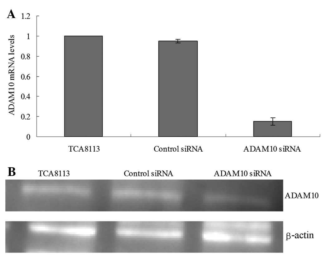
Knockdown of ADAM10 in TCA8113 cells
The expression of ADAM10 was examined using RT-qPCR
and western blot analysis to validate the silencing efficiency of
the target gene following RNA interference. Stably ADAM10
siRNA-transfected TCA8113 cells (ADAM10-siRNA) and a
mock-transfected control cell line (control siRNA) were
established, as described above. Compared with the parental TCA8113
cells and the control siRNA cells, ADAM10 mRNA and protein
expression was significantly reduced in the ADAM10 siRNA cells 24 h
following siRNA transfection (P<0.05; Fig. 1A and B), which persisted for at
least 96 h (data not shown).
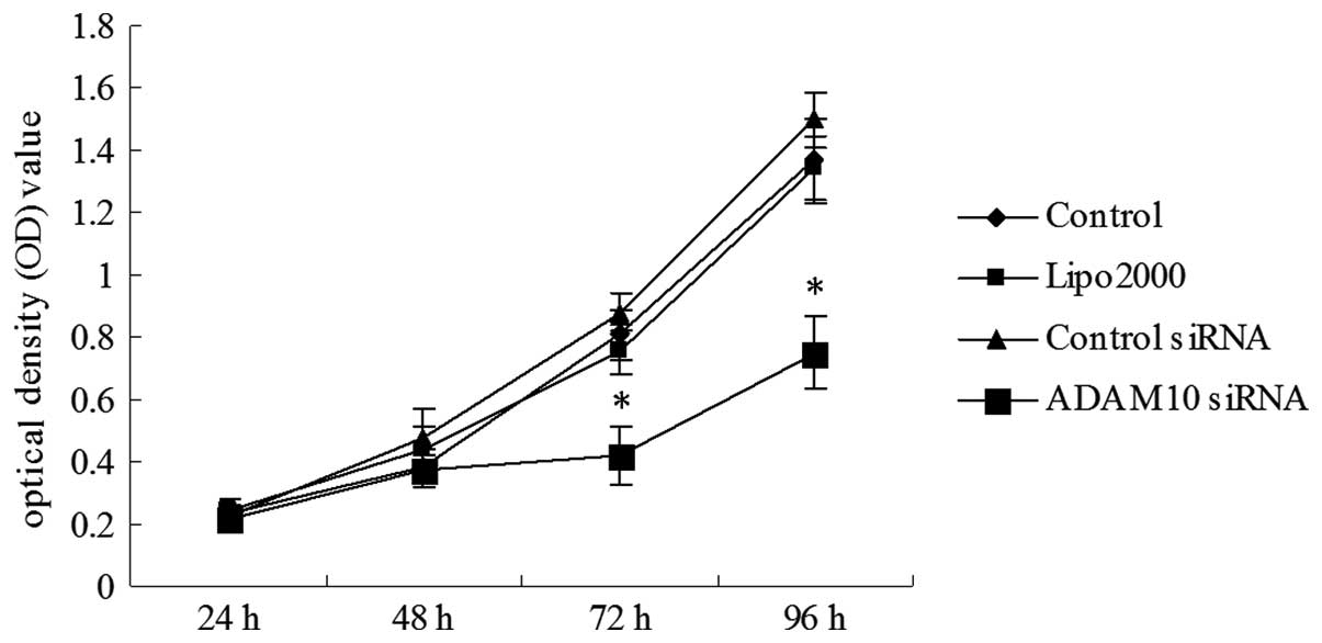
Gene silencing of ADAM10 reduces cell
proliferation and cell colony formation in TCA8113 cells
To examine whether the knockdown of ADAM10
expression affected cell growth, an MTT cell proliferation assay
was performed. Compared with the blank control, Lipo2000 and
control siRNA group cells, a decrease in cell proliferation in the
ADAM10-siRNA group was observed. These results suggested that
ADAM10 promoted TCA8113 cell growth (P<0.05; Fig. 2). In addition, the soft agar assay
revealed that the cell colony number significantly decreased in the
ADAM10-siRNA group compared with that in the blank control group,
Lipo2000 group and control siRNA group (P<0.05, Fig. 3A and B).
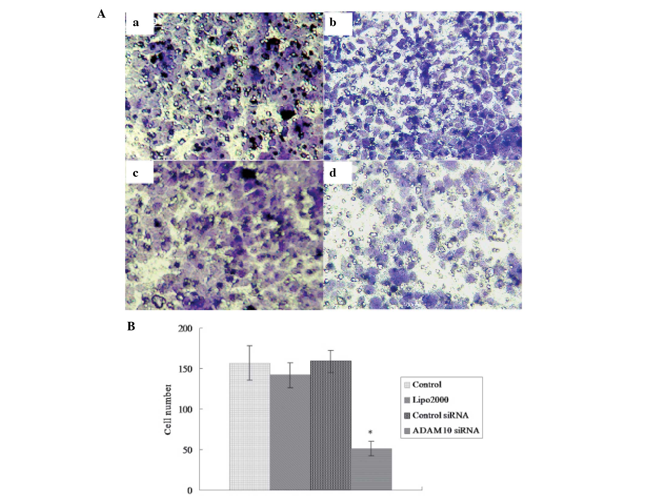
Gene silencing of ADAM10 reduces cell
migration in TCA8113 cells
The effect of ADAM10 gene silencing on the migration
of TCA8113 cells was investigated using a Transwell invasion assay
(Fig. 4A). The results
demonstrated that transfecting cells with ADAM10 siRNA led to a
marked reduction in the capability of cells to pass through the
basement membrane, compared with that of the other groups (all
P<0.05; Fig. 4B). These results
suggested that the expression of ADAM10 may be associated with cell
migration.
Gene silencing of ADAM10 reduces the
invasive ability of TCA8113 cells
A Matrigel invasion assay was used to determine the
invasive potential of TCA8113 cells transfected with ADAM10 siRNA.
The assay results demonstrated that the capability of the treated
TCA8113 cells to pass through the basement membrane decreased
markedly compared with that of the other groups (all P<0.05;
Fig. 5A and B). These results
indicated the importance of ADAM10 in oral cancer cell
invasion.
Silencing of ADAM10 by siRNA stimulates
the activation of E-cadherin and suppresses the activation of EGFR
in TCA8113 cells
In the present study, changes in the protein levels
of EGFR and E-cadherin were detected by western blot analysis. The
results revealed that at 72 h after transfection, the protein
levels of EGFR were significantly decreased in the TCA8113 cells
treated with ADAM10 siRNA compared with those in the other groups
(all P<0.05; Fig. 6), while
levels of E-cadherin were significantly increased in the ADAM10
siRNA-treated cells compared with those in the cells in the other
groups (all P<0.05; Fig.
7).
Discussion
Various members of the ADAM family, including
ADAM10, are overexpressed in different types of malignant tumor and
may be associated with the biological behavior of the latter
(9–11). The expression of ADAM10 is
significantly increased in non-small cell lung cancer (NSCLC)
tissues, particularly in metastatic tissues (12). Downregulation of the expression of
ADAM10 with short hairpin RNA against ADAM10 has been shown to
inhibit the migration and invasion of NSCLC cells (12). The present study hypothesized that
the downregulation of ADAM10 may affect the biological behavior of
TCA8113 cells. However, as there are few previous studies
associated with this hypothesis, the purpose of the present study
was to analyze the association between the gene silencing of ADAM10
and the proliferation, invasion and migration capability of TCA8113
cells in vitro. In addition, the association between the
expression of ADAM10 and the expression of EGFR and E-cadherin
proteins in TCA8113 cells was examined.
The results of the present study demonstrated that
downregulation of ADAM10 resulted in the suppression of
TCA8113-cell proliferation and a significant reduction in cellular
invasion and migration, which indicated that ADAM10 was involved in
processes of tumor development and metastasis. These findings were
consistent with previous studies on the expression and functional
roles of ADAM10 (9,18,20).
Armanious et al (20)
demonstrated that the active/mature form of ADAM10 is expressed in
mantle cell lymphoma (MCL) cell lines and was observed in all 12
patient samples examined. The MCL cells transfected with ADAM10
siRNA demonstrated growth inhibition and cell-cycle arrest, while
addition of recombinant ADAM10 to MCL cells induced a significant
increase in cell growth. Yuan et al (9) revealed that the siRNA-mediated
knockdown of ADAM10 significantly inhibited the growth, migration
and invasion of HepG2 cells. Lee et al (18) demonstrated that the expression of
ADAM10 increased markedly in melanoma metastasis compared with that
in primary lesions. Downregulation of ADAM10 with siRNA led to a
decrease in the growth and migration of melanoma cells. In
addition, the migration of melanoma cells was induced by
overexpression of ADAM10.
The effect of ADAM10 on the biological behaviors of
tumor cells may be associated with its protease activity. The
biological behavior of cancer cells is regulated by multiple growth
factors and cytokines, a number of which are present in a
membrane-bound form and which undergo proteolytic shedding in order
to be activated (21).
ADAM10-mediated proteolytic cleavage of substrate proteins is
considered to be involved in the pathophysiology of multiple
life-threatening diseases, including cancer (7). Important substrates of ADAM proteases
include growth factors, cytokines and their receptors and adhesion
proteins (22).
EGFR is a member of the epidermal growth factor
family of receptors, a subfamily which comprises four receptor
tyrosine kinases which are closely associated (23). Activation of EGFR leads to signal
transduction cascades that promote cell proliferation and cell
growth (23). Yan et al
(24) demonstrated that ADAM10
stimulated the G protein-coupled receptor (GPCR) transactivation of
EGFR. The effect of ADAM10 on EGFR transactivation depends on its
metalloprotease activity and is due to the activation of the EGFR
ligand heparin-binding EGF by GPCR signaling. In the present study,
the expression of EGFR was significantly downregulated in the
ADAM10 siRNA-treated cells compared with that in the control group.
This indicated a positive correlation between ADAM10 and EGFR and
suggested that ADAM10 may regulate the proliferation, migration and
invasion of TCA8113 cells via modulating the EGFR signaling
pathway.
ADAM10 may also promote tumor invasion and
metastasis by degrading the extracellular matrix and affecting
cell-cell signaling (25–27). Destruction of the basement
membranes has been implicated as an early event in tumor
metastasis. Millichip et al (25) revealed that cleavage of the
basement membrane type IV collagen resulted from bovine ADAM10. Pan
et al (26) demonstrated
that, in the pituitary adenoma cell line AtT-20, ADAM10 facilitated
cell migration via affecting the cleavage of CD44 and L1.
E-cadherin is a transmembrane molecule which functions as an
adhesion molecule. Increased expression of ADAM10 may lead to
elevated shedding of E-cadherin and loss of cell-cell contact
(18). Solanas et al
(27) demonstrated that in
epithelial cells, ephrin (Eph) B receptors interact with E-cadherin
and ADAM10 at sites of adhesion and Eph/ephrin activation induces
E-cadherin shedding by ADAM10. In the present study, the expression
of E-cadherin was significantly upregulated in the ADAM10
siRNA-treated cells compared with that in the control group, which
indicated a negative correlation between ADAM10 and E-cadherin and
suggested that ADAM10 may promote the proliferation, migration and
invasion of TCA8113 cells through modulation of cell adhesion and
cell contact.
In conclusion, the cellular proliferation, migration
and invasion abilities of the ADAM10 siRNA-transfected TCA8113
cells were significantly decreased, indicating that ADAM10 may be
involved in the genesis and progression of oral cancer. Therefore,
ADAM10 may serve as a potential marker and therapeutic target in
the treatment of oral cancer. Activating the EGFR signaling pathway
and modulating cell-cell interactions via inducing the shedding of
E-cadherin may contribute to the mechanisms of action of ADAM10.
Future studies are required to further investigate the molecular
mechanisms underlying the involvement of ADAM10 in tumorigenesis
and progression of human oral cancer cells.
References
|
1
|
Myers JN, Elkins T, Roberts D and Byers
RM: Squamous cell carcinoma of the tongue in young adults:
Increasing incidence and factors that predict treatment outcomes.
Otolaryngol Head Neck Surg. 122:44–51. 2000. View Article : Google Scholar : PubMed/NCBI
|
|
2
|
Hernández-Guerrero JC, Jacinto-Alemán LF,
Jiménez-Farfán MD, Macario-Hernández A, Hernández-Flores F and
Alcántara-Vázquez A: Prevalence trends of oral squamous cell
carcinoma. Mexico City’s General Hospital experience. Med Oral
Patol Oral Cir Bucal. 18:e306–e311. 2013.PubMed/NCBI
|
|
3
|
Bagan J, Sarrion G and Jimenez Y: Oral
cancer: clinical features. Oral Oncol. 46:414–417. 2010. View Article : Google Scholar
|
|
4
|
Haddad RI and Shin DM: Recent advances in
head and neck cancer. N Engl J Med. 359:1143–1154. 2008. View Article : Google Scholar : PubMed/NCBI
|
|
5
|
Duffy MJ, Mullooly M, O’Donovan N, Sukor
S, Crown J, Pierce A and McGowan PM: The ADAMs family of proteases:
new biomarkers and therapeutic targets for cancer? Clin Proteomics.
8:92011. View Article : Google Scholar : PubMed/NCBI
|
|
6
|
Lin J, Luo J and Redies C: Differential
expression of five members of the ADAM family in the developing
chicken brain. Neuroscience. 157:360–375. 2008. View Article : Google Scholar : PubMed/NCBI
|
|
7
|
Murphy G: The ADAMs: signalling scissors
in the tumour microenvironment. Nat Rev Cancer. 8:929–941. 2008.
View Article : Google Scholar : PubMed/NCBI
|
|
8
|
Lu X, Lu D, Scully M and Kakkar V: ADAM
proteins - therapeutic potential in cancer. Curr Cancer Drug
Targets. 8:720–732. 2008. View Article : Google Scholar : PubMed/NCBI
|
|
9
|
Yuan S, Lei S and Wu S: ADAM10 is
overexpressed in human hepatocellular carcinoma and contributes to
the proliferation, invasion and migration of HepG2 cells. Oncol
Rep. 30:1715–1722. 2013.PubMed/NCBI
|
|
10
|
Doberstein K, Pfeilschifter J and Gutwein
P: The transcription factor PAX2 regulates ADAM10 expression in
renal cell carcinoma. Carcinogenesis. 32:1713–1723. 2011.
View Article : Google Scholar : PubMed/NCBI
|
|
11
|
Gaida MM, Haag N, Günther F, et al:
Expression of A disintegrin and metalloprotease 10 in pancreatic
carcinoma. Int J Mol Med. 26:281–288. 2010.PubMed/NCBI
|
|
12
|
Guo J, He L, Yuan P, et al: ADAM10
overexpression in human non-small cell lung cancer correlates with
cell migration and invasion through the activation of the Notch1
signaling pathway. Oncol Rep. 28:1709–1718. 2012.PubMed/NCBI
|
|
13
|
Wang YY, Ye ZY, Li L, Zhao ZS, Shao QS and
Tao HQ: ADAM 10 is associated with gastric cancer progression and
prognosis of patients. J Surg Oncol. 103:116–123. 2011. View Article : Google Scholar : PubMed/NCBI
|
|
14
|
Prox J, Willenbrock M, Weber S, et al:
Tetraspanin15 regulates cellular trafficking and activity of the
ectodomain sheddase ADAM10. Cell Mol Life Sci. 69:2919–2932. 2012.
View Article : Google Scholar : PubMed/NCBI
|
|
15
|
Turner SL, Blair-Zajdel ME and Bunning RA:
ADAMs and ADAMTSs in cancer. Br J Biomed Sci. 66:117–128.
2009.PubMed/NCBI
|
|
16
|
Jones AV, Lambert DW, Speight PM and
Whawell SA: ADAM 10 is over expressed in oral squamous cell
carcinoma and contributes to invasive behaviour through a
functional association with αvβ6 integrin. FEBS Lett.
587:3529–3534. 2013.PubMed/NCBI
|
|
17
|
Shao Y, Zhang SQ, Quan F, Zhang PF and Wu
SL: MicroRNA-145 inhibits the proliferation, migration and invasion
of the human TCA8113 oral cancer line. Oncol Lett. 6:1636–1640.
2013.PubMed/NCBI
|
|
18
|
Lee SB, Schramme A, Doberstein K, et al:
ADAM10 is upregulated in melanoma metastasis compared with primary
melanoma. J Invest Dermatol. 130:763–773. 2010. View Article : Google Scholar : PubMed/NCBI
|
|
19
|
Yu Y, Chen W, Zhang Y, Hamburger AW, Pan H
and Zhang Z: Suppression of salivary adenoid cystic carcinoma
growth and metastasis by ErbB3 binding protein Ebp1 gene transfer.
Int J Cancer. 120:1909–1913. 2007. View Article : Google Scholar : PubMed/NCBI
|
|
20
|
Armanious H, Gelebart P, Anand M, Belch A
and Lai R: Constitutive activation of metalloproteinase ADAM10 in
mantle cell lymphoma promotes cell growth and activates the
TNFα/NFκB pathway. Blood. 117:6237–6246. 2011.PubMed/NCBI
|
|
21
|
Zheng X, Jiang F, Katakowski M, Lu Y and
Chopp M: ADAM17 promotes glioma cell malignant phenotype. Mol
Carcinog. 51:150–164. 2012. View
Article : Google Scholar : PubMed/NCBI
|
|
22
|
Ebsen H, Schröder A, Kabelitz D and
Janssen O: Differential surface expression of ADAM10 and ADAM17 on
human T lymphocytes and tumor cells. PLoS One. 8:e768532013.
View Article : Google Scholar : PubMed/NCBI
|
|
23
|
Normanno N, De Luca A, Bianco C, et al:
Epidermal growth factor receptor (EGFR) signaling in cancer. Gene.
366:2–16. 2006. View Article : Google Scholar : PubMed/NCBI
|
|
24
|
Yan Y, Shirakabe K and Werb Z: The
metalloprotease Kuzbanian (ADAM10) mediates the transactivation of
EGF receptor by G protein-coupled receptors. J Cell Biol.
158:221–226. 2002. View Article : Google Scholar : PubMed/NCBI
|
|
25
|
Millichip MI, Dallas DJ, Wu E, Dale S and
McKie N: The metallo-disintegrin ADAM10 (MADM) from bovine kidney
has type IV collagenase activity in vitro. Biochem Biophys Res
Commun. 245:594–598. 1998. View Article : Google Scholar : PubMed/NCBI
|
|
26
|
Pan Y, Han C, Wang C, et al: ADAM10
promotes pituitary adenoma cell migration by regulating cleavage of
CD44 and L1. J Mol Endocrinol. 49:21–33. 2012. View Article : Google Scholar : PubMed/NCBI
|
|
27
|
Solanas G, Cortina C, Sevillano M and
Batlle E: Cleavage of E-cadherin by ADAM10 mediates epithelial cell
sorting downstream of EphB signalling. Nat Cell Biol. 13:1100–1107.
2011. View
Article : Google Scholar : PubMed/NCBI
|