Introduction
Breast cancer is the most frequently diagnosed
cancer among women, accounting for 23% of the total cancer cases
(1). Over the past two decades,
the death rate due to breast cancer has decreased by <30% due to
the improvement of therapeutic strategies. However, breast cancer
remains the leading cause of cancer-related death in women
worldwide (1,2). Furthermore, breast cancer accounted
for 29% of all new cancer cases among women in 2013 (2). Therefore, in vitro
investigations on the molecular mechanisms underlying breast cancer
may provide important data for the development of therapeutic
strategies for breast cancer.
microRNAs (miRNAs) are a type of endogenous
non-coding RNAs. They act as gene expression suppressors via
binding to 3′ untranslated region (3′ UTR) of their target mRNAs,
and further leading to either translational repression or mRNA
degradation (3). Recently,
accumulating evidence has demonstrated that downregulation of
miR-335 is involved in tumorigenesis. Xiong et al (4) showed that miR-335 is downregulated in
prostate cancer tissues and cells, and that its low expression is
significantly associated with a high Gleason score, advanced
clinical stage, and metastasis in prostate cancer patients.
Moreover, miR-335 also plays a role in the regulation of cancer
progression. For instance, miR-335 was recently found to suppress
osteosarcoma cell migration and invasion by targeting ROCK1
(5). In addition, miR-335 was
reported to inhibit small cell lung cancer bone metastases
(6). It has been also demonstrated
that miR-335 acts as a tumor suppressor in breast cancer. Heyn
et al (7) showed that
overexpression of miR-335 leads to decreased cell viability and an
increase in apoptosis in breast cancer cells. Moreover, miR-335 has
been suggested to suppress breast cancer metastasis (8,9).
However, the underlying molecular mechanism by which miR-335
affects these biological processes, especially cell-cycle
progression, in breast cancer cells remains largely unknown.
Paired box 6 (PAX6), a highly conserved
transcription factor, plays a crucial role in the development of
the eyes, central nervous system, and pancreas (10,11).
The role of PAX6 in malignant tumors has been elucidated in recent
studies. PAX6 was demonstrated to act as an oncogene in
various cancers, including breast cancer (12–14).
Therefore, PAX6 may constitute a promising therapeutic target for
breast cancer. However, whether miR-335 regulates breast cancer by
targeting the PAX6 gene has not been studied to date.
The present study mainly aimed to investigate the
molecular mechanism by which miR-335 regulates breast cancer in
vitro. Our findings suggest that miR-335 inhibits cellular
proliferation, cell-cycle progression, colony formation and
invasion, via directly targeting PAX6 in breast cancer
cells.
Materials and methods
Tissue collection
All protocols in this study were approved by the
Ethics Committees of the Guangxi Medical University and the
People’s Hospital of Guangxi Zhuang Autonomous Region. Each patient
in this study signed an informed consent. The breast cancer and the
adjacent healthy tissues were collected from 24 patients from the
Department of General Surgery, the First Affiliated Hospital of
Guangxi Medical University, and the Department of Hepatobiliary and
Endocrine Surgery, the People’s Hospital of Guangxi Zhuang
Autonomous Region, between March 2012 and September 2012. All
samples were stored in liquid nitrogen until further use.
Cell culture
Three human breast cancer cell lines, MCF-7, Bcap-37
and CWR22-RV1, and the mammary epithelial cell line MCF-10A were
purchased from the American Type Culture Collection (ATCC;
Manassas, VA, USA). Cells were cultured in Dulbecco’s modified
Eagle’s medium (DMEM) supplemented with 10% fetal bovine serum
(FBS) and 1% penicillin/streptomycin (all from Life Technologies,
Grand Island, NY, USA) at 37°C in a humidified incubator containing
5% CO2.
RNA extraction and reverse
trancription-quantitative polymerase chain reaction (RT-qPCR)
Total RNA was extracted from tissues or cells using
the Invitrogen™ TRIzol reagent (Thermo Fisher Scientific, Waltham,
MA, USA) according to the manufacturer’s instructions. The
RevertAid First Strand cDNA Synthesis kit (#K1621; Thermo Fisher
Scientific) was used to reverse transcribe the RNA into cDNA, which
was further used as the PCR template. Expression of the PAX6
mRNA was detected using a SYBR-Green qPCR assay kit (Bio-Rad,
Hercules, CA, USA). The glyceraldehyde 3-phosphate dehydrogenase
gene (GAPDH) was used as an endogenous control. The specific
primers for amplification were the following: PAX6 forward (F),
5′-AACGATAACATACCAAGC GTGT-3′, and reverse (R),
5′-GGTCTGCCCGTTCAACATC-3′; GAPDH F, 5′-ACAACT TTGGTATCGTGGAAGG-3′,
and R, 5′-GCCATCACGCCA CAGTTTC-3′. The cycling conditions were as
follows: 95°C for 15 sec, then 40 cycles at the following
conditions: 94°C for 15 sec, 55°C for 15 sec, 68°C for 30 sec. The
relative expression of miR-335 was measured using an All-in-One™
miRNA qRT-PCR Detection kit (GeneCopoeia, Rockville, MD, USA).
Expression of U6 was used as an endogenous control. Data from qPCR
were analyzed using the 2−ΔΔCt method.
Western blot assay
Breast cancer tissues or MCF-7 cells were
solubilized in cold RIPA lysis buffer (20 mM Hepes-KOH, pH 7.5, 210
mM sucrose, 70 mM mannitol, 1.5 mM MgCl2, 10 mM
KCl2, 10 mg/ml leupeptin, and 10 mM digitonin). Next,
proteins were extracted using the Nuclear-Cytosol Extraction kit
(Applygen Technologies, Inc., Beijing, China), separated by 5%
sodium dodecyl sulfate-polyacrylamide gel electrophoresis
(SDS-PAGE), and transferred onto a polyvinylidene difluoride
membrane. The membrane was blocked in 5% nonfat dried milk in
phosphate-buffered saline (PBS) with Tween-20 for 4 h, and next
incubated overnight at 4°C with mouse anti-PAX6, -p27, -cyclin D1
or -GAPDH primary antibodies (Abcam, Cambridge, UK). Following
incubation with the rabbit anti-mouse secondary antibody (Abcam),
the proteins were visualized using an enhanced chemiluminescence
(ECL) substrate (Millipore, Billerica, MA, USA). GAPDH was used as
an endogenous control for normalization.
Transfection
For the transfection experiments, the miR-335 mimic,
scramble miRNA, miR-335 inhibitor, PAX6 siRNA or the PAX6 plasmid
pcDNA3.1-PAX3 (Nlunbio, Changsha, China) were transfected into the
cells with Invitrogen™ Lipofectamine™ 2000 (Thermo Fisher
Scientific), according to the manufacturer’s instructions. The PAX6
plasmid was transfected into the cells resulting in an
overexpression of PAX6.
Luciferase reporter assays
A fragment of the 3′ UTR of PAX6 containing
the putative miR-335 binding site was amplified by PCR using the
Takara LA PCR Amplification kit (Takara Biotechnology Co., Ltd.,
Dalian, China) and the following primers: F,
ATACGCGTCTTCAACAGACCATGCTCCC, and R, GCACTAGTGTTCCATCTTCGGACGTTGA.
The cycling conditions were as follows: 95°C for 5 mins, then 35
cycles at the following conditions: 94°C for 30 sec, 60°C for 30
sec, 72°C for 30 sec, then 72°C for 5 min. Then, the PCR product
was ligated into a psiCHECK™-2 vector (Promega, Madison, WI, USA)
downstream of the luciferase gene sequence. A psiCHECK-2 construct
containing the mutant 3′ UTR of PAX6, that contains mutant
binding sites to miR-335, was synthesized using the Mut-PAX6
primers (FulenGen Co., Ltd., Guangzhou, China). MCF-7 cells were
plated in 96-well plates, and the wild-type (Wt)-PAX6-3′
UTR-psiCHECK-2 or the Mut-PAX6-3′ UTR-psiCHECK-2 vector was
co-transfected with the miR-335 mimic, the miR-335 inhibitor
(FulenGen Co., Ltd.), the negative control (NC) miRNA mimic, or the
NC miRNA inhibitor. Untransfected cells were used as the control.
At 48 h after transfection, luciferase activity was detected using
the Dual-Luciferase® Reporter Assay system (Promega) and
normalized to the activity of Renilla.
Cell proliferation assay
A cell proliferation assay was performed to
determine the effects of miR-335 and PAX6 on MCF-7 cell
proliferation. Five thousand cells in each group were plated into a
96-well plate. Following incubation for 12, 24, 48 and 72 h, 20 μl
of MTT (5 mg/ml; Sigma-Aldrich, St. Louis, MO, USA) were added to
each well. The cells were then incubated at 37°C for 4 h, the
reaction was terminated by adding 150 μl of dimethyl sulfoxide, and
the cells were left for 10 min at room temperature. Formazan
production was detected by measuring the optical density at 570 nm
using a Multiskan FC enzyme immunoassay analyzer (Thermo Fisher
Scientific).
Cell-cycle progression assay
For each group, 106 cells were collected
in 1X PBS, resuspended in 70% ethanol, and allowed to fix overnight
at −20°C. Cells were pelleted at 1,000 × g for 5 min, washed in 1X
PBS, and then pelleted at 1,000 × g for 5 min. Following
resuspension in 300 μl of propidium iodide staining buffer, the
cells were incubated for 30 min at room temperature, and the DNA
content was analyzed using a FACSCalibur flow cytometer (Beckman
Coulter, Brea, CA, USA).
Colony formation assay
For each group, 4 ml of complete medium (DMEM + 10%
FBS) containing 200 cells were added to a 60-mm dish. Following a
14-day cell culture at 37°C with 5% CO2, the supernatant
was discarded, and cells were washed with PBS 3 times. Cells were
then fixed with 4% paraformaldehyde for 15 min, and stained with
Giemsa (Solarbio Science & Technology, Co., Ltd., Beijing,
China) for 20 min. Colonies were counted under an inverted
microscope (Nikon, Tokyo, Japan). This assay was repeated 3
times.
Cell invasion assay
For the cell invasion assay, 24-well Transwell
chambers (Chemicon International, Temecula, CA, USA) with a layer
of matrix gel were used. For each group, a cell suspension
(5×10⁵ cells/ml) was prepared in serum-free DMEM, and
500 μl of DMEM with 10% FBS were added into the lower chamber,
while 300 μl of the cell suspension were added into the upper
chamber. Following incubation at 37°C with 5% CO2 for 24
h, the non-invading cells and the matrix gel were removed, and
cells that had invaded through the membrane were stained for 20 min
with 0.1% of crystal violet (Nlunbio, Changsha, China). The cells
were next rinsed with water, and dried in the air. The number of
stained cells was counted in six random fields under an inverted
microscope (ECLIPSE TE2000-S; Nikon). This assay was repeated 3
times.
Statistical analysis
Statistical analysis was performed using the
statistical software SPSS 19.0 (SPSS Inc., Chicago, IL, USA). All
data were expressed as mean values ± standard deviation (SD) of
triplicate experiments, and all experiments were repeated at least
3 times. The data were analyzed by one-way analysis of variance
(ANOVA) and Student’s t-tests. P-values <0.05 were considered to
indicate statistically significant differences.
Results
miR-335 is downregulated in breast cancer
tissues and cell lines
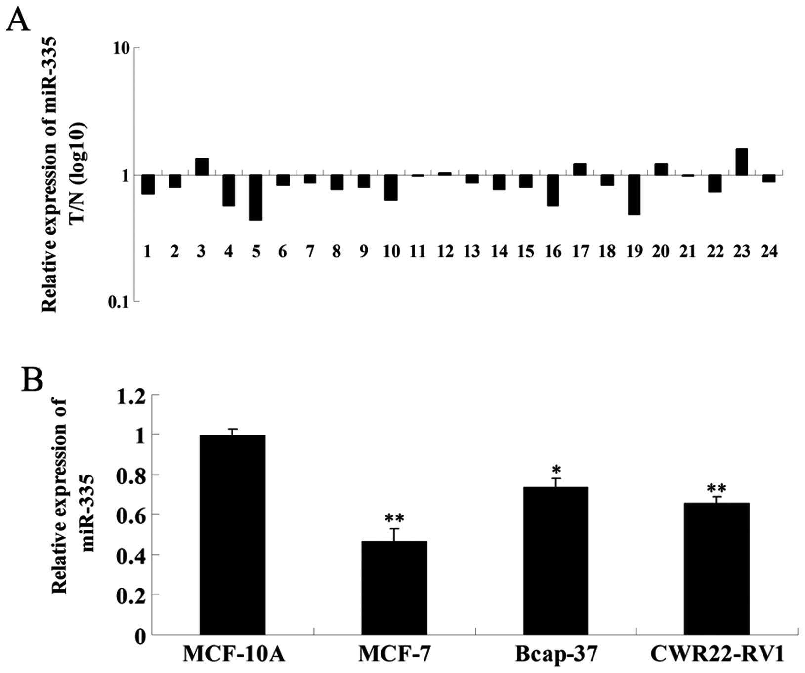
We first examined the expression of miR-335 in
breast cancer and adjacent tissues using RT-qPCR. As shown in
Fig. 1A, the expression level of
miR-335 in breast cancer tissues was markedly reduced compared to
adjacent healthy (normal) tissues. We further examined the
expression of miR-335 in three breast cancer cell lines, MCF-7,
Bcap-37 and CWR22-RV1. The normal human mammary epithelial cell
line MCF-10A was used as a control. miR-335 expression was
significantly reduced in breast cancer cell lines compared to
MCF-10A cells (Fig. 1B). These
findings suggest that miR-335 may be involved in breast cancer.
miR-335 negatively regulates PAX6 by
binding to its 3′ UTR in MCF-7 cells
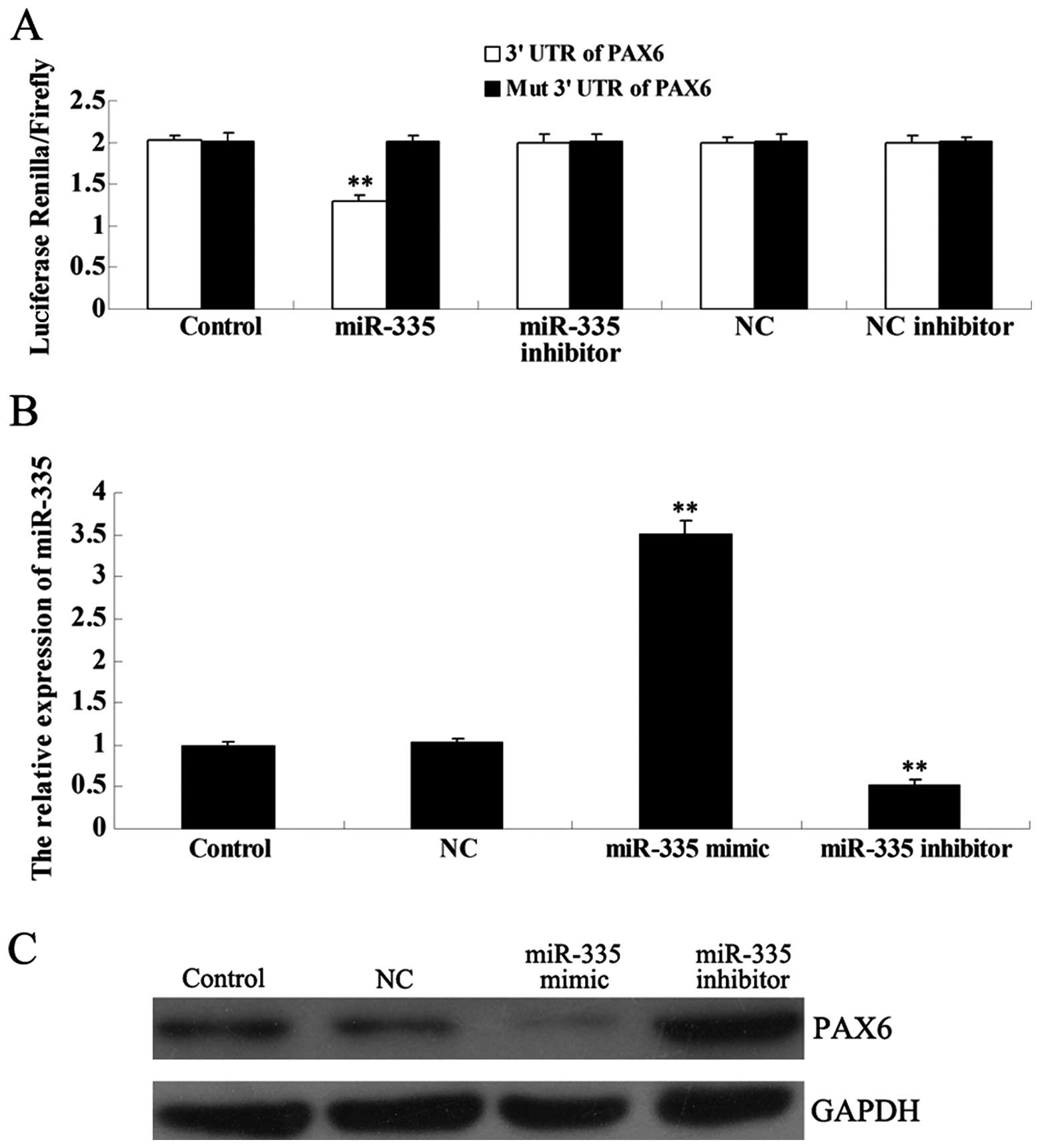
As predicted by the online software miRWalk
(15), the PAX6 gene is a
putative target of miR-335. To determine whether PAX6 is a
direct target of miR-335, a luciferase reporter assay was further
performed. In MCF-7 cells co-transfected with miR-335 and the 3′
UTR of PAX6, the Renilla/Firefly value was
significantly reduced, when compared to the control group
(P<0.01), and this effect was reversed by the miR-335 inhibitor
(Fig. 2A). However, in MCF-7 cells
co-transfected with miR-335 and the mutant 3′ UTR of PAX6,
the Renilla/Firefly value did not decrease, indicating that
miR-335 can not bind to the mutant 3′ UTR of PAX6. In
addition, the NC miRNA and the NC inhibitor also had no effect on
luciferase activity. Based on these findings, we suggest that
PAX6 is a novel target of miR-335.
Since miRNAs generally inhibit the expression of
their target genes at the post-transcriptional level, we next
investigated the effects of miR-335 over- or underexpression at the
PAX6 protein level. Following transfection of MCF-7 cells with the
miR-335 mimic or inhibitor, we first determined the efficiency of
transfection, which was satisfactory (Fig. 2B). Next, western blot analysis was
performed to examine the protein level of PAX6 in each group. The
protein level of PAX6 was reduced when miR-335 was overexpressed,
but increased when miR-335 expression was inhibited, suggesting
that miR-335 negatively regulates the protein expression of PAX6
(Fig. 2C).
Overexpression of miR-335 inhibits MCF-7
cell proliferation by targeting PAX6
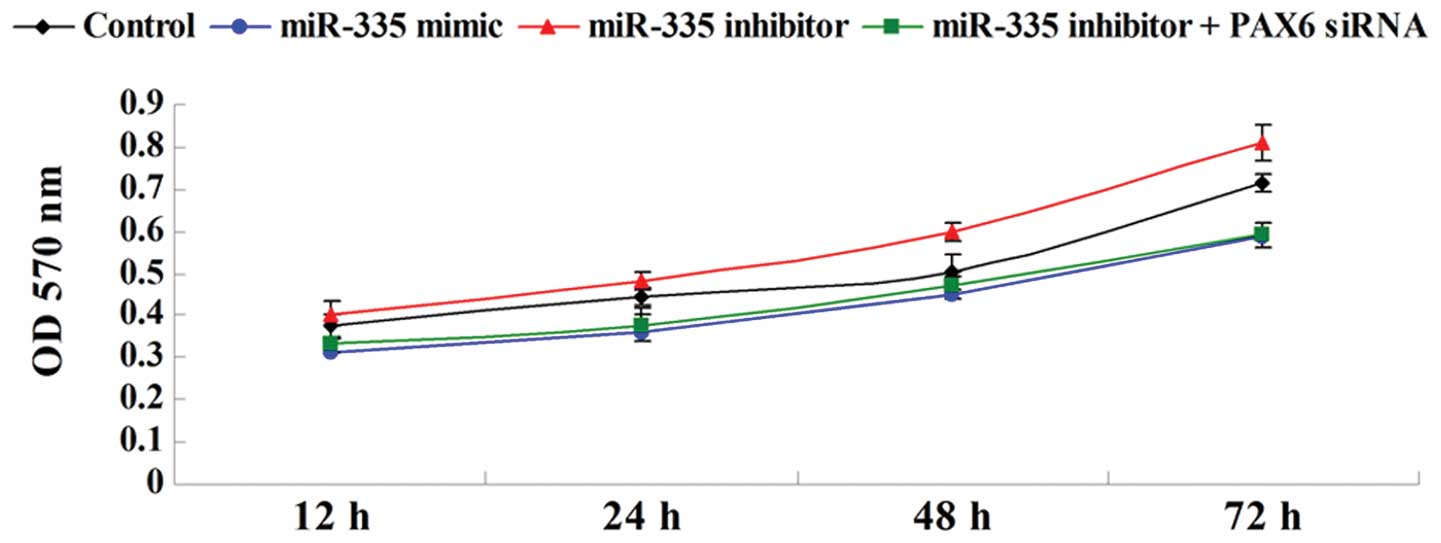
Cell proliferation was examined in breast cancer
MCF-7 cells following transfection with the miR-335 mimic or the
miR-335 inhibitor, or co-transfection with the miR-335 inhibitor
and the PAX6 small interfering RNA (siRNA). miR-335
overexpression markedly inhibited MCF-7 cell proliferation;
however, inhibition of miR-335 expression promoted cell
proliferation, an effect that was reversed by PAX6 silencing
(Fig. 3). These data indicate that
miR-335 inhibits MCF-7 cell proliferation via targeting
PAX6.
miR-335 upregulation induces cell-cycle
arrest at the G1 phase via PAX6 targeting in MCF-7 cells
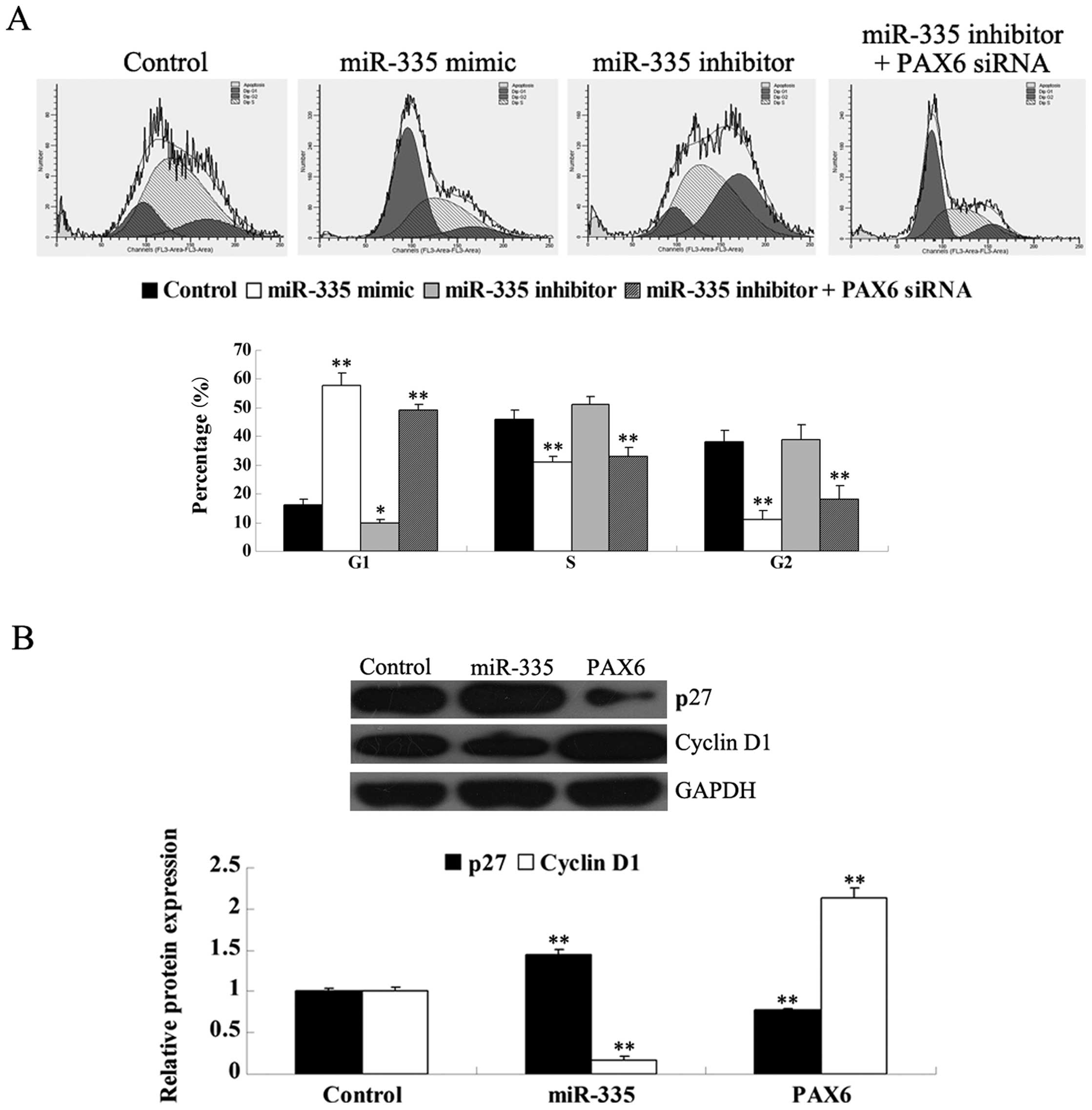
Cell-cycle distribution was examined following
transfection of MCF-7 cells with the miR-335 mimic or the miR-335
inhibitor, or co-transfection with the miR-335 inhibitor and the
PAX6 siRNA. miR-335 overexpression induced cell-cycle arrest
at the G1 phase; however, inhibition of miR-335 expression
significantly promoted cell-cycle progression, an effect that was
reversed by PAX6 silencing (Fig. 4). These data indicate that miR-335
inhibits cell-cycle progression of human cancer MCF-7 cells by
inducing cell-cycle arrest at the G1 phase via PAX6.
We further investigated the underlying molecular
mechanism, and found that miR-335 overexpression upregulates the
protein p27, while it downregulates cyclin D1. However, PAX6
overexpression decreased the protein level of p27 but increased the
protein level of cyclin D1. These findings suggest that miR-335 can
induce cell-cycle arrest at the G1 phase at least partially via
targeting PAX6, which is in accordance with the upregulation
of p27 and the downregulation of cyclin D1.
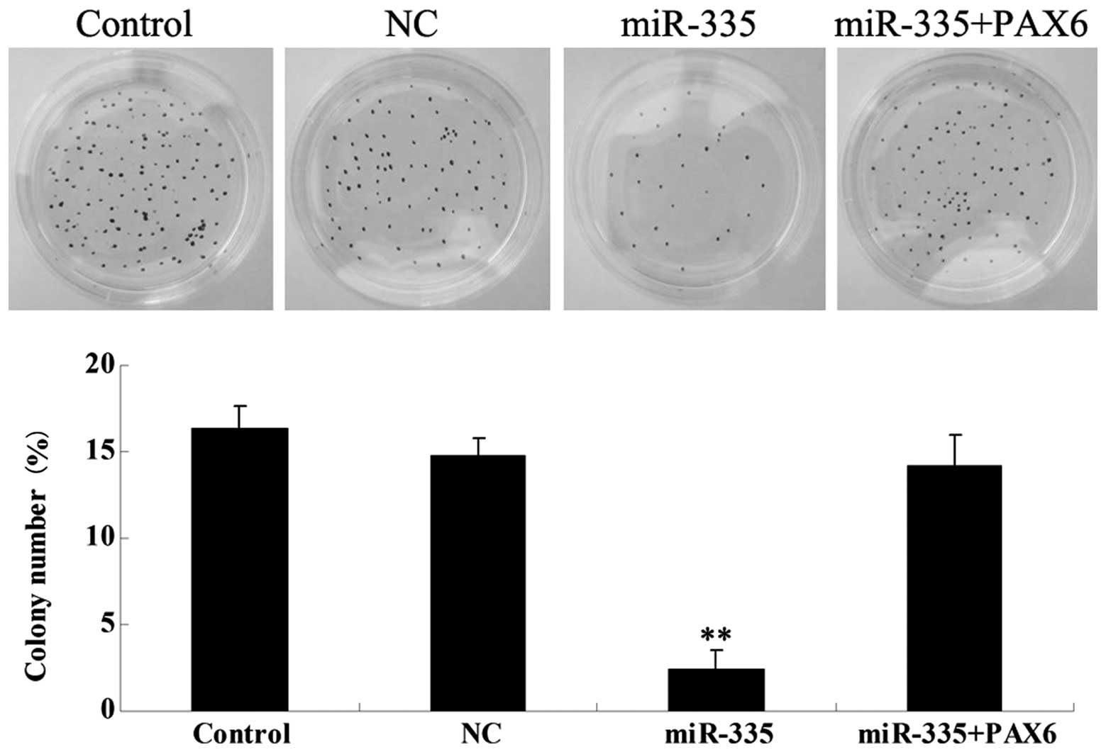
Overexpression of miR-335 reduces the
ability of MCF-7 cells to form colonies by inhibiting PAX6
We further examined the roles of miR-335 and
PAX6 in the regulation of colony formation in breast cancer
MCF-7 cells. Overexpression of miR-335 significantly inhibited
colony formation in MCF-7 cells (Fig.
5). However, the inhibitory effect of miR-335 on MCF-7 cell
colony formation was markedly attenuated by overexpression of
PAX6. These findings suggest that the PAX6 protein may be
involved in the miR-335-induced inhibition of colony formation in
breast cancer MCF-7 cells.
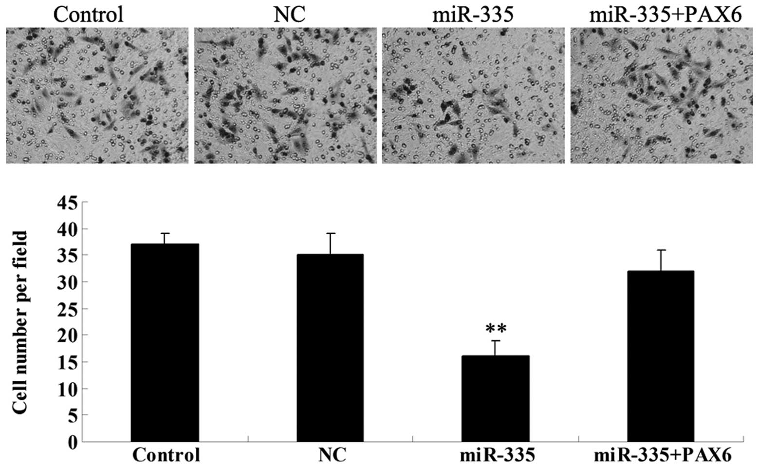
miR-335 overexpression reduces MCF-7 cell
invasion by inhibiting PAX6
Since tumor cell invasion is a key index of tumor
malignancy, we further determined the effects of miR-335 and PAX6
on MCF-7 cell invasion by performing a Transwell assay.
Overexpression of miR-335 significantly reduced MCF-7 cell invasion
(Fig. 6). However, the inhibitory
effect of miR-335 on MCF-7 cell invasion was markedly attenuated by
PAX6 overexpression. These data suggest that miR-335
inhibits MCF-7 cell invasion at least partially via targeting
PAX6.
Discussion
Recently, accumulating evidence has suggested that
miRNAs play a vital role in human cancer via directly targeting
oncogenes or tumor suppressors (16). Among miRNAs, miR-335 has been
demonstrated to be involved in various human malignancies,
including ovarian, small cell lung, prostate and gastric cancer,
meningioma, osteosarcoma, and hepatocellular carcinoma (4–6,17–22).
Furthermore, miR-335 was shown to suppress cell viability and
induce cell apoptosis in breast cancer cells (7), while it may also play a role in
breast cancer metastasis (8,9).
However, the effect of miR-335 on breast cancer cell proliferation,
as well as the underlying molecular mechanism remain largely
unknown. In this study, we showed that miR-335 can inhibit cellular
proliferation by inducing cell-cycle arrest at the G1 phase in
breast cancer cells.
To further investigate the molecular mechanism by
which miR-335 exerts its effects on breast cancer cells, we focused
on its target genes, and identified, for the first time to the best
of our knowledge, PAX6 as a novel target of miR-335.
PAX6, a member of the PAX gene family, encodes a
transcription factor highly conserved in both flies and mammals,
which has been demonstrated to play a role in the development of
the eyes, central nervous system, and pancreas (11,23,24).
More recently, PAX6 was shown to act as a key regulator in various
cancers, including breast cancer. Zong et al (14) suggested a potential role of PAX6 in
promoting breast cancer in vitro and in vivo. They
found that knockdown of PAX6 leads to decreased cell
viability, DNA synthesis and colony formation, as well as to
significantly reduced tumorigenesis in xenograft nude mice. In the
present study, we found that miR-335 negatively regulates PAX6
protein expression by directly binding to the 3′ UTR of the
PAX6 gene in breast cancer cells. Moreover, we showed that
miR-335 inhibits breast cancer cell proliferation and induces
cell-cycle arrest by targeting PAX6. PAX6 has been
shown to be regulated by additional miRNAs in cancer. For instance,
Huang et al (25) showed
that PAX6 is involved in miR-223-mediated glioblastoma cell
growth and invasion, while Wang et al (12) reported that miR-365b-3p regulates
cell-cycle progression and apoptosis in human retinoblastoma cells
by targeting PAX6.
Several molecules have been shown to play vital
roles in the regulation of cell-cycle progression, including p27
and cyclin D1. p27, a key protein for the G0/G1 checkpoint, can
block the transition from the G1 to the S phase. Cyclin D1 is
essential for the progression through the cell cycle, and
inhibition of cyclin D1 can lead to cell-cycle arrest at the G1
phase. Accordingly, we further investigated the underlying
molecular mechanism by which miR-335 regulates cell-cycle
progression in breast cancer cells, and found that overexpression
of miR-335 causes an increase in p27 and a decrease in cyclin D1
protein levels. These changes in response to miR-335 can explain
the cell-cycle arrest observed upon overexpresion of miR-335. The
PAX6 protein has also been demonstrated to participate in the
regulation of cell-cycle progression. Wang et al (12) reported that PAX6 is involved in the
miR-365b-3p-induced cell-cycle arrest via affecting the protein
expression of p21, p27, Cdc2 and cyclin D1. Moreover, Zong et
al (14) showed that knockdown
of PAX6 induces cell-cycle arrest at the G0/G1 phase in
breast cancer cells. Here, we showed that the effects of miR-335
overexpression on p27 and cyclin D1 are abolished by restoring
PAX6 expression, which further supports the hypothesis that
miR-335 regulates cell-cycle progression via targeting
PAX6.
PAX6 silencing was shown to significantly
inhibit colony formation in breast cancer cells (14). Since we demonstrated that miR-335
negatively regulates the protein expression of PAX6, we
hypothesized that miR-335 may have an inhibitory effect on colony
formation in MCF-7 cells. The colony formation assay confirmed that
overexpression of miR-335 inhibits colony formation at least
partially via directly targeting PAX6 in MCF-7 cells. The
effect of miR-335 on colony formation has been reported in several
types of cancer. For instance, Gong et al (6) reported that overexpression of miR-335
suppresses colony formation in small cell lung cancer SBC-5 cells.
Martin et al (26) reported
reduced colony survival following irradiation in HeLa cells
overexpressing miR-335.
Furthermore, miR-335 has been associated with cancer
cell invasion in several types of cancer. Wang et al
(27) found that miR-335
overexpression significantly inhibits cellular invasion in
non-small cell lung cancer A549 and H1299 cells. Xu et al
(28) found that overexpression of
miR-335 suppresses gastric cancer cell invasion. Moreover, miR-35
was suggested to act as a suppressor of breast cancer metastasis
(8). In this study, miR-335
overexpression significantly reduced MCF-7 cell invasion, an effect
that was attenuated by overexpression of PAX6. Our findings
suggest that miR-335 inhibits breast cancer cell invasion by
directly downregulating PAX6.
In conclusion, this study provides novel insights
into the molecular mechanism by which miRNA-335 and PAX6 regulate
cellular proliferation, cell-cycle progression, colony formation
and cellular invasion in breast cancer in vitro, and
suggests that miRNA-335 may constitute a promising candidate for
the treatment of breast cancer.
References
|
1
|
Jemal A, Bray F, Center MM, Ferlay J, Ward
E and Forman D: Global cancer statistics. CA Cancer J Clin.
61:69–90. 2011. View Article : Google Scholar
|
|
2
|
Siegel R, Naishadham D and Jemal A: Cancer
statistics, 2013. CA Cancer J Clin. 63:11–30. 2013. View Article : Google Scholar
|
|
3
|
Cullen BR: MicroRNAs as mediators of viral
evasion of the immune system. Nat Immunol. 14:205–210. 2013.
View Article : Google Scholar : PubMed/NCBI
|
|
4
|
Xiong SW, Lin TX, Xu KW, et al:
MicroRNA-335 acts as a candidate tumor suppressor in prostate
cancer. Pathol Oncol Res. 19:529–537. 2013. View Article : Google Scholar : PubMed/NCBI
|
|
5
|
Wang Y, Zhao W and Fu Q: miR-335
suppresses migration and invasion by targeting ROCK1 in
osteosarcoma cells. Mol Cell Biochem. 384:105–111. 2013. View Article : Google Scholar : PubMed/NCBI
|
|
6
|
Gong M, Ma J, Guillemette R, et al:
miR-335 inhibits small cell lung cancer bone metastases via IGF-1R
and RANKL pathways. Mol Cancer Res. 12:101–110. 2014. View Article : Google Scholar : PubMed/NCBI
|
|
7
|
Heyn H, Engelmann M, Schreek S, et al:
MicroRNA miR-335 is crucial for the BRCA1 regulatory cascade in
breast cancer development. Int J Cancer. 129:2797–2806. 2011.
View Article : Google Scholar : PubMed/NCBI
|
|
8
|
Negrini M and Calin GA: Breast cancer
metastasis: a microRNA story. Breast Cancer Res. 10:2032008.
View Article : Google Scholar : PubMed/NCBI
|
|
9
|
Tavazoie SF, Alarcón C, Oskarsson T, et
al: Endogenous human microRNAs that suppress breast cancer
metastasis. Nature. 451:147–152. 2008. View Article : Google Scholar : PubMed/NCBI
|
|
10
|
Yamaoka T and Itakura M: Development of
pancreatic islets (Review). Int J Mol Med. 3:247–261. 1999.
|
|
11
|
Elso C, Lu X, Weisner PA, et al: A
reciprocal translocation dissects roles of Pax6 alternative
promoters and upstream regulatory elements in the development of
pancreas, brain, and eye. Genesis. 51:630–646. 2013.
|
|
12
|
Wang J, Wang X, Wu G, Hou D and Hu Q:
MiR-365b-3p, down-regulated in retinoblastoma, regulates cell cycle
progression and apoptosis of human retinoblastoma cells by
targeting PAX6. FEBS Lett. 587:1779–1786. 2013. View Article : Google Scholar : PubMed/NCBI
|
|
13
|
Li L, Li B, Zhang H, et al: Lentiviral
vector-mediated PAX6 overexpression promotes growth and inhibits
apoptosis of human retinoblastoma cells. Invest Ophthalmol Vis Sci.
52:8393–8400. 2011. View Article : Google Scholar : PubMed/NCBI
|
|
14
|
Zong X, Yang H, Yu Y, et al: Possible role
of Pax-6 in promoting breast cancer cell proliferation and
tumorigenesis. BMB Rep. 44:595–600. 2011. View Article : Google Scholar : PubMed/NCBI
|
|
15
|
Dweep H, Sticht C, Pandey P and Gretz N:
miRWalk - database: prediction of possible miRNA binding sites by
“walking” the genes of 3 genomes. Journal Biomed Inform.
44:839–847. 2011.
|
|
16
|
Yates LA, Norbury CJ and Gilbert RJ: The
long and short of microRNA. Cell. 153:516–519. 2013. View Article : Google Scholar : PubMed/NCBI
|
|
17
|
Walter BA, Valera VA, Pinto PA and Merino
MJ: Comprehensive microRNA profiling of prostate cancer. J Cancer.
4:350–357. 2013. View
Article : Google Scholar
|
|
18
|
Cao J, Cai J, Huang D, et al: miR-335
represents an invasion suppressor gene in ovarian cancer by
targeting Bcl-w. Oncol Rep. 30:701–706. 2013.PubMed/NCBI
|
|
19
|
Liu J, Mao Q, Liu Y, Hao X, Zhang S and
Zhang J: Analysis of miR-205 and miR-155 expression in the blood of
breast cancer patients. Chin J Cancer Res. 25:46–54.
2013.PubMed/NCBI
|
|
20
|
Dohi O, Yasui K, Gen Y, et al: Epigenetic
silencing of miR-335 and its host gene MEST in hepatocellular
carcinoma. Int J Oncol. 42:411–418. 2013.PubMed/NCBI
|
|
21
|
Shi L, Jiang D, Sun G, et al: miR-335
promotes cell proliferation by directly targeting Rb1 in
meningiomas. J Neurooncol. 110:155–162. 2012. View Article : Google Scholar : PubMed/NCBI
|
|
22
|
Yan Z, Xiong Y, Xu W, et al:
Identification of hsa-miR-335 as a prognostic signature in gastric
cancer. PLoS One. 7:e400372012. View Article : Google Scholar : PubMed/NCBI
|
|
23
|
Georgala PA, Carr CB and Price DJ: The
role of Pax6 in forebrain development. Dev Neurobiol. 71:690–709.
2011. View Article : Google Scholar : PubMed/NCBI
|
|
24
|
Hanson IM: PAX6 and congenital eye
malformations. Pediatr Res. 54:791–796. 2003. View Article : Google Scholar : PubMed/NCBI
|
|
25
|
Huang BS, Luo QZ, Han Y, Li XB, Cao LJ and
Wu LX: microRNA-223 promotes the growth and invasion of
glioblastoma cells by targeting tumor suppressor PAX6. Oncol Rep.
30:2263–2269. 2013.PubMed/NCBI
|
|
26
|
Martin NT, Nakamura K, Davies R, et al:
ATM-dependent MiR-335 targets CtIP and modulates the DNA damage
response. PLoS Genet. 9:e10035052013. View Article : Google Scholar : PubMed/NCBI
|
|
27
|
Wang H, Li M, Zhang R, et al: Effect of
miR-335 upregulation on the apoptosis and invasion of lung cancer
cell A549 and H1299. Tumour Biol. 34:3101–3109. 2013. View Article : Google Scholar : PubMed/NCBI
|
|
28
|
Xu Y, Zhao F, Wang Z, et al: MicroRNA-335
acts as a metastasis suppressor in gastric cancer by targeting
Bcl-w and specificity protein 1. Oncogene. 31:1398–1407. 2012.
View Article : Google Scholar : PubMed/NCBI
|