Introduction
At present, lung cancer is the most commonly
diagnosed cancer and is the most common cause of cancer-related
mortality, with an increasing number of diagnoses every day
worldwide (1). Among the subtypes
of lung cancer, non-small cell lung cancer (NSCLC) is the most
common type, accounting for 85% of newly diagnosed cases (2). The majority of patients with NSCLC
exhibit metastases in local lymph nodes or distant sites (3).
Epithelial to mesenchymal transition (EMT) has been
hypothesized as a key step in determining the metastatic potential
of cancer cells (4). During
embryonic development, epithelial cells undergo EMT, in which
epithelial cells transform into mesenchymal cells. During this
transition, epithelial cells lose their epithelial characteristics,
including cell polarity and specialized cell-to-cell contacts, and
acquire mesenchymal characteristics. These characteristics include
change from a cobblestone to elongated morphology and individual
growth (5–7). At the molecular level, several
specific markers of EMT have been identified with significantly
altered levels of expression during EMT progression (8). For example, loss or gain of the cell
adhesion molecules, E-cadherin, N-cadherin and vimentin, is
considered to be the most important molecular marker of EMT.
Several other EMT markers, including the transcription factors
Snail (Snai1), Slug (Snai2), ZEB1 and ZEB2/Sip1 have been
demonstrated to inhibit E-cadherin expression (9,10).
In in vitro studies, EMT is induced in cancer
cells by transforming growth factor-β1 (TGF-β1). Induced cells
exhibit altered expression of EMT markers, undergo morphological
transitions and acquire characteristics of stem cells (11) and drug resistance (12,13).
Previous studies have revealed that EMT is a common
cell behavior in NSCLC (14–17).
When EMT occurs, multiple changes at the molecular and cellular
levels occur to alter transcription profiling, the cell cycle or
cell energy metabolism. Mitochondria are known as the energy
producers of the cell and are able to adapt to conditions in
various cellular contexts (18–20).
The aim of the present study was to determine whether mitochondria
number and mitochondrial DNA (mtDNA) copy number is modified during
EMT in NSCLC cells.
Materials and methods
Cell culture
The A549 NSCLC cell line was obtained from the
American Type Culture Collection (Manassas, VA, USA) and cultured
in RPMI-1640 medium (Gibco-BRL, Carlsbad, CA, USA) supplemented
with 10% fetal bovine serum (Gibco-BRL), 100 IU/ml penicillin
(Sigma-Aldrich, St Louis, MO, USA), 100 μg/ml streptomycin
(Sigma-Aldrich), 2 mM glutamine (Gibco-BRL) and 1 mM sodium
pyruvate (Gibco-BRL) in a humidified incubator at 37°C and 5%
CO2 atmosphere in 100-mm culture dishes.
Induction of EMT using TGF-β1 in culture
cells
EMT was induced by TGF-β1 as described previously
(21). Briefly, at 70–80%
confluence, A549 cells were trypsinized and seeded into 6-well
plates in duplicate (4×10⁵ cells/well). At 24 h, cells
were cultured in EMT-induction medium [serum free, 10 ng/ml TGF-β1
and 100 ng/ml epithelial growth factor (EGF)] in a humidified
incubator for an additional 48–72 h. Cells were monitored for
morphological changes, including loss of cell-to-cell contact and
transition from a cobblestone to elongated morphology, using a
CKX31 microscope (Olympus Corporation, Tokyo, Japan).
RNA isolation and reverse
transcription-quantitative polymerase chain reaction (RT-qPCR)
analysis
Following induction of EMT, total RNA was extracted
using TRIzol reagent (Life Technologies, Grand Island, NY, USA)
according to the manufacturer’s instructions. RNA concentration and
quality were determined using a NanoDrop-2000 spectrophotometer
(Thermo Fisher Scientific, Waltham, MA, USA). A reverse
transcription kit (Toyobo Co., Ltd., Osaka Japan) was used to
generate cDNA from total RNA. qPCR was performed using a StepOne
instrument (Applied Biosystems, Bedford, MA, USA) in a final volume
of 25 μl (1 μl cDNA, 10.5 μl SYBR green PCR Master mix, 3 μl
forward and reverse primers mix, 0.4 μl ROX dye and 10.1 μl
distilled deionized water) using a Premix Ex Taq™ PCR kit (Perfect
Real Time) (Takara Biotechnology, Co., Ltd., Dalian, China). The
PCR conditions were set as follows: 95°C for 5 min, and 40 cycles
of 95°C for 5 sec and 60°C for 1 min. A melting curve analysis was
then performed, with the temperature increasing from 60–90°C, in
increments of 0.3°C. GAPDH was used as internal control. Primer
sequences were as follows: Forward: 5′-ACCCAGAAGACTGTGGATGG-3′ and
reverse: 5′-TCTAGACGGCAGGTCAGGTC-3′ for GAPDH; forward:
5′-TGCCCAGAAAATGAAAAAGG-3′ and reverse 5′-GTGTATGTGGCAATGCGTTC-3′
for E-cadherin; forward: 5′-ACAGTGGCCACCTACAAAGG-3′ and reverse:
5′-CCGAGATGGGGTTGATAATGN-3′ for N-cadherin; forward:
5′-CAGTGGGAGACCTCGAGAAG-3′ and reverse: 5′-TCCCTCGGAACATCAGAAAC-3′
for fibronectin; forward 5′-GAGAACTTTGCCGTTGAAGC-3′ and reverse
5′-GCTTCCTGTAGGTGGCAATC-3′ for vimentin; forward:
5′-CCTCCCTGTCAGATGAGGAC-3′ and reverse 5′-CCAGGCTGAGGTATTCCTTG-3′
for Snail; forward: 5′-GGAGTCCGCAGTCTTACGAG-3′ and reverse:
5′-TCTGGAGGACCTGGTAGAGG-3′ for Twist; and forward:
5′-GGGGAGAAGCCTTTTTCTTG-3′ and reverse: 5′-TCCTCATGTTTGTGCAGGAG-3′
for Slug (11).
Western blot analysis
EMT-induced A549 cells were washed twice in ice-cold
PBS and lysed on ice using RIPA buffer (50 mM Tris-HCl, 150 mM
NaCl, 1% NP-40, 0.1% SDS, 0.5% sodium deoxycholate, 2 mM sodium
fluoride, 2 mM Na3VO4, 1 mM EDTA, 1 mM EDTA
and protease inhibitor PMSF; Beyotime Institute of Biotechnology,
Jiangsu, China). Protein concentration of lysates was determined
using a bicinchoninic acid assay kit (Beyotime Institute of
Biotechnology) and then several marker proteins of EMT were
detected using SDS-PAGE electrophoresis. Antibodies against
N-cadherin, E-cadherin, vimentin (11) and cytochrome c (Cyt
c) were purchased from Abcam (Cambridge, UK) and Cell
Signaling Technologies, Inc. (Danvers, MA, USA). Finally, protein
bands were detected using the chemiluminescence detection kit
(Beyotime Institute of Biotechnology).
Mitochondrial density of A549 EMT cells
determined by MitoTracker green staining
A549 cells were seeded in 24-well plates (Corning
Incorporated, Corning, NY, USA) and treated with EMT induction
medium. Following induction of EMT, cells were stained with a
MitoTracker Green kit (Beyotime Institute of Biotechnology)
according to the manufacturer’s instructions to determine
mitochondrial density (22).
Briefly, cells were washed twice in PBS and incubated at 37°C with
50 nM MitoTracker Green probe for 30 min. Next, the staining buffer
was removed and changed for fresh complete medium. Fluorescence was
detected under a fluorescence microscope (Olympus, Tokyo,
Japan).
Genome DNA extraction and qPCR analysis
of mtDNA
mtDNA copy number in EMT-induced A549 cells was
determined as described previously (23). Total genomic DNA was isolated using
the PureGene kit (Qiagen, Hilden, Germany) according to the
manufacturer’s instructions. mtDNA content was determined in cells
by qPCR using an SYBR green assay. The method for detection of
mtDNA copy number was based on qPCR and utilized a 107 bp amplicon
of mtDNA tRNALeu (UUR) (forward:
5′-CACCCAAGAACAGGGTTTGT-3′ and reverse:
5′-TGGCCATGGGTATGTTGTTA-3′). An 86 bp amplicon of β2-microglobulin
(forward: 5′-TGCTGTCTCCATGTTTGATGTATCT-3′ and reverse:
5′-TCTCTGCTCCCCACCTCTAAGT-3′) was used to determine nuclear DNA
(nDNA) as an internal control (23). qPCR was performed as follows: 1
cycle of 95°C for 10 min; followed by 40 cycles of 95°C 15 sec and
62°C 30 sec; and melting curve acquisition at 50–95°C and measuring
points at 0.5°C intervals and performed using a StepOne system
(Applied Biosystems). qPCR analysis was performed in triplicate for
each DNA sample. The expression of mtDNA copy number relative to
nDNA was determined using the formula: 2×2ΔCT with ΔCT
representing the difference in CT values between the
β2-microglobulin gene and tRNALeu (UUR).
Statistical analysis
Data are expressed as the mean ± standard deviation.
Statistical significance of differences was evaluated using an
unpaired, non-parametric Student’s t-test. P<0.05 was considered
to indicate a statistically significant difference.
Results
Induction of EMT in A549 NSCLC cells by
TGF-β1
A number of previous studies have reported that
exposure of the A549 NSCLC cell line to appropriate concentrations
of TGF-β1 induces transition from an epithelial to mesenchymal
phenotype (17,24,25).
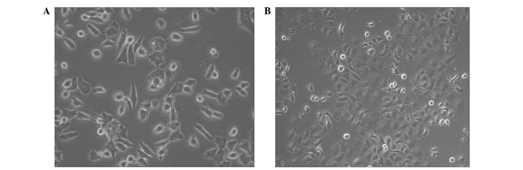
In the present study, A549 cells were cultured in serum free medium
containing 10 ng/ml TGF-β1 and 100 ng/ml EGF. Following 48–72 h
treatment, morphological changes of A549 cells were observed,
including loss of epithelial morphology and gain of mesenchymal
phenotype, i.e., elongated and individual appearance (Fig. 1A and B).
TGF-β1-induced A549 cells exhibit altered
expression of EMT markers at the mRNA and protein level
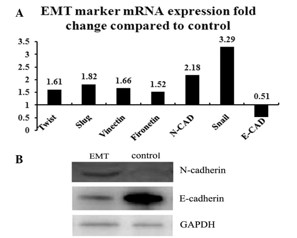
In addition to morphological changes, epithelial
cells undergoing EMT lose expression of epithelial markers,
including E-cadherin and gain expression of mesenchymal markers,
such as N-cadherin, fibronectin and vimentin. Simultaneously,
transitioned epithelial cells lose their polarity and become more
fibroblast-like (26).
Confirmation of A549 cell EMT was established at the molecular
level. RT-qPCR and western blot analysis were performed to identify
the changes in EMT markers. TGF-β1 induced expression of Snail,
Slug and Twist transcription factors, as well as a decrease in
E-cadherin expression and upregulation of vimentin and N-cadherin
through direct suppression or indirect regulation (Fig. 2A). Results of western blot analysis
were consistent with these observations (Fig. 2B). These results indicate that EMT
was successfully induced in A549 cells.
MitoTracker Green staining of EMT A549
cells
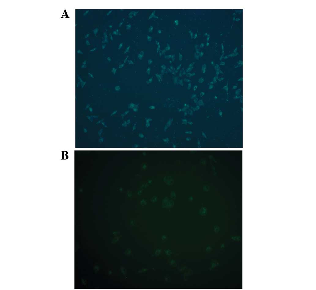
A549 cells were incubated with TGF-β1 for EMT
induction and then stained with MitoTracker Green, a specific
mitochondrial dye. Fluorescence microscopy was used to observe and
capture images of mitochondria in EMT and control A549 cells
(Fig. 3). EMT cells were observed
to exhibit increased fluorescence intensity compared with control.
MitoTracker Green probe stains all mitochondria regardless of
competency (27). Image analysis
revealed that mitochondrial number increased during EMT in A549
cells.
Mitochondrial content determination: DNA
copy number and Cyt c protein
Mitochondrial disorders are a reflection of
complicated heterogeneous diseases, which may be caused by
molecular or cellular defects. It is clear that a constant number
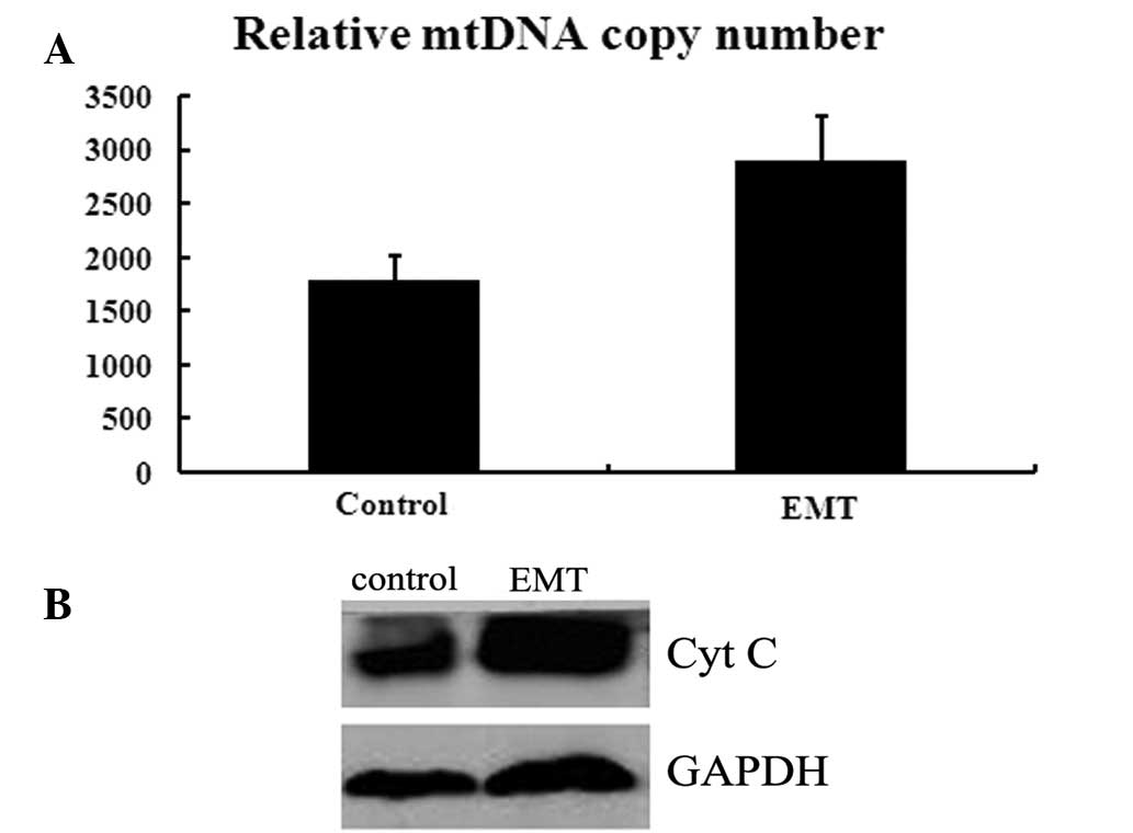
of mtDNA copies is essential for homeostasis in cells. In the
present study, changes in the copy number of mtDNA during EMT were
determined. Total genomic DNA was extracted from cells and the
relative ratio of mtDNA/nDNA was investigated by qPCR. Using nDNA
as an internal control, the relative mtDNA copy number was
identified as significantly increased from 1,700 to 2,800 compared
with control A549 cells (P<0.01; Fig. 4A). To further determine variations
in mitochondrial components, protein expression of the
mitochondrial protein, Cyt c, was analyzed by SDS-PAGE. As
demonstrated in Fig. 4B, Cyt
c levels were augmented following induction of EMT,
consistent with observations of mtDNA copy number. These results
quantitatively demonstrate that mitochondrial components (mtDNA
copy number and Cyt c protein) increase during EMT in the
A549 NSCLC cell line.
Discussion
EMT induced by TGF-β1 is essential for stem cell
differentiation and tissue and organ generation during normal
mammalian development (6,7). In the tumor microenvironment, TGF-β1
may function as an autocrine or paracrine factor. An increasing
number of studies have reported that EMT is involved in the
mobility, invasion and migratory ability of cancer cells, providing
these cells with enhanced metastatic properties (5,28).
When the condition or state of cancer cells is altered, changes in
the energy metabolism of these cells occur to adapt to the new
status (29,30). Mitochondria are directly involved
in cellular energy metabolism and mtDNA copy number and other
components affect mitochondrial function (31,32).
In the present study, the A549 NSCLC cell line was
induced to undergo EMT by exposure to TGF-β1 and EGF, which was
accompanied with increased expression of mesenchymal specific
protein markers and decreased expression of epithelial specific
protein markers. Compared with control cells, A549 cells treated
with TGF-β1 and EGF were successfully induced to undergo EMT, as
determined by RT-qPCR and western blot analysis to identify the
expression of EMT-specific markers.
Mitochondria are pivotal in cellular and subcellular
mechanisms as they function as energy-generating factories. Human
mtDNA has a circular double-stranded structure of ~16.6 kbp and
codes for 13 proteins of the mammalian mitochondrial respiratory
chain (Co I–III, Cytb, ND1–6, 4L, ATP6 and ATP8), 22 tRNAs and 2
rRNAs (23,33). A somatic mammalian cell contains
1,000–10,000 copies of mtDNA. Depletion or deficiency of mtDNA is
known to cause several genetic diseases, including deficiencies in
SUCLG1 in encephalomyopathy and thymidine phosphorylase in
mitochondrial neurogastrointestinal encephalomyopathy and mutations
in POLG, DGUOK, MPV17 and TWINKLE in the hepatocerebral form of
mtDNA depletion syndrome (23).
In the current study, mitochondrial copy number and
the number of mitochrondria were increased following induction of
EMT in A549 cells. To the best of our knowledge, no studies have
been performed on EMT and the mitochondria. When the cell phenotype
is altered, coordinated internal mechanisms also occur in specific
systems, including energy metabolism. mtDNA copy number is not
random and is specific to the developmental stage of cells,
particularly cancer cells (34).
To date, the mechanism by which mtDNA copy number is regulated at
various stages of the cell life cycle has remained unclear. In the
current study, EMT was hypothesized to be accompanied by changes in
energy metabolism. Therefore, copy number and Cyt c protein
expression in A549 cells undergoing EMT were determined. Copy
number decreased by ~50% compared with control A549 cells and
mitochondrial content in EMT cells increased and Cyt c
protein was observed to increase significantly.
Variations in mtDNA copy number are associated with
a number of diseases (35). Xing
et al (36) investigated
the mtDNA content of patients with renal cell carcinoma and
concluded that mtDNA content appeared to exhibit heritability and
low mtDNA content was associated with increased risk of renal cell
carcinoma. Blokhin et al (37) analyzed pathology-related variations
in mtDNA copy number in the brains of patients with multiple
sclerosis and found significantly higher mtDNA copy number values
in neurons of normal-appearing gray matter than in cells of other
multiple brain regions. In addition, numerous diseases are
associated with a decreased mtDNA copy number, including liver
diseases (38), biliary atresia
(39), type 2 diabetes (40), cardiomyopathy (41) and breast cancer (41).
In the present study, EMT-induced NSCLC cells were
observed to exhibit increases in mtDNA copy number and other
mitochondrial contents. These events may represent energy
preparation for EMT, a process hypothesized to be important for
cancer cell migration, invasion and metastasis (12). In this study, mitochondrial content
was altered, particularly the mtDNA copy number. However, the
mechanisms involved in this change in mitochondrial content during
EMT remain unclear and further studies are required. Although
several models of the mechanism by which mtDNA copy number is
regulated have been hypothesized, understanding of this process
remains extremely limited (34).
Results of the present study indicate that increased content of
mitochondria may contribute to the increasing energy requirements
of cancer cells undergoing EMT and that EMT-mediated metastasis of
malignant cells demands increased energy availability which would
affect the subsequent cell behavior.
In the current study, the A549 NSCLC cell line, was
induced to undergo EMT and mitochondrial content was found to
increase significantly. These observations indicate that epithelial
cells undergoing transition to mesenchymal-like cells undergo
changes in the energy metabolism system, in additional to the
well-known morphological changes. The mechanisms by which the
energy system and mitochondria is altered during EMT requires
further investigation and novel therapeutic targets for cancer may
be identified.
Acknowledgements
The authors of the present study would like to thank
colleagues at the Shanghai Lung Cancer Center for their
recommendations on experimental design and writing.
References
|
1
|
Jemal A, Bray F, Center MM, Ferlay J, Ward
E and Forman D: Global cancer statistics. CA Cancer J Clin.
61:69–90. 2011. View Article : Google Scholar
|
|
2
|
Dela Cruz CS, Tanoue LT and Matthay RA:
Lung cancer: epidemiology, etiology and prevention. Clin Chest Med.
32:605–644. 2011.PubMed/NCBI
|
|
3
|
Travis WD, Travis LB and Devesa SS: Lung
cancer. Cancer. 75:191–202. 1995. View Article : Google Scholar : PubMed/NCBI
|
|
4
|
Kudo-Saito C, Shirako H, Takeuchi T and
Kawakami Y: Cancer metastasis is accelerated through
immunosuppression during Snail-induced EMT of cancer cells. Cancer
Cell. 15:195–206. 2009. View Article : Google Scholar
|
|
5
|
Kalluri R and Weinberg RA: The basics of
epithelial-mesenchymal transition. J Clin Invest. 119:1420–1428.
2009. View
Article : Google Scholar : PubMed/NCBI
|
|
6
|
Acloque H, Adams MS, Fishwick K,
Bronner-Fraser M and Nieto MA: Epithelial-mesenchymal transitions:
the importance of changing cell state in development and disease. J
Clin Invest. 119:1438–1449. 2009. View
Article : Google Scholar : PubMed/NCBI
|
|
7
|
Thiery JP, Acloque H, Huang RY and Nieto
MA: Epithelial-mesenchymal transitions in development and disease.
Cell. 139:871–890. 2009. View Article : Google Scholar : PubMed/NCBI
|
|
8
|
Zeisberg M and Neilson EG: Biomarkers for
epithelial-mesenchymal transitions. J Clin Invest. 119:1429–1437.
2009. View
Article : Google Scholar : PubMed/NCBI
|
|
9
|
Peinado H, Olmeda D and Cano A: Snail, Zeb
and bHLH factors in tumour progression: an alliance against the
epithelial phenotype? Nat Rev Cancer. 7:415–428. 2007. View Article : Google Scholar : PubMed/NCBI
|
|
10
|
Kumarswamy R, Mudduluru G, Ceppi P, et al:
MicroRNA-30a inhibits epithelial-to-mesenchymal transition by
targeting Snai1 and is downregulated in non-small cell lung cancer.
Int J Cancer. 130:2044–2053. 2012. View Article : Google Scholar : PubMed/NCBI
|
|
11
|
Mani SA, Guo W, Liao MJ, et al: The
epithelial-mesenchymal transition generates cells with properties
of stem cells. Cell. 133:704–715. 2008. View Article : Google Scholar : PubMed/NCBI
|
|
12
|
Voulgari A and Pintzas A:
Epithelial-mesenchymal transition in cancer metastasis: mechanisms,
markers and strategies to overcome drug resistance in the clinic.
Biochim Biophys Acta. 1796:75–90. 2009.PubMed/NCBI
|
|
13
|
Arumugam T, Ramachandran V, Fournier KF,
et al: Epithelial to mesenchymal transition contributes to drug
resistance in pancreatic cancer. Cancer Res. 69:5820–5828. 2009.
View Article : Google Scholar : PubMed/NCBI
|
|
14
|
Thomson S, Buck E, Petti F, et al:
Epithelial to mesenchymal transition is a determinant of
sensitivity of non-small-cell lung carcinoma cell lines and
xenografts to epidermal growth factor receptor inhibition. Cancer
Res. 65:9455–9462. 2005. View Article : Google Scholar : PubMed/NCBI
|
|
15
|
Yauch RL, Januario T, Eberhard DA, et al:
Epithelial versus mesenchymal phenotype determines in vitro
sensitivity and predicts clinical activity of erlotinib in lung
cancer patients. Clin Cancer Res. 11:8686–8698. 2005. View Article : Google Scholar
|
|
16
|
Witta SE, Gemmill RM, Hirsch FR, et al:
Restoring E-cadherin expression increases sensitivity to epidermal
growth factor receptor inhibitors in lung cancer cell lines. Cancer
Res. 66:944–950. 2006. View Article : Google Scholar : PubMed/NCBI
|
|
17
|
Maitah MY, Ali S, Ahmad A, Gadgeel S and
Sarkar FH: Up-regulation of sonic hedgehog contributes to
TGF-beta1-induced epithelial to mesenchymal transition in NSCLC
cells. PLoS One. 6:e160682011. View Article : Google Scholar : PubMed/NCBI
|
|
18
|
Wang D, Su LY, Zhang AM, et al:
Mitochondrial DNA copy number, but not haplogroup, confers a
genetic susceptibility to leprosy in han chinese from southwest
china. PLoS One. 7:e388482012. View Article : Google Scholar : PubMed/NCBI
|
|
19
|
Gianotti TF, Castano G, Gemma C, et al:
Mitochondrial DNA copy number is modulated by genetic variation in
the signal transducer and activator of transcription 3 (STAT3).
Metabolism. 60:1142–1149. 2011. View Article : Google Scholar : PubMed/NCBI
|
|
20
|
Carling PJ, Cree LM and Chinnery PF: The
implications of mitochondrial DNA copy number regulation during
embryogenesis. Mitochondrion. 11:686–692. 2011. View Article : Google Scholar : PubMed/NCBI
|
|
21
|
Moreno-Bueno G, Peinado H, Molina P, et
al: The morphological and molecular features of the
epithelial-to-mesenchymal transition. Nat Protoc. 4:1591–1613.
2009. View Article : Google Scholar : PubMed/NCBI
|
|
22
|
Fetisova EK, Avetisyan AV, Izyumov DS,
Korotetskaya MV, Chernyak BV and Skulachev VP:
Mitochondria-targeted antioxidant SkQR1 selectively protects MDR
(Pgp 170)-negative cells against oxidative stress. FEBS letters.
584:562–566. 2010. View Article : Google Scholar : PubMed/NCBI
|
|
23
|
Venegas V, Wang J, Dimmock D and Wong LJ:
Real-time quantitative PCR analysis of mitochondrial DNA content.
Curr Protoc Hum Genet. 68:19.7.1–19.7.12. 2011.
|
|
24
|
Kasai H, Allen JT, Mason RM, Kamimura T
and Zhang Z: TGF-beta1 induces human alveolar epithelial to
mesenchymal cell transition (EMT). Respir Res. 6:562005. View Article : Google Scholar : PubMed/NCBI
|
|
25
|
Pirozzi G, Tirino V, Camerlingo R, et al:
Epithelial to mesenchymal transition by TGFbeta-1 induction
increases stemness characteristics in primary non small cell lung
cancer cell line. PLoS One. 6:e215482011. View Article : Google Scholar : PubMed/NCBI
|
|
26
|
Tiwari N, Gheldof A, Tatari M and
Christofori G: EMT as the ultimate survival mechanism of cancer
cells. Semin Cancer Biol. 22:194–207. 2012. View Article : Google Scholar : PubMed/NCBI
|
|
27
|
Dingley S, Chapman KA and Falk MJ:
Fluorescence-activated cell sorting analysis of mitochondrial
content, membrane potential and matrix oxidant burden in human
lymphoblastoid cell lines. Methods Mol Biol. 837:231–239. 2012.
View Article : Google Scholar
|
|
28
|
Thiery JP: Epithelial-mesenchymal
transitions in tumour progression. Nat Rev Cancer. 2:442–454. 2002.
View Article : Google Scholar : PubMed/NCBI
|
|
29
|
Pietila M, Palomaki S, Lehtonen S, et al:
Mitochondrial function and energy metabolism in umbilical cord
blood- and bone marrow-derived mesenchymal stem cells. Stem Cells
Dev. 21:575–588. 2012. View Article : Google Scholar : PubMed/NCBI
|
|
30
|
Folmes CD, Nelson TJ, Dzeja PP and Terzic
A: Energy metabolism plasticity enables stemness programs. Ann NY
Acad Sci. 1254:82–89. 2012. View Article : Google Scholar : PubMed/NCBI
|
|
31
|
Diaz-Ruiz R, Uribe-Carvajal S, Devin A and
Rigoulet M: Tumor cell energy metabolism and its common features
with yeast metabolism. Biochim Biophys Acta. 1796:252–265.
2009.PubMed/NCBI
|
|
32
|
Westermann B: Mitochondrial fusion and
fission in cell life and death. Nat Rev Mol Cell Biol. 11:872–884.
2010. View
Article : Google Scholar : PubMed/NCBI
|
|
33
|
Falkenberg M, Larsson NG and Gustafsson
CM: DNA replication and transcription in mammalian mitochondria.
Annu Rev Biochem. 76:679–699. 2007. View Article : Google Scholar : PubMed/NCBI
|
|
34
|
Clay Montier LL, Deng JJ and Bai Y: Number
matters: control of mammalian mitochondrial DNA copy number. J
Genet Genomics. 36:125–131. 2009.PubMed/NCBI
|
|
35
|
Bai RK, Perng CL, Hsu CH and Wong LJ:
Quantitative PCR analysis of mitochondrial DNA content in patients
with mitochondrial disease. Ann NY Acad Sci. 1011:304–309. 2004.
View Article : Google Scholar : PubMed/NCBI
|
|
36
|
Xing J, Chen M, Wood CG, et al:
Mitochondrial DNA content: its genetic heritability and association
with renal cell carcinoma. J Natl Cancer Inst. 100:1104–1112. 2008.
View Article : Google Scholar : PubMed/NCBI
|
|
37
|
Blokhin A, Vyshkina T, Komoly S and Kalman
B: Variations in mitochondrial DNA copy numbers in MS brains. J Mol
Neurosci. 35:283–287. 2008. View Article : Google Scholar : PubMed/NCBI
|
|
38
|
Morten KJ, Ashley N, Wijburg F, et al:
Liver mtDNA content increases during development: a comparison of
methods and the importance of age- and tissue-specific controls for
the diagnosis of mtDNA depletion. Mitochondrion. 7:386–395. 2007.
View Article : Google Scholar
|
|
39
|
Tiao MM, Lin TK, Kuo FY, et al: Early
stage of biliary atresia is associated with significant changes in
8-hydroxydeoxyguanosine and mitochondrial copy number. J Pediatr
Gastroenterol Nutr. 45:329–334. 2007. View Article : Google Scholar : PubMed/NCBI
|
|
40
|
Choi YS, Kim S and Pak YK: Mitochondrial
transcription factor A (mtTFA) and diabetes. Diabetes Res Clin
Pract. 54(Suppl 2): S3–S9. 2001. View Article : Google Scholar : PubMed/NCBI
|
|
41
|
Bai RK and Wong LJ: Simultaneous detection
and quantification of mitochondrial DNA deletion(s), depletion and
over-replication in patients with mitochondrial disease. J Mol
Diagn. 7:613–622. 2005. View Article : Google Scholar : PubMed/NCBI
|