Introduction
Neuroblastoma is a common extracranial pediatric
solid tumor, accounting for up to 10% of pediatric cancers and
ultimately resulting in 15% of cancer-related mortality in children
(1,2). Histologically, neuroblastoma is a
heterogeneous group of tumors ranging from benign changes in
sympathetic neurons to tumors that cause critical illness as a
result of extensive invasion and metastasis (3–5). The
clinical presentation of neuroblastoma is variable and advanced
cases are often found to be highly resistant to conventional
treatment modalities based on surgery, chemotherapy and
radiotherapy (6). Therefore,
recent studies have primarily focused on these particularly
aggressive cases, with the goal of identifying additional
therapeutic options (7).
Natural compounds extracted from herbs, such as
Taxol, have been widely used in cancer therapy (8). A number of active compounds isolated
from Chinese herbs have been shown to have antitumor properties
(9,10). Thus, traditional Chinese medicine
provides an important additional option for the development of
novel cancer treatments. Triptolide is a diterpene triepoxide and
is extracted from the Chinese herb Tripterygium wilfordii
Hook F, which has been used to treat inflammation and autoimmune
diseases in Chinese medicine (11,12).
Recently, evidence has shown that triptolide has a potent
immunosuppressive effect and antineoplastic activity in certain
types of cancer, including breast cancer, pancreatic cancer,
melanoma and prostate cancer (13–16).
In addition, triptolide has been shown to exert its antitumor
properties through induction of apoptosis and inhibition of cell
proliferation, angiogenesis, cell invasion and metastasis (17–20).
This study investigated the effects of triptolide on malignant
neuroblastoma cell growth and cell proliferation with the aim of
providing evidence that may support the use of triptolide as a
novel drug for the treatment of neuroblastoma.
Materials and methods
Cell culture
BE(2)-C neuroblastoma cells were obtained from the
American Type Culture Collection (Manassus, VA, USA), and was
cultured in a 1:1 mixture of Dulbecco’s modified Eagle’s medium and
Ham’s nutrient mixture F12 (DMEM/F-12; Invitrogen, Carlsbad, CA,
USA) plus 10% fetal bovine serum (Invitrogen) and 1% penicillin and
streptomycin (Invitrogen), and was incubated at 37°C in a 5%
CO2 humidified incubator. Purified triptolide (>98%)
was purchased from Sigma-Aldrich (T3652; St. Louis, MO, USA), which
was dissolved in dimethyl sulfoxide (DMSO; Sigma-Aldrich) at a
stock concentration of 50 mM and stored at 4°C.
Cell growth and viability assays
BE(2)-C cells grown in 96-well culture plates were
treated either with various doses of triptolide (5, 10, 25, 50 or
100 nM), or DMSO. The cell growth rate was analyzed with the cell
counting kit-8 (CCK-8) growth assay after 24 h culture. Briefly,
cells in each well were incubated with 10 μl CCK-8 reagent
at 37°C for 2 h. The optical density was measured at a wavelength
of 450 nm using a microplate reader (Model 550, Bio-Rad, Hercules,
CA, USA). In addition, BE(2)-C cells were treated with 25 or 50 nM
for 24 h, photographed using an Olympus 1X71 (Olympus Corporation,
Tokyo, Japan) and counted with a TC10™ Automated Cell
Counter (Bio-Rad).
5-Bromo-2-deoxyuridine (BrdU) staining
assay
For BrdU immunofluorescent staining, cells were
grown on coverslips. After treatment with 25 or 50 nM triptolide
for 24 h, cells were incubated with 10 μg/ml BrdU (Sigma)
for 30 min, then washed with phosphate-buffered saline (PBS) and
fixed in 4% paraformaldehyde for 20 min. Subsequently, cells were
treated with 1 mol/L HCl, and blocked with 10% goat serum for 1 h,
followed by a monoclonal rat primary antibody against BrdU (1:200,
ab6326, Abcam, Cambridge, MA, USA) for 1 h and Alexa
FluorR® 594 goat anti-rat IgG secondary antibody, (H+L;
Invitrogen). DAPI (300 nM) was used for nuclear staining, after
which the percentage BrdU uptake in 10 microscopic fields was
calculated (Nikon 80i, Nikon Corporation, Tokyo, Japan).
Cell cycle assay
After treatment with triptolide (25 nM) for 24 h,
cells were collected by centrifugation at 211 × g for 5 min, washed
with ice-cold PBS, fixed with 70% ethanol, stained with 20
μg/ml propidium iodide (Invitrogen) and analyzed by flow
cytometry (BD FACSVerse™, BD BioSciences, Franklin Lakes, NJ, USA).
The data were analyzed with CellQuest Pro software, version 5.0 (BD
BioSciences).
Cell death and apoptosis assays
Cells were either untreated or treated with
triptolide. DMSO was used as control. After 24 h treatment,
adherent and floating cells were pooled, collected by
centrifugation at 211 × g for 5 min, and washed once with ice-cold
PBS. The cell death rate was detected with 0.2% trypan blue dye
(Bio-Rad). Apoptotic cells were determined by the Annexin
V-fluorescein isothiocyanate (FITC) kit (Sigma-Aldrich), using flow
cytometry according to the manufacturer’s instructions.
Reverse transcription-quantitative
polymerase chain reactions (RT-qPCR) assay
After treatment with triptolide for 24 h, cells were
harvested and lysed with TRIzol (Invitrogen) for total RNA
purification. RNA was reverse transcribed into cDNA using M-MLV
reverse transcriptase (Promega Corporation, Madison, WI, USA). The
caspase-3 and caspase-9 mRNA transcripts were determined using the
SYBRR Green PCR Master mix (Takara Bio, Inc., Shiga, Japan) by
RT-qPCR. RT-qPCR reactions in triplicate were conducted using the
OneStep plus7500 real-time PCR system (Bio-Rad). The individual
values were normalized to that of the GAPDH control. Primer
sequences were as follows: Forward: 5′-AGCGAATCAATGGACTCTGGA-3′ and
reverse: 5′-CTGAATGTTTCCCTGAGGTTTG-3′ for caspase-3, forward:
5′-GCTCTTCCTTTGTTCATCTCC-3′ and reverse:
5′-CATCTGGCTCGGGGTTACTGC-3′ for caspase-9, and forward:
5′-ACGGATTTGGTCGTATTGGG-3′ and reverse: 5′-TCCTGGAAGATGGTGATGGG-3′
for GAPDH.
Soft agar clonogenic assay
Cells (1×10³) were mixed in 0.3% Noble
agar in a growth medium containing vehicle or triptolide, and
plated into six-well plates containing a solidified bottom layer
(0.6% Noble agar in the same growth medium). Colonies were
photographed after 14 days (Olympus 1X71) and cell numbers were
counted from at least five randomly selected fields.
In vivo tumorigenic assay
Six female non-obese diabetic (NOD)/severe combined
immunodeficiency (SCID) mice (4 weeks old) were used and maintained
under specific pathogen-free conditions. Cells (1×10⁶)
were suspended in 200 μl serum-free DMEM/F12, and injected
subcutaneously into both flanks. After tumor formation, mice were
divided randomly into treatment and control groups. Mice in the
treatment group were injected with triptolide at 0.4 mg/kg daily
for seven days (21), while
control mice were injected with DMSO. Tumor size was measured using
calipers and tumor volume was calculated using the formula
4/3πr3, where r is the radius of the tumor. Mouse body
weight was monitored daily. Three weeks after tumor growth,
xenograft tumors were removed and weighed immediately after the
mice were sacrificed by cervical dislocation. All animal
experiments were approved by the Institutional Animal Care and Use
Committee of Southwest University (Chonqing, China).
Statistical analysis
All observations were confirmed by at least three
independent experiments. Quantitative data are expressed as the
mean ± standard deviation. Two-tailed Student’s t-test was
performed for paired samples using GraphPad Prism version 6.0
(GraphPad Software, Inc., La Jolla, CA, USA). P<0.05 was
considered to indicate a statistically significant difference.
Results
Triptolide inhibits neuroblastoma cell
growth and viability
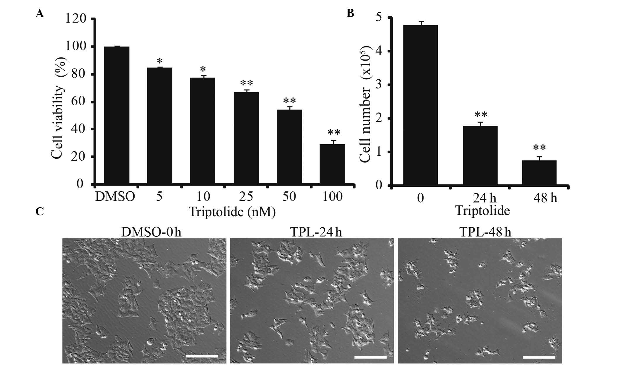
BE(2)-C cells were treated with increasing doses of
triptolide for 24 h. A concentration-dependent response to
triptolide in the BE(2)-C cells was observed. As shown in Fig. 1A, triptolide inhibited cell growth
even at a low dose of 5 nM. The cell viability was significantly
reduced to 50% at 50 nM of triptolide. Triptolide also inhibited
cell growth in a time dependent manner (Fig. 1B and C). Moreover,
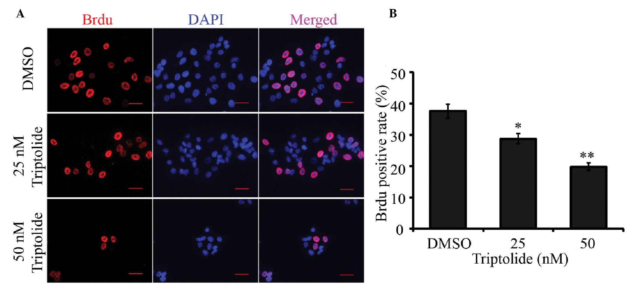
immunofluorescent staining using a BrdU label confirmed that
triptolide markedly inhibited cell proliferation (Fig. 2A and B).
Triptolide induces neuroblastoma cell
cycle arrest and apoptosis
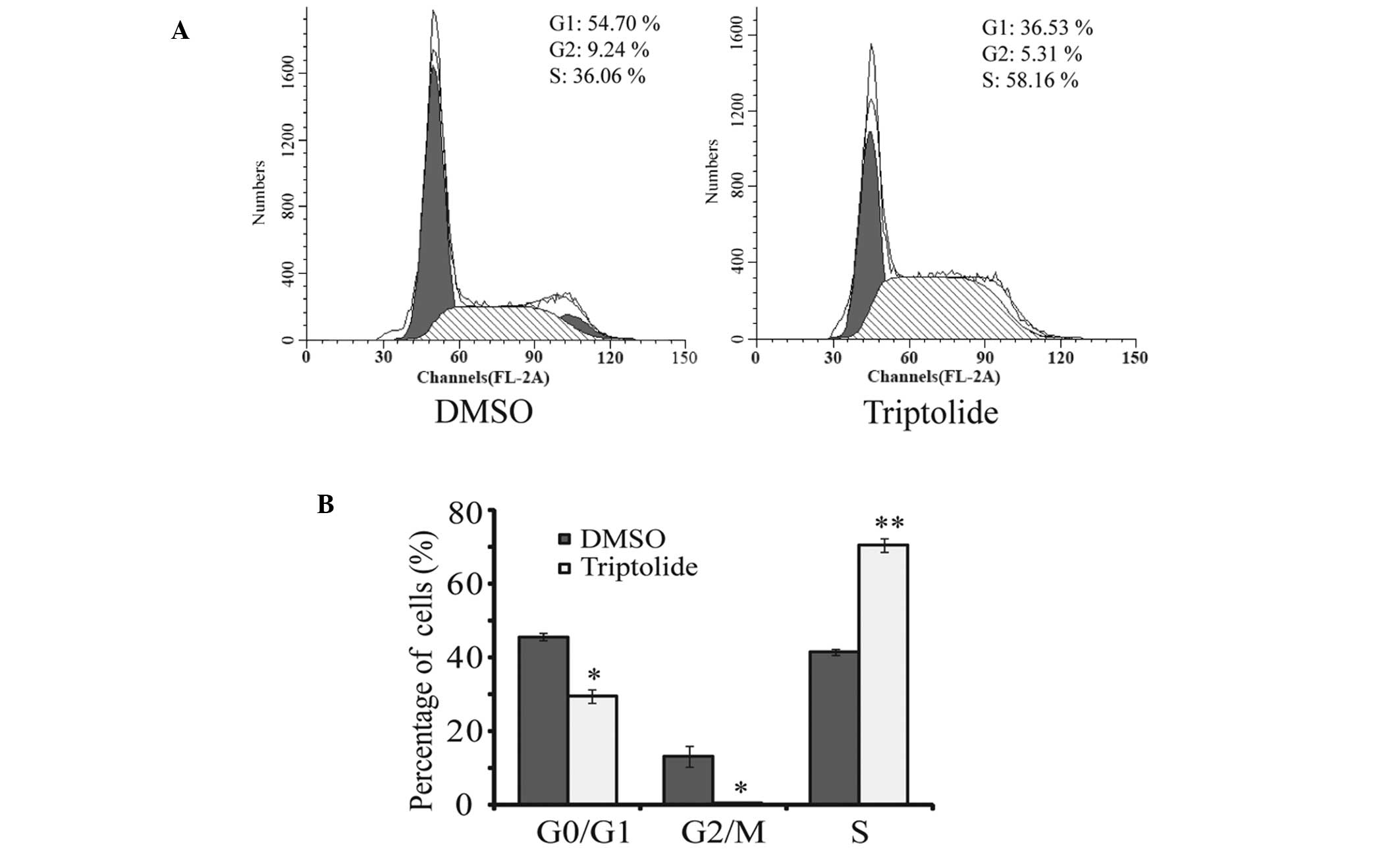
The effect of triptolide on cell cycle was
investigated. It was found that the percentage of cells in S phase
increased from 36.06 to 58.16% (Fig.
3A and B). This result suggests that triptolide induces cell
cycle arrest in the S phase, which may contribute to inhibition of
cell proliferation.
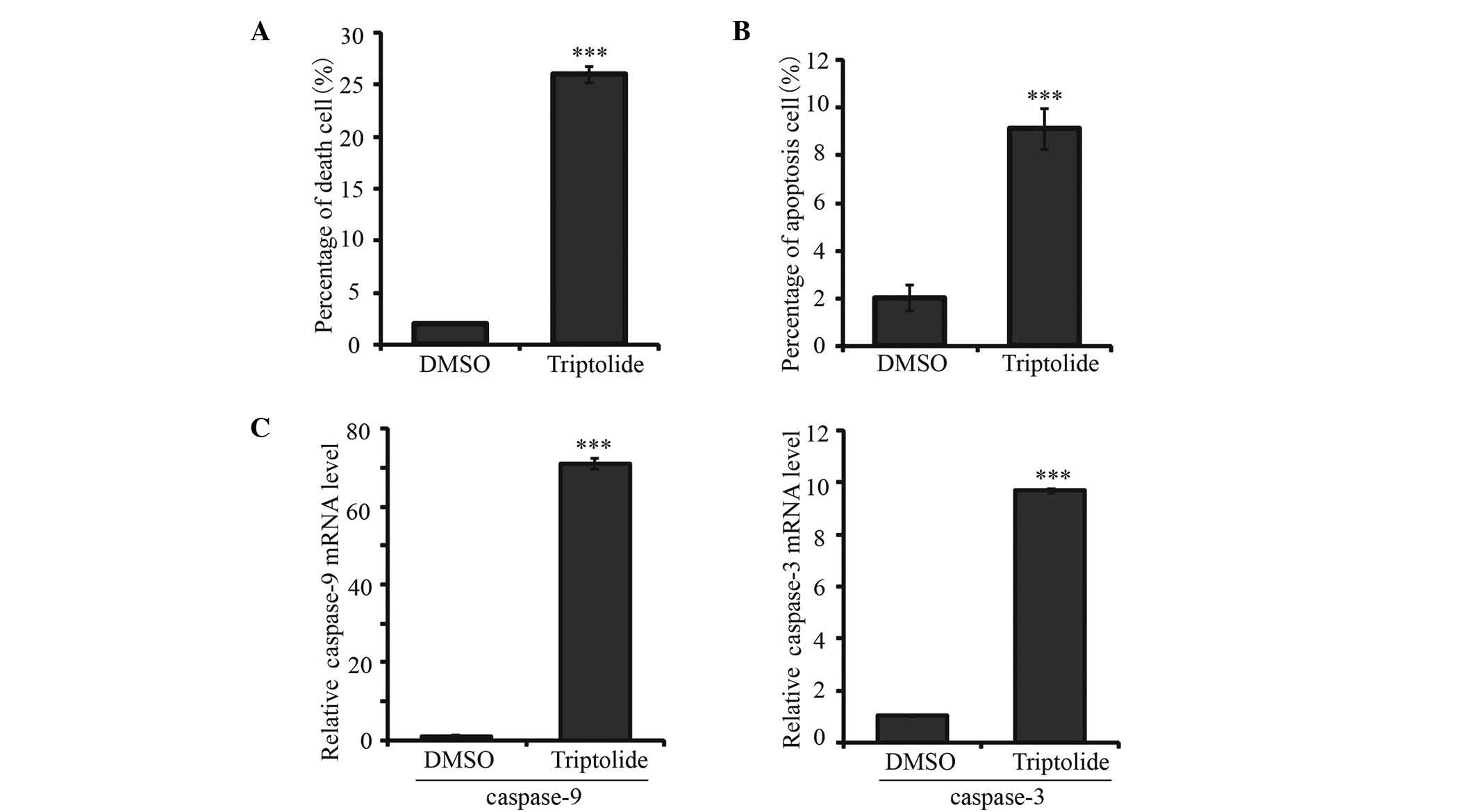
It was also observed that exposure of BE(2)-C cells
to triptolide could induce cell death and apoptosis. Triptolide
significantly increased cell death from 1.88 in the control group
to 25.9% in the triptolide 25 nM group (P<0.001; Fig. 4A). The apoptosis rate was also
increased after treatment with 25 nM triptolide for 24 h (Fig. 4B). As shown in Fig. 4C and D, following triptolide
treatment mRNA expression levels of caspase-9 and caspase-3 were
increased 70.9 and 9.7 fold, respectively, compared with control.
These results indicate that triptolide induces cell death and
apoptosis through caspase-9 and caspase-3 activation.
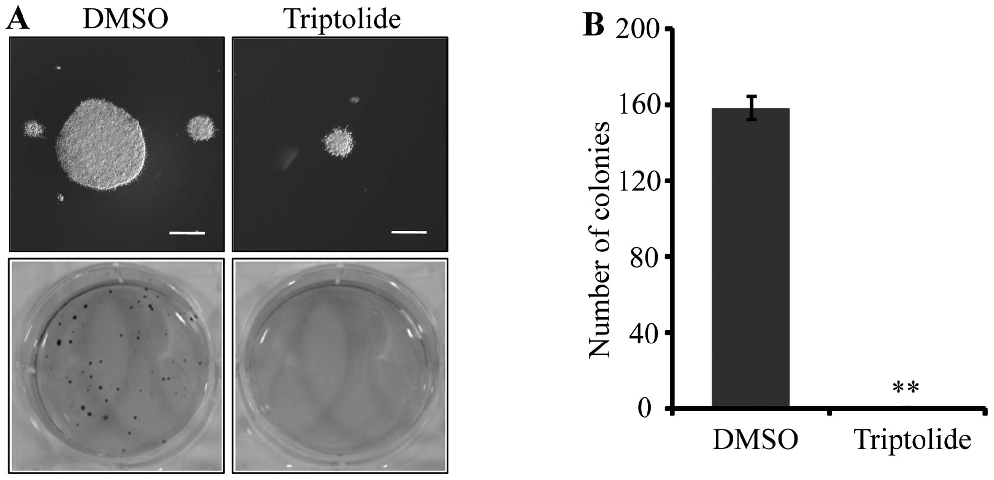
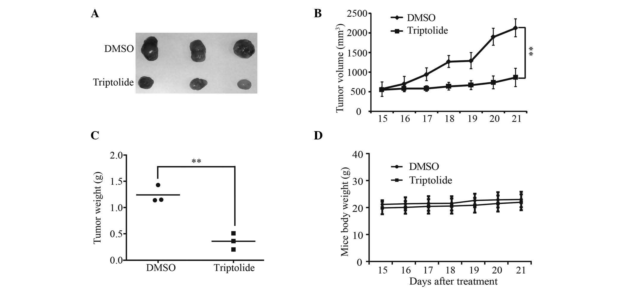
Triptolide suppresses neuroblastoma cell
colony formation in vitro and tumorigenicity in vivo
The role of triptolide in neuroblastoma
tumorigenesis was examined. BE(2)-C cells treated with 25 nM
triptolide gave rise to smaller and and sparser colonies in soft
agar, compared with cells treated with DMSO (Fig. 5A and B). The xenograft study in
NOD/SCID mice showed that the volume and weight of xenograft tumors
in the triptolide treatment group were lower than those in the DMSO
group (Fig. 6). These data
indicate that triptolide may inhibit neuroblastoma cell
self-renewal and tumorigenesis. In addition, there was no
significant difference in mouse body weight after triptolide
treatment (Fig. 6D), which
suggests that the administered dose of triptolide may have minimal
toxic side effects.
Discussion
Recently, Chinese herbs have attracted attention
from researchers worldwide due to their potential efficacy in the
treatment of a number of diseases (22,23).
A large number of active compounds have been extracted from Chinese
herbs. Tripterygium wilfordii Hook F has been used in
traditional Chinese medicine for centuries for the treatment of
fever, chills, carbuncles and edema (24,25).
The diterpenoid epoxide triptolide is one of the two main bioactive
components of Tripterygium wilfordii Hook F, which exhibits
antitumor activity (26,27). However, there is little data
regarding the efficacy of triptolide against neuroblastoma cells.
This study aimed to investigate the effect of triptolide on
neuroblastoma cell growth and tumor development, with the aim of
providing more information for the development of novel
neuroblastoma treatments.
The current study demonstrated that triptolide not
only induced neuroblastoma cell death and apoptosis via
caspase-9/caspase-3 pathway activation, but also inhibited cell
growth and viability by inducing cell cycle arrest at the S phase.
Furthermore, the results showed that triptolide inhibited
neuroblastoma cell colony-forming capability in vitro and
tumor progression in vivo. In conclusion, triptolide may be
a potent natural candidate for neuroblastoma treatment.
Acknowledgements
This study was supported by the National Basic
Research Program of China (grant no. 2012cb114603); the National
Natural Science Foundation of China (grant no. 81201551); the
Natural Science Foundation of Chongqing (grant no.
cstc2013jcyjys0007); and the Fundamental Research Funds for the
Central Universities (grant nos. SWU111014 and SWU112033).
References
|
1
|
Castleberry RP, Pritchard J, Ambros P, et
al: The International Neuroblastoma Risk Groups (INRG): a
preliminary report. Eur J Cancer. 33:2113–2116. 1997. View Article : Google Scholar
|
|
2
|
Li T, Wang L, Ke XX, et al: DNA-damaging
drug-induced apoptosis sensitized by N-myc in neuroblastoma cells.
Cell Biol Int. 36:331–337. 2012. View Article : Google Scholar
|
|
3
|
Shimada H, Ambros IM, Dehner LP, et al:
The International Neuroblastoma Pathology Classification (the
Shimada system). Cancer. 86:364–372. 1999. View Article : Google Scholar : PubMed/NCBI
|
|
4
|
Brodeur GM: Neuroblastoma: biological
insights into a clinical enigma. Nat Rev Cancer. 3:203–216. 2003.
View Article : Google Scholar : PubMed/NCBI
|
|
5
|
Cui H, Ma J, Ding J, et al: Bmi-1
regulates the differentiation and clonogenic self-renewal of I-type
neuroblastoma cells in a concentration-dependent manner. J Biol
Chem. 281:34696–34704. 2006. View Article : Google Scholar : PubMed/NCBI
|
|
6
|
Cheung NK and Dyer MA: Neuroblastoma:
developmental biology, cancer genomics and immunotherapy. Nat Rev
Cancer. 13:397–411. 2013. View
Article : Google Scholar : PubMed/NCBI
|
|
7
|
Morgenstern DA, Baruchel S and Irwin MS:
Current and future strategies for relapsed neuroblastoma:
challenges on the road to precision therapy. J Pediatr Hematol
Oncol. 35:337–347. 2013. View Article : Google Scholar : PubMed/NCBI
|
|
8
|
Camirand A, Fadhil I, Luco AL, et al:
Enhancement of taxol, doxorubicin and zoledronate
anti-proliferation action on triple-negative breast cancer cells by
a PTHrP blocking monoclonal antibody. Am J Cancer Res. 3:500–508.
2013.PubMed/NCBI
|
|
9
|
Jia L, Ma S, Hou X, et al: The synergistic
effects of traditional Chinese herbs and radiotherapy for cancer
treatment. Oncol Lett. 5:1439–1447. 2013.PubMed/NCBI
|
|
10
|
Kavandi L, Lee LR, Bokhari AA, et al: The
Chinese herbs Scutellaria baicalensis and Fritillaria cirrhosa
target NFκB to inhibit proliferation of ovarian and endometrial
cancer cells. Mol Carcinog. 2013. View
Article : Google Scholar
|
|
11
|
Hailong G, Yujie Z, Hanying M, et al:
Effectiveness of triptolide-coated stent on decreasing inflammation
and attenuation of intimal hyperplasia in a pig after coronary
angioplasty. Angiology. 62:265–269. 2011. View Article : Google Scholar
|
|
12
|
Wu R, Li Y, Guo Z, et al: Triptolide
ameliorates ileocolonic anastomosis inflammation in IL-10 deficient
mice by mechanism involving suppression of miR-155/SHIP-1 signaling
pathway. Mol Immunol. 56:340–346. 2013. View Article : Google Scholar : PubMed/NCBI
|
|
13
|
Owa C, Messina ME Jr and Halaby R:
Triptolide induces lysosomal-mediated programmed cell death in
MCF-7 breast cancer cells. Int J Womens Health. 5:557–569.
2013.PubMed/NCBI
|
|
14
|
Banerjee S, Sangwan V, McGinn O, et al:
Triptolide-induced cell death in pancreatic cancer is mediated by
O-GlcNAc modification of transcription factor Sp1. J Biol Chem.
288:33927–33938. 2013. View Article : Google Scholar : PubMed/NCBI
|
|
15
|
Chueh FS, Chen YL, Hsu SC, et al:
Triptolide induced DNA damage in A375.S2 human malignant melanoma
cells is mediated via reduction of DNA repair genes. Oncol Rep.
29:613–618. 2013.
|
|
16
|
Chen YW, Lin GJ, Hueng DY, et al: Enhanced
anti-tumor activity of triptolide in combination with irradiation
for the treatment of oral cancer. Planta Med. 80:255–261. 2014.
View Article : Google Scholar : PubMed/NCBI
|
|
17
|
Wang XF, Zhao YB, Wu Q, et al: Triptolide
induces apoptosis in endometrial cancer via a p53-independent
mitochondrial pathway. Mol Med Rep. 9:39–44. 2014.
|
|
18
|
Krizanova O, Markova J, Pacak K, et al:
Triptolide induces apoptosis through the SERCA 3 upregulation in
PC12 cells. Gen Physiol Biophys. 33:137–144. 2014. View Article : Google Scholar : PubMed/NCBI
|
|
19
|
Johnson SM, Wang X and Evers BM:
Triptolide inhibits proliferation and migration of colon cancer
cells by inhibition of cell cycle regulators and cytokine
receptors. J Surg Res. 168:197–205. 2011. View Article : Google Scholar
|
|
20
|
Ma JX, Sun YL, Wang YQ, et al: Triptolide
induces apoptosis and inhibits the growth and angiogenesis of human
pancreatic cancer cells by downregulating COX-2 and VEGF. Oncol
Res. 20:359–368. 2013. View Article : Google Scholar : PubMed/NCBI
|
|
21
|
Antonoff MB, Chugh R, Borja-Cacho D, et
al: Triptolide therapy for neuroblastoma decreases cell viability
in vitro and inhibits tumor growth in vivo. Surgery. 146:282–290.
2009. View Article : Google Scholar : PubMed/NCBI
|
|
22
|
Wan YG, Che XY, Sun W, et al: Low-dose of
multi-glycoside of Tripterygium wilfordii Hook. f., a natural
regulator of TGF-β1/Smad signaling activity improves
adriamycin-induced glomerulosclerosis in vivo. J Ethnopharmacol.
151:1079–1089. 2014. View Article : Google Scholar
|
|
23
|
Ge Y, Xie H, Li S, et al: Treatment of
diabetic nephropathy with Tripterygium wilfordii Hook F extract: a
prospective, randomized, controlled clinical trial. J Transl Med.
11:1342013. View Article : Google Scholar : PubMed/NCBI
|
|
24
|
Chen Y, Gong Z, Chen X, et al:
Tripterygium wilfordii Hook F (a traditional Chinese medicine) for
primary nephrotic syndrome. Cochrane Database Syst Rev.
8:CD0085682013.PubMed/NCBI
|
|
25
|
Helmstädter A: Tripterygium wilfordii
Hook. f. - how a traditional Taiwanese medicinal plant found its
way to the West. Pharmazie. 68:643–646. 2013.PubMed/NCBI
|
|
26
|
Huang W, He T, Chai C, et al: Triptolide
inhibits the proliferation of prostate cancer cells and
down-regulates SUMO-specific protease 1 expression. PLoS One.
7:e376932012. View Article : Google Scholar : PubMed/NCBI
|
|
27
|
Liu Z, Ma L, Wen ZS, et al: Cancerous
inhibitor of PP2A is targeted by natural compound celastrol for
degradation in non-small-cell lung cancer. Carcinogenesis.
35:905–914. 2014. View Article : Google Scholar
|