Introduction
Several glomerular diseases, including focal
segmental glomerulosclerosis (FSGS) variants, immunoglobulin A
(IgA)nephropathy and lupus nephritis are associated with mesangial
cell proliferation and expansion (1). Thus, immunosuppressive agents that
have an inhibitory effect on mesangial cell expansion and
proliferation are of considerable interest. Patients with
glomerulonephritis involving mesangial proliferation are often
treated with agents including corticosteroids,
calcineurin-inhibitors, cyclophosphamide (CyA) and anti-metabolites
such as mycophenolic acid (MMF). These agents have narrow
therapeutic windows and serious side-effects (2,3).
Combination and sequential therapy using various immunosuppressive
agents have been used to successfully treat kidney transplant
recipients and myelogenous leukemia patients (4–6).
Therefore, it was hypothesized that a complementary or sequential
immunosuppressant treatment strategy may be capable of effectively
suppressing human mesangial cell proliferation. The aim of the
present study was to acquire more information regarding the effects
of these immunosuppressive agents on the cell cycle progression of
human mesangial cells and to investigate whether a combination of
these agents may result in a more effective suppression of
mesangial cell proliferation.
Inflammation or cell injury triggers mesangial cell
proliferation, which causes activation and progression of the cell
cycle. Interfering with processes at any stage of the cell cycle
can arrest proliferation or promote apoptosis (7). Drugs commonly used to treat
glomerulonephritis include tacrolimus (Tac), cyclosporine A (CsA),
methylprednisone (MP) and MMF. Several studies have demonstrated
that these agents can inhibit the proliferation of mesangial cells
and may therefore be effectively used to treat glomerular disease
(1,8–11).
However, a detailed explanation regarding the effect that these
drugs exert on the human mesangial cell cycle is lacking. Knowledge
of the mechanism of the effects of these drugs on the cell cycle is
of potential use in disease monitoring and treatment of glomerular
disorders. The present study investigated how each of these agents
influenced the proliferation, apoptosis and cell cycle progression
of human mesangial cells using a dose-escalation and sequential
approach.
Materials and methods
Cell cultures
A human mesangial cell line T-SV40, provided by Dr
Li Xuewang at Peking Union Medical College Hospital (Beijing,
China) (12,13), was cultured at 37°C in a humidified
5% CO2 atmosphere with RPMI-1640 medium (Sigma, St.
Louis, MO, USA) containing 10% fetal calf serum (FCS; Sijiqing
Biological Engineering Materials Co., Ltd., Hangzhou, China). Prior
to stimulating proliferation, 60%-confluent cells were starved in
serum-free medium for 24 h and then treated with medium containing
10% FCS and various immunosuppressive agents. Cells were used at
passage 17 and no mycoplasmic infection was detected.
MTT assay
Human mesangial cells were seeded at a density of
1×10⁵/ml into 96-well plates for 24 h. Each plate
contained three wells of each experimental condition and three
control wells. Following treatment with various immunosuppressive
agents for 24, 48 or 72 h, cells were incubated with MTT (0.5%,
Sigma) for 4 h at 37°C. The medium was subsequently removed and 150
μl dimethyl sulfoxide (Sigma-Aldrich, Beijing, China) was added to
each well prior to measuring the absorbance (490 nm, model 550,
Bio-Rad, Hercules, CA, USA).
Cell cycle analysis
Cell cycle progression was assessed by flow
cytometry (FCM). Human mesangial cells were seeded at a density of
~1×10⁵/ml in six-well plates for 24 h prior to the
addition of various immunosuppressive agents, including TAC, CsA,
MP and MMF (all Sigma-Aldrich) for 24, 48 or 72 h. Cells were
collected, fixed in 1% methanol-free formaldehyde (Sigma-Aldrich)
for 20 min and suspended in 70% ethanol solution to dehydrate for
24 h at −20°C. Cells were washed with phosphate-buffered saline
(PBS; Sigma-Aldrich) and incubated in PBS containing RNAse for 10
min at room temperature. Finally, 200 μl propidium iodide solution
was added to each well for 10 min on ice to stain the nuclei.
Samples were immediately examined by FCM using a FACstar Plus
cytometer (Becton-Dickinson, Mountain View, CA, USA) and the
results analyzed by Cell Quest software (Becton-Dickinson). Each
experiment was performed three times, and the ratio of cells in the
G0/G1, S and G2/M phases was
determined and expressed as the mean ± standard deviation (SD).
Cell apoptosis analysis
Apoptotic cells were detected by FCM. Human
mesangial cells were seeded at a density of ~1×10⁵/ml in
six-well plates for 24 h. Following administration of various
immunosuppressive agents for 24, 48 or 72 h, cells were collected,
washed with PBS and adjusted to a density of ~1×10⁶/ml
with PBS. 100 μl cell suspension was transferred into tubes
containing 5 μl Annexin V/fluorescein isothiocyanate (Life
Technologies, Grand Island, NY, USA) and 10 μl propidium iodide
solution. The cells were fixed for 15 min in the dark. Finally, 400
μl PBS was added to each tube and the contents immediately analyzed
with the flow cytometer (Becton-Dickinson) to detect apoptosis.
Statistical analysis
All experiments were repeated three times and
results were presented as the mean ± SD. The treatment effects were
analyzed by one-way analysis of variance using Sigma stat 3.5
(Systat Software, San Jose, CA, USA) to test differences amongst
the groups. P<0.05 was considered to indicate a statistically
significant difference between values.
Results
Tacrolimus
The effects of Tac on the cell cycle of human
mesangial cells were examined, firstly by treating human mesangial
cells with Tac (1–5 μmol/l) and assessing their proliferation by an
MTT assay. Cellular proliferation was significantly decreased
following Tac treatment. This inhibitory effect occurred in a dose-
and time-dependent manner (Fig.
1A). The effects of Tac on cell cycle progression were then
examined (Fig. 1B–D). Upon
exposure to 5 μmol/l Tac for 48 h, the percentage of cells in the S
phase decreased by 41%, while the percentage of cells in
G0/G1 phase increased by 30%. These results
indicated that Tac prevented the progression of human mesangial
cells into S phase (Fig. 1E). The
effects of Tac on apoptosis of human mesangial cells were also
examined. Tac (at 1 and 5 μmol/l) did not significantly alter the
apoptotic rate of human mesangial cells following 48 h of treatment
(Fig. 1F).
Cyclosporine A
As in the case of Tac, when human mesangial cells
were exposed to CsA (1 and 5 μmol/l) in a dose- and time-dependent
manner, cellular proliferation was inhibited (Fig. 2A). Following 48 h of exposure to
CsA (1 and 5 μmol/l), the percentage of cells in S phase was
significantly decreased and there was a significant increase in the
percentage of cells in the G0/G1 phase
(Fig. 2B–D). This indicated that
CsA arrested human mesangial cells prior to their entry into S
phase (Fig. 2E). Finally, the
effects of CsA on apoptosis of human mesangial cells were assessed.
When cells were exposed to CsA (1 and 5 μmol/l) for 48 h, the
percentage of apoptotic cells significantly increased in a
dose-dependent manner (Fig.
2F).
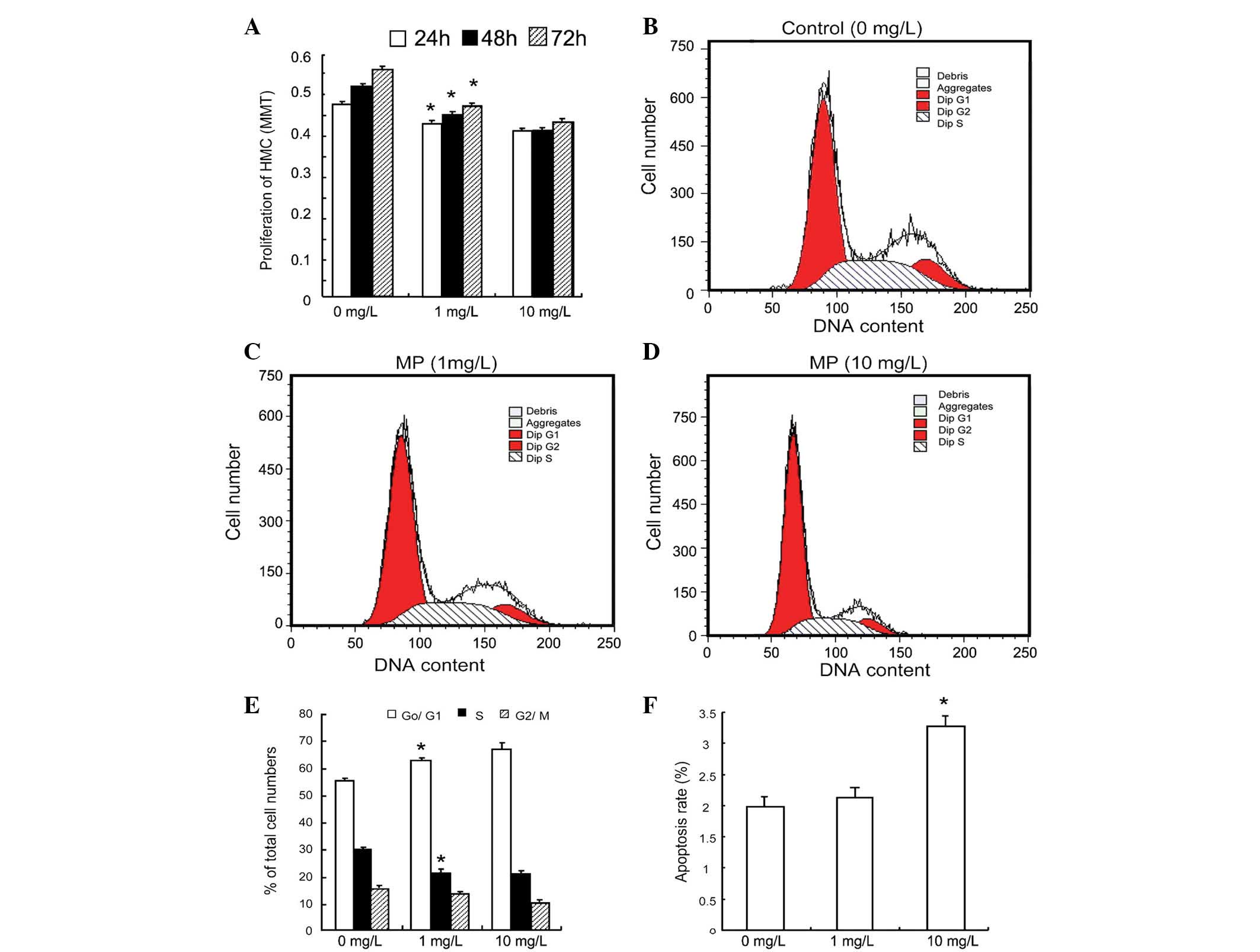
Methylprednisolone
The influence of MP on human mesangial cell growth
has not previously been studied, to the best of our knowledge. At
concentrations of 1 and 10 mg/l, MP inhibited the proliferation of
human mesangial cells in a dose- and time-dependent manner
(Fig. 3A). It was also determined
that at concentrations of 1 and 10 mg/l, MP significantly decreased
the percentage of cells in S phase, while increasing the percentage
of cells in G0/G1 phase (Fig. 3B–E). Similarly to CsA, MP (1–10
mg/l) significantly increased the apoptotic rate in human mesangial
cells following 48 h of treatment (Fig. 3F).
Mycophenolic acid
The present study investigated how MMF influenced
the proliferation of human mesangial cells and found that 0.25–10
μmol/l MMF significantly inhibited the proliferation of human
mesangial cells following 24, 48 or 72 h of treatment (Fig. 4A). MMF also significantly
suppressed the entry of cells into G2/M phase, causing
cell cycle arrest in the S phase (Fig.
4B–E). As shown in Fig. 4F,
there was a significant increase in the early apoptotic rate of
human mesangial cells that were treated for 48 h with MMF.
Immunosuppressants inhibit proliferation
and cell cycle progression of human mesangial cells
The effects of Tac, CsA, MP and MMF on human
mesangial cell cycle progression are summarized in Fig. 5A. Since MMF is often used with
adjunctive immunosuppressants, human mesangial cells were treated
with 2.5 mmol/l MMF in order to block cells in the S phase from
entering into G2/M phase and 1 mg/l MP to block cells
progressing from G0 phase to S phase. This combination
of drugs inhibited the proliferation of mesangial cells more
efficiently than each drug separately (Fig. 5B). This combination also interfered
with the progression of mesangial cells in the
G0/G1 and S phase (Fig. 5C).
Discussion
Mesangial cells serve a number of functions in the
renal glomerulus, including structural support of the capillary
tuft, modulation of glomerular hemodynamics and phagocytic removal
of macromolecules and immune complexes. These cells also have
complex interactions with infiltrating inflammatory cells,
responding and contributing to the amplification of inflammation,
fibrosis and the development of glomerulosclerosis (14). The proliferation of mesangial cells
is a common pathological feature of glomerular diseases, including
IgA nephropathy and lupus nephritis (11). For these reasons, numerous studies
have investigated the contribution of mesangial cells to the
development of glomerulosclerosis (15). However, these studies have
concentrated on cultured cells or animal models of glomerular
injury and there have been few studies of human mesangial cells.
Specific targeting of mesangial cell proliferation may more
effectively retard the progress of glomerular disease.
In mouse renal tubular epithelial cells, CsA caused
cell cycle arrest in the G0/G1 phase and
inhibited DNA synthesis (16).
These results are similar to those the present study obtained on
human mesangial cells. Compared to Tac, CsA caused a marked
increase in apoptosis in human mesangial cells. This response may
be linked to the activation of pre-apoptotic pathways or to the
release of cytochrome c into the cytosol (17–20).
The present study found that similarly to Tac and
CsA, MP caused mesangial cell cycle arrest in the
G0/G1 phase and prevented cells from entering
the S phase. This is in agreement with a study by Bladh et
al (21), who reported that
glucocorticoids can decrease the percentage of cells in
S/G2/M phase and impair the proliferation of human
embryonic kidney 293 cells by suppressing nuclear factor
κ-light-chain-enhancer of activated B-cell activity.
Glucocorticoids exert an antiproliferative effect in numerous cell
types (22–26); therefore, it was hypothesized that
the anti-proliferative effect of MP may be due to induction of
cyclin-dependent kinase inhibitors such as p21Cip1 or p57Kip2
(27,28). Alternatively, MP may suppress c-myc
or cyclins, which are capable of stimulating cell cycle progression
(19). In contrast to Tac, CsA and
MP, MMF significantly inhibited mesangial cell growth by preventing
cells from entering G2/M phase. This increased the
percentage of cells in the S phase and decreased the percentage of
cells in G2/M phase.
The present study suggested a theoretical basis for
sequential therapy with various immunosuppressive agents to treat
glomerular diseases featuring mesangial proliferation. Sequential
therapy with various immunosuppressive agents may limit the
complications associated with steroid treatment or dependency and
potentially provide an alternative treatment for steroid-resistant
disease. It was found that the combination of MP and MMF was more
effective at inhibiting mesangial cell proliferation.
In conclusion, Tac, CsA, MP and MMF suppressed human
mesangial cell proliferation by targeting different phases of the
cell cycle. A sequential therapy based on these differences may
potentially be used as a strategy to treat proliferative glomerular
diseases. Further studies to assess the in vivo responses of
human mesangial cells to sequential therapy in
mesangioproliferative disease models are required.
Acknowledgements
This study was supported by the Key Scientific and
Technological Research Project Grant, Department of Public Health,
Shanxi Province, China (grant no. 200919). The authors would like
to thank Dr Xuewang Li for providing the human mesangial cells.
References
|
1
|
Akool el-S, Doller A, Babelova A,
Tsalastra W, Moreth K, Schaefer L, Pfeilschifter J and Eberhardt W:
Molecular mechanisms of TGF beta receptor-triggered signaling
cascades rapidly induced by the calcineurin inhibitors cyclosporin
A and FK506. J Immunol. 181:2831–2845. 2008. View Article : Google Scholar
|
|
2
|
Trachtman H, Vento s, Gipson D, et al:
Novel therapies for resistant focal segmental glomerulosclerosis
(FONT) phase II clinical trial: study design. BMC Nephrol.
12:82011. View Article : Google Scholar : PubMed/NCBI
|
|
3
|
Ponticelli C and Passerini P: Other
immunosuppressive agents for focal segmental glomerulosclerosis.
Semin Nephrol. 23:242–248. 2003. View Article : Google Scholar : PubMed/NCBI
|
|
4
|
Sabuda-Widemann D, Grabensee B, Schwandt C
and Blume C: Mycophenolic acid inhibits the autocrine PDGF-B
synthesis and PDGF-BB-induced mRNA expression of Egr-1 in rat
mesangial cells. Nephrol Dial Transplant. 24:52–61. 2009.
View Article : Google Scholar
|
|
5
|
Radeke HH, Kuster S, Kaever V and Resch K:
Effects of cyclosporin and FK-506 on glomerular mesangial cells.
Evidence for direct inhibition of thromboxane synthase by low
cyclosporin concentrations. Eur J Clin Pharmacol. 44(Suppl 1):
S11–S16. 1993. View Article : Google Scholar : PubMed/NCBI
|
|
6
|
Miao L, Sun J, Yuan H, Jia Y and Xu Z:
Combined therapy of low-dose tacrolimus and prednisone in nephrotic
syndrome with slight mesangial proliferation. Nephrology (Carlton).
11:449–454. 2006. View Article : Google Scholar
|
|
7
|
Pastukhov O, Schwalm S, Römer I,
Zangemeister-Wittke U, Pfeilschifter J and Huwiler A: Ceramide
kinase contributes to proliferation but not to prostaglandin E2
formation in renal mesangial cells and fibroblasts. Cell Physiol
Biochem. 34:119–133. 2014. View Article : Google Scholar : PubMed/NCBI
|
|
8
|
Anil KMS, Irfan SM, Ranganna K, Malat G,
Sustento-Reodica N, Kumar AM and Meyers WC: Comparison of four
different immunosuppression protocols without long-term steroid
therapy in kidney recipients monitored by surveillance biopsy:
five-year outcomes. Transpl Immunol. 20:32–42. 2008. View Article : Google Scholar
|
|
9
|
Boletis J, Balitsari A, Filiopoulos V,
Stamataki E, Lionaki S, Zavos G and Kostakis A: Delayed renal graft
function: the influence of immunosuppression. Transplant Proc.
37:2054–2059. 2005. View Article : Google Scholar : PubMed/NCBI
|
|
10
|
Ren H, Guo N and Lu D: Successful
engraftment of HLA-identical sibling cord blood transplantation in
an adult with chronic myelogenous leukemia. Chinese Journal of
Hematology. 22:621–624. 2001.(In Chinese).
|
|
11
|
Kurogi Y: Mesangial cell proliferation
inhibitors for the treatment of proliferative glomerular disease.
Med Res Rev. 23:15–31. 2003. View Article : Google Scholar
|
|
12
|
Delarue F, Virone A, Hagege J, Lacave R,
Peraldi MN, Adida C, Rondeau E, Feunteun J and Sraer JD: Stable
cell line of T-SV40 immortalized human glomerular visceral
epithelial cells. Kidney Int. 40:906–912. 1991. View Article : Google Scholar : PubMed/NCBI
|
|
13
|
Ruan XZ, Varghese Z, Fernando R and
Moorhead JF: Cytokine regulation of low-density lipoprotein
receptor gene transcription in human mesangial cells. Nephrol Dial
Transplant. 13:1391–1397. 1998. View Article : Google Scholar : PubMed/NCBI
|
|
14
|
Pereira RL, Felizardo RJ, Cenedeze MA, et
al: Balance between the two kinin receptors in the progression of
experimental focal and segmental glomerulosclerosis in mice. Dis
Mod Mech. 7:701–710. 2014. View Article : Google Scholar
|
|
15
|
Liu CY, Zhou LL, Cheng Q, Jiang SN, Sheng
J, Sun JD and Zhao JY: Effect of bradykinin on renal mesangial cell
proliferation and extracellular matrix secretion. Genet Mol Res.
13:490–498. 2014. View Article : Google Scholar : PubMed/NCBI
|
|
16
|
Jennings P, Koppelstaetter C, Aydin S,
Abberger T, Wolf AM, Mayer G and Pfaller W: Cyclosporine A induces
senescence in renal tubular epithelial cells. Am J Physiol Renal
Physiol. 293:F831–F838. 2007. View Article : Google Scholar : PubMed/NCBI
|
|
17
|
Choi SJ, You HS and Chung SY:
Tacrolimus-induced apoptotic signal transduction pathway.
Transplant Proc. 40:2734–2736. 2008. View Article : Google Scholar : PubMed/NCBI
|
|
18
|
Migita K and Eguchi K: FK 506-mediated
T-cell apoptosis induction. Transplant Proc. 33:2292–2293. 2001.
View Article : Google Scholar : PubMed/NCBI
|
|
19
|
Park JW, Bae EH, Kim IJ, Ma SK, Choi C,
Lee J and Kim SW: Paricalcitol attenuates cyclosporine-induced
kidney injury in rats. Kidney Int. 77:1076–1085. 2010. View Article : Google Scholar : PubMed/NCBI
|
|
20
|
de Arriba G, de Hornedo JP, Rubio SR,
Fernández MC, Martinez SB, Camarero MM and Cid TP: Vitamin E
protects against the mitochondrial damage caused by cyclosporin A
in LLC-PK1 cells. Toxicol Appl Pharmacol. 239:241–250. 2009.
View Article : Google Scholar : PubMed/NCBI
|
|
21
|
Bladh LG, Lidén J, Pazirandeh A, Rafter I,
Dahlman-Wright K, Nilsson S and Okret S: Identification of target
genes involved in the antiproliferative effect of glucocorticoids
reveals a role for nuclear factor-(kappa)B repression. Mol
Endocrinol. 19:632–643. 2005. View Article : Google Scholar
|
|
22
|
Rogatsky I, Trowbridge JM and Garabedian
MJ: Glucocorticoid receptor-mediated cell cycle arrest is achieved
through distinct cell-specific transcriptional regulatory
mechanisms. Mol Cell Biol. 17:3181–3193. 1997.PubMed/NCBI
|
|
23
|
Smith E, Redman RA, Logg CR, Coetzee GA,
Kasahara N and Frenkel B: Glucocorticoids inhibit developmental
stage-specific osteoblast cell cycle. Dissociation of cyclin
A-cyclin-dependent kinase 2 from E2F4-p130 complexes. J Biol Chem.
275:19992–20001. 2000. View Article : Google Scholar : PubMed/NCBI
|
|
24
|
Rhee K, Reisman D, Bresnahan W and
Thompson EA: Glucocorticoid regulation of G1 cyclin-dependent
kinase genes in lymphoid cells. Cell Growth Differ. 6:691–698.
1995.PubMed/NCBI
|
|
25
|
Helmberg A, Auphan N, Caelles C and Karin
M: Glucocorticoid-induced apoptosis of human leukemic cells is
caused by the repressive function of the glucocorticoid receptor.
EMBO J. 14:452–460. 1995.PubMed/NCBI
|
|
26
|
Sánchez I, Goya L, Vallerga AK and
Firestone GL: Glucocorticoids reversibly arrest rat hepatoma cell
growth by inducing an early G1 block in cell cycle progression.
Cell Growth Differ. 4:215–225. 1993.PubMed/NCBI
|
|
27
|
Corroyer S, Nabeyrat E and Clement A:
Involvement of the cell cycle inhibitor CIP1/WAF1 in lung alveolar
epithelial cell growth arrest induced by glucocorticoids.
Endocrinology. 138:3677–3685. 1997.PubMed/NCBI
|
|
28
|
Cha HH, Cram EJ, Wang EC, Huang AJ, Kasler
HG and Firestone GL: Glucocorticoids stimulate p21 gene expression
by targeting multiple transcriptional elements within a steroid
responsive region of the p21waf1/cip1 promoter in rat hepatoma
cells. J Biol Chem. 273:1998–2007. 1998. View Article : Google Scholar : PubMed/NCBI
|