Introduction
Breast cancer is the most common malignancy amongst
females worldwide (1).
Experimental data strongly suggest that estrogens have an important
role in the development and progression of hormone-dependent breast
cancer (2). Approximately two
thirds of postmenopausal breast cancer cases are hormone-dependent,
which means that they are estrogen receptor (ER)-positive and
require estrogens for tumor growth (3). Aromatase is the rate-limiting enzyme
in the synthesis of estrogens from androgenic substrates (4). Aromatase inhibitors (AIs) were found
to markedly suppress plasma estrogen levels, as well as
intratumoral aromatase activity in postmenopausal females with
breast cancer by inhibiting or inactivating the aromatase enzyme
(4–7). As a result, less estrogen becomes
available to stimulate the growth of hormone-dependent breast
cancer cells. Large, adjuvant randomized trials have demonstrated
that AIs exhibited significant improvement in disease-free
survival, time to recurrence and time to distant recurrence
compared with tamoxifen (8–10).
For postmenopausal females with receptor-positive breast cancers,
AIs have emerged as an alternative to tamoxifen due to its superior
efficacy and reduced incidence of side effects, including
endometrial cancer and thromboembolism (11–13).
In addition to stimulating ER-positive breast cancer
cell growth, estrogens also have an important role in maintaining
normal bone mass (14). Bone
remodeling, which comprises bone formation by osteoblasts and bone
resorption by osteoclasts, is a dynamic metabolic process that
occurs throughout life (15).
Estrogens are known to regulate bone homeostasis by inhibiting
osteoclast activity, as well as enhancing osteoblast proliferation
and osteoblast-related collagen formation (16,17).
Bone mineral density (BMD) declines in females concurrently with
the onset of menopause and may be accompanied by an increased risk
of fracture due to the rapid decrease in serum estrogen levels
(18). AI treatment markedly
reduces these low circulating estrogen levels by a further 80–90%
in postmenopausal patients with breast cancer (19). Therefore, AIs lead to an increase
in bone loss (osteoporosis), known as aromatase
inhibitor-associated bone loss (AIBL), and a higher bone fracture
rate than tamoxifen (11–13). Therefore, accelerated bone loss due
to long-term estrogen deprivation has become a major concern
underlying the safety of AI treatment.
Clinical evidence has shown that bisphosphonates may
maintain BMD and decrease fracture risk for patients with breast
cancer receiving adjuvant AI therapy (20–23).
However, side effects, including renal dysfunction and
osteonecrosis of the jaw, have been reported following
bisphosphonate treatment (24–26).
Therefore, the combination of AI treatment with alternative
approaches with long-term efficacy and safety profiles, including
the use of Traditional Chinese Medicine, for the management of AIBL
requires further analysis.
Shu-Gan-Liang-Xue decoction (SGLXD), a clinical
prescription, has been used extensively for ameliorating hot flush
symptoms in patients with breast cancer receiving endocrine therapy
(27). SGLXD inhibited breast
tumor growth in tumor-bearing nude mice (28), and none of the component herbs
exhibited estrogenic activity (29). Modified Shu-Gan-Liang-Xue decoction
(mSGLXD) has been used to prevent AIBL and has achieved good
clinical efficacy. SGLXD was supplemented with Rhizoma Drynariae
(RD; Gu-Sui-Bu in Chinese), Caulis Piperis Kadsurae (Hai-Feng-Teng
in Chinese) and Caulis Trachelospermi (Luo-Shi-Teng in Chinese),
and Radix Cynanchum Strati (Baiwei in Chinese) and Fructus
Schisandrae (Wuweizi in Chinese) were removed, to produce
mSGLXD.
RD, the dried rhizome of Drynaria fortunei
(Kunze) J. Sm., has been known as a kidney-tonifying and
anti-osteoporosis herb for the treatment of osteoporosis and bone
fractures for thousands of years in China (30,31).
The natural product RD, which contains phenolic compounds, was
suggested to possess estrogenic activity (32) and the methanolic extract of RD was
able to increase the growth of MCF-7 cells at low concentrations
(33). Although SGLXD has
demonstrated anti-tumor efficacy, it was necessary to test the
effects of mSGLXD on breast cancer cell proliferation due to the
supplementation of RD. Therefore, the effects of mSGLXD and RD
alone on the proliferation of ER-positive breast cancer cell line
MCF-7 were investigated and their estrogenic activities were also
evaluated. To further elucidate the role of mSGLXD in alleviating
AIBL, the effects of mSGLXD alone or in combination with an AI
(anastrozole) on the proliferation and differentiation of
osteoblastic cell lines in vitro were investigated using
MC3T3-E1 cells, a mouse calvaria osteoblast-like cell line
(34).
Materials and methods
Cell culture
MCF-7 (HTB-22, a human breast cancer cell line) was
purchased from the American Type Culture Collection (Rockville, MD,
USA) and MC3T3-E1 (3111C0001CCC000012), an osteoblast-like cell
line from the C57BL/6 mouse calvaria, was obtained from the Cell
Resource Center (IBMS, CAMS/PUMC, Beijing, China). The MCF-7 cell
line was grown in Dulbecco’s modified Eagle’s medium (DMEM; Bioroc,
Tianjin, China) and the MC3T3-E1 cell line was cultured in
α-modified minimal essential medium (α-MEM) with 292 mg/ml
L-glutamine, 10 mg/l ribonucleosides and 10 mg/l
deoxyribonucleosides (Bioroc Pharmaceutical & Biotech Co., Ltd,
Tianjin, China). Unless specified, the medium contained 10%
heat-inactivated fetal bovine serum (FBS; Gibco-BRL, Invitrogen
Life Technologies, Carlsbad, CA, USA), 100 U/ml penicillin and 100
μg/ml streptomycin (Solarbio Science & Technology Co., Ltd.,
Beijing, China). Cells were incubated at 37°C in a humidified
atmosphere with 5% CO2. For all experiments, routine
cell culture procedures were strictly followed to maintain cell
density and all subcultures were used prior to passage 20.
Preparation of drugs
The components of SGLXD and mSGLXD are exhibited in
Table I. The Chinese herbs were
processed into formula granules by Beijing Tcmages Pharmaceutical
Co., Ltd (Beijing, China). The quality of formula granules was
monitored by Fourier transform infrared spectroscopy (FTIR) (Model
IRPRestige-21; Shiamdzu Corporation, Kyoto, Japan). Prior to use,
the formula granules were dissolved in deionized distilled water to
achieve a concentration of 1 g/ml crude drug. The solutions were
sterilized by filtration through a 0.22-μm pore-sized membrane (EMD
Millipore, Billerica, MA, USA) and stored at −80°C. The
concentrations of mSGLXD and RD in the present study refer to the
crude drug concentrations.
 | Table IComponents of Shu-Gan-Liang-Xue
decoction (SGLXD) and modified SGLXD (mSGLXD). |
Table I
Components of Shu-Gan-Liang-Xue
decoction (SGLXD) and modified SGLXD (mSGLXD).
| Chinese name | English name | Botanical name | mSGLXD (g) | SGLXD (g) |
|---|
| Baiwei | Radix Cynanchum
Strati | Cynanchum
atratum Bunge, in Asclepiadaceae | - | 15 |
| Mudanpi | Tree peony
bark | Paeonia
suffruticosa Andr. | 15 | 15 |
| Baishao | White peony
root | Paeonia
lactiflora Pall. | 15 | 15 |
| Chaihu | Chinese thorowax
root | Bupleurum
chinense DC. | 10 | 10 |
| Yujin | Wenchow turmeric
root tuber | Curcuma
aromatic Salisb. | 10 | 10 |
| Wuweizi | Fructus
Schisandrae | Schisandra
chinensis (Turcz.) Baill. | - | 15 |
| Gusuibu | Rhizoma
Drynariae | Drynaria
fortunei (Kunze ex Mett.) J. Sm. | 15 | - |
| Haifengteng | Caulis Piperis
Kadsurae | Piper
kadsura (Choisy) Ohwi. | 15 | - |
| Luoshiteng | Caulis
Trachelospermi | Trachelospermum
jasminoides (Lindl.) Lem. | 15 | - |
Estrogenic activity of mSGLXD and RD
Estrogenic activity was evaluated using a
Dual-Luciferase® reporter assay (Promega Corp., Beijing,
China) based bioluminescent measurement method. The
p(estrogen-responsive element)-TK-Luciferase and p(Renilla
luciferase)-TK plasmids were provided by Professor Wen-Ling
Han (Center for Human Disease Genomics, Peking University, Peking,
China). Following transfection for 24 h, MCF-7 cells were treated
with various concentrations of mSGLXD (0.625, 2.5 or 10 mg/ml),
17β-estradiol (E2; 10 nmol/ml; Sigma-Aldrich, St Louis,
MO, USA) or RD (10 mg/ml) for 48 h, and the control group was
treated with equal drug dissolved solute only, prior to being lysed
for the measurement of luciferase activity. The
Dual-Luciferase® reporter assay system contains Passive
Lysis Buffer (PLB), which can directly lyse cells. Briefly, growth
media was removed from the cultured cells, which were then washed
with 1X phosphate-buffered saline. Following washing 100 μl 1X PLB
was added to each well and the culture plates were gently agitated
for 15 min at room temperature. The lysates were then transferred
to tubes and centrifuged at 12,000 × g for 10 min at 4°C.
Luciferase activity was detected using chemiluminescence apparatus
(Model LMax II; Molecular Devices, Sunnyvale, CA, USA).
Cell proliferation assays
MCF-7 and MC3T3-E1 cells were suspended in DMEM and
α-MEM culture media and plated at a density of 5.0×10³
cells/well in 96-well culture dishes (Costar, Cambridge, MA, USA).
Following 24 h of culture, the medium was replaced with complete
culture medium supplemented with various concentrations of drugs.
To assess the effects of mSGLXD and RD alone on MCF-7 cell
proliferation, MCF-7 cells were treated with mSGLXD (1.25–50 mg/ml)
or RD (1.25–50 mg/ml). To assess the effects of mSGLXD and
anastrozole alone or in combination on MC3T3-E1 cell proliferation,
MC3T3-E1 cells were treated with mSGLXD (0.625–10 mg/ml),
anastrozole (0.01–100 μmol/l) or mSGLXD (0.625–10 mg/ml) as well as
10 or 100 μmol/l anastrozole. Following 48 h of drug treatment, the
cells were incubated with cell counting kit-8 solution (CCK-8;
Dojindo Molecular Technologies, Inc., Kumamoto, Japan) for 2 h.
Subsequently, the absorbance (optical density, OD) at 450 nm was
measured using a microplate reader (Model 680; Bio-Rad
Laboratories, Hercules, CA, USA) and cell viability was calculated
according to the following formula:
(ODsample−ODblank)/(ODcontrol−ODblank)×100%.
Reverse transcription polymerase chain
reaction (PCR) analysis
For analysis of alkaline phosphatase (ALP) and
osteocalcin (OCN) gene expression, MC3T3-E1 cells were treated with
mSGLXD (10 mg/ml) and anastrozole (10 μmol/l), alone or in
combination, for 48 h. Total RNA was extracted from cells using
TRIzol reagent (Invitrogen Life Technologies). The concentration
and quality of the extracted RNA were measured with a NanoDrop 2000
(Thermo Fisher Scientific, Wilmington, DE, USA). The first-strand
cDNA was generated using the TransScript first-strand cDNA
synthesis supermix (Transgen, Beijing, China) according to the
manufacturer’s instructions. Primers designed for PCR were
synthesized by Sangon Biotech Co., Ltd (Shanghai, China) and are
shown in Table II. The PCR assay
was performed using SYBR green qPCR supermix (Applied Biosystems
Life Technologies, Foster City, CA, USA) and performed in an ABI
prism 7500 sequence detection system (Applied Biosystems Life
Technologies). The PCR was carried out using the following
conditions: 35°C for 10 min, followed by 40 cycles of 95°C for 30
sec, and 72°C for 32 sec. The amount of mRNA for each gene was
calculated using the delta-delta CT (cycle threshold) method
(35), and gene expression levels
were normalized to GAPDH.
| Table IIPrimers for polymerase chain
reaction. |
Table II
Primers for polymerase chain
reaction.
| Target gene | Primers
(5′-3′) | Annealing
temperature | Amplification
length (bp) |
|---|
| Alkaline
phosphatase |
TCCTGACCAAAAACCTCAAAGG
TGCTTCATGCAGAGCCTGC | 60°C | 101 |
| Osteocalcin |
CTCACAGATGCCAAGCCCA
CCAAGGTAGCGCCGGAGTCT | 60°C | 98 |
| GAPDH |
GGTGAAGGTCGGTGTGAACG
CTCGCTCCTGGAAGATGGTG | 62°C | 233 |
Biochemical markers
MC3T3-E1 cells were cultured in a six-well culture
plate (Costar) at a density of 4×10⁴ cells/well for 24
h. Following treatment with mSGLXD (10 mg/ml) and anastrozole (10
μmol/l), alone or in combination, for 48 h, cell ALP activity was
determined using an ALP Assay kit (Jiancheng, Nanjing, China)
according to the manufacturer’s instructions. ALP activity was
normalized to total protein, as determined by bicinchoninic acid
protein assay (Thermo Fisher Scientific). The OCN content in
MC3T3-E1 cells was measured using a sandwich ELISA assay kit from
Beijing Ke Ying Mei Technology Co. Ltd (Beijing, China).
Mineralization assay
Bone mineralization was determined by alizarin red S
(AR-S) staining. Calcium was bound selectively to AR-S and stained
dark red. MC3T3-E1 cells were cultured in differentiation medium
[α-MEM supplemented with 10% FBS, 10 mmol/l β-glycerophosphate and
50 μg/ml ascorbic acid (Sigma-Aldrich)] with or without mSGLXD (10
mg/ml) and anastrozole (10 μmol/l), alone or in combination, for 21
days in six-well plates (4×10⁴ cells/well). The treated
cells were subsequently stained using an AR-S cell staining kit
(Genmed, Shanghai, China) according to the manufacturer’s
instructions. Images of the stained matrix were observed under an
inverted microscope (CKX41; Olympus Corporation, Tokyo, Japan) and
captured using a digital camera (Canon, Inc., Tokyo, Japan). To
quantify matrix mineralization, AR-S staining was released from the
cell matrix by incubation with 10% cetylpyridinium chloride in 10
mmol/l sodium phosphate (pH 7.0; Sigma-Aldrich) for 20 min. The
AR-S concentration was determined by measuring the absorbance at
562 nm (36), using a microplate
reader (Model 680; Bio-Rad Laboratories).
Statistical analysis
All experiments were repeated three to five times
and values are expressed as the mean ± standard deviation. All data
were analyzed using one-way analysis of variance followed by least
significant difference comparison using SPSS statistical software
16.0 (SPSS, Inc., Chicago, IL, USA). P<0.05 was considered to
indicate a statistically significant difference between values.
Results
mSGLXD does not possess estrogenic
activity, whereas RD alone does
Following treatment for 48 h, the luciferase
activities in MCF-7 cells treated with various concentrations of
mSGLXD were significantly lower than those induced by E2
(10 nmol/l; P<0.01). There was no significant difference for
luciferase activities between cells treated with mSGLXD and
negative control groups (P>0.05), which suggested that mSGLXD
did not possess estrogenic activity (Fig. 1). The luciferase activity induced
by 10 mg/ml RD was ~2.5-fold that of the negative control and
mSGLXD groups (P<0.01), indicating that RD exerted estrogenic
activity.
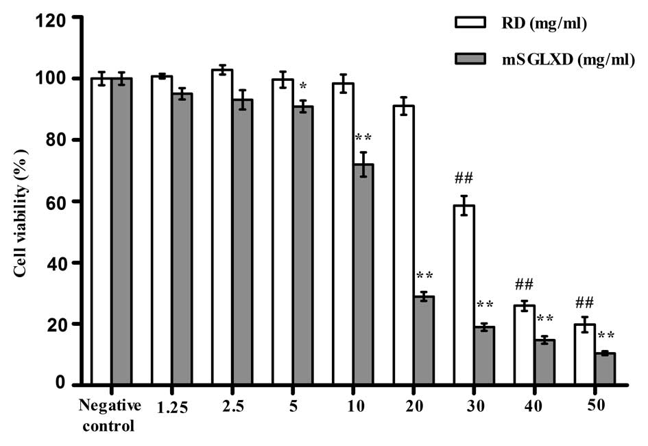
mSGLXD inhibits MCF-7 cell
proliferation
Following treatment for 48 h, mSGLXD significantly
inhibited MCF-7 cell proliferation in a dose-dependent manner
compared to that of the control group (P<0.01), while low
concentrations of RD (1.25, 2.5 mg/ml) slightly promoted MCF-7 cell
proliferation (P>0.05). However, RD dose-dependently inhibited
MCF-7 cell proliferation in the range of 5–50 mg/ml (Fig. 2).
mSGLXD enhances MC3T3-E1 cell
proliferation and attenuates anastrozole-induced inhibition of
proliferation
mSGLXD dose-dependently stimulated MC3T3-E1 cell
proliferation in the range of 0.625–10 mg/ml following treatment
for 48 h (P<0.01) (Fig. 3A).
Compared with the negative control, cell viability was increased by
22.49% in the 10 mg/ml mSGLXD treatment group.
Low concentrations of anastrozole (0.01–1 μmol/l)
did not influence MC3T3-E1 cell proliferation following treatment
for 48 h, while high concentrations of anastrozole (10 and 100
μmol/l) inhibited MC3T3-E1 cell proliferation by 12.31 and 28.38%,
respectively, compared with that of the negative control group
(P<0.01; Fig. 3B).
Furthermore, mSGLXD was able to prevent 10 and 100
μmol/l anastrozole-induced MC3T3-E1 cell death, and had a more
marked effect on proliferation in the 10 μmol/l anastrozole-treated
group. Cells treated with combined 10 μmol/l anastrozole and 10
mg/ml mSGLXD demonstrated a significant increase in cell viability
by 15.81%, as compared to cells treated with 10 μmol/l anastrozole
alone (P<0.05). Lower concentrations of mSGLXD (0.625, 2.5
mg/ml) demonstrated certain protective effects against
anastrozole-induced cell viability inhibition but without
significant difference (Fig. 3C and
D).
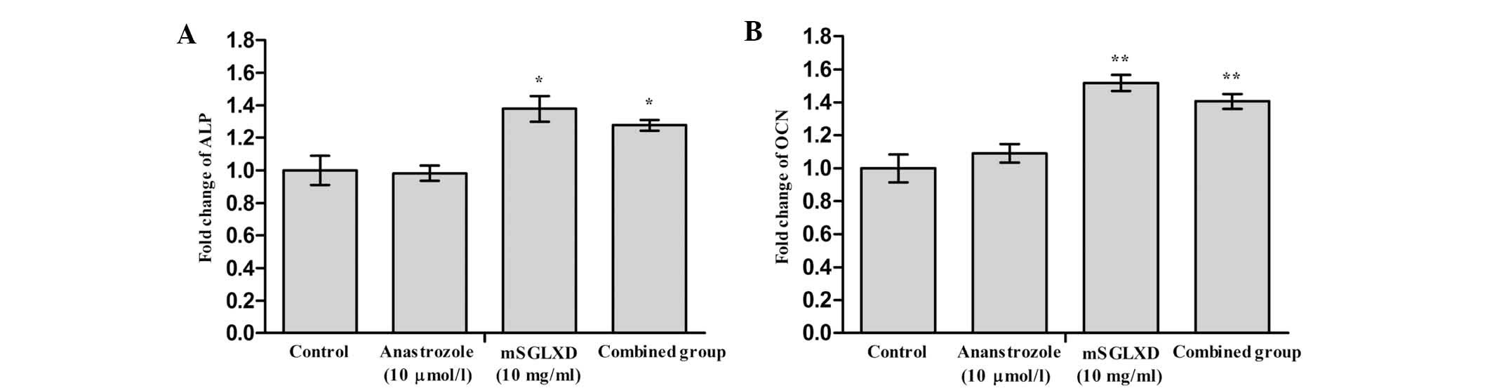
mSGLXD alone or in combination with
anastrozole enhances ALP and OCN mRNA expression
Based on the results of the aforementioned
experiments, 10 μmol/l anastrozole and 10 mg/ml mSGLXD were used
for the following experiments. The PCR analysis results indicated
that ALP and OCN mRNA expression levels were increased following
treatment with mSGLXD alone or combined with anastrozole, in
comparison with those of the control group (P<0.05 and
P<0.01, respectively; Fig. 4).
No significant change was observed in the anastrozole only
treatment group.
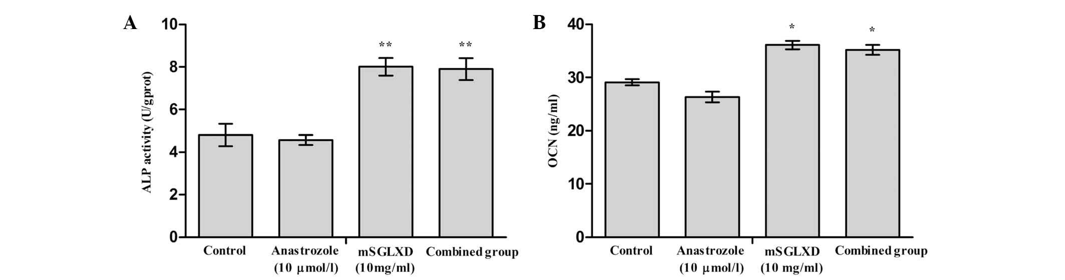
mSGLXD alone or in combination with
anastrozole enhances ALP activity and OCN protein expression
The effects of mSGLXD and anastrozole alone or in
combination on ALP activity and OCN content in MC3T3-E1 cells are
exhibited in Fig. 5A and B. In the
presence of 10 mg/ml mSGLXD alone or combined with 10 μmol/l
anastrozole, ALP activity and OCN content were significantly
increased following 48 h of culture (P<0.01 and P<0.05,
respectively), while 10 μmol/l anastrozole had no significant
effect.
mSGLXD alone or in combination with
anastrozole enhances bone mineralization of MC3T3-E1 cells
AR-S staining is a standard method used for the
visualization of nodular patterns and calcium deposition in
MC3T3-E1 cell cultures in vitro. As shown in Fig. 6A, following culture in differential
medium supplemented with β-glycerophosphate and ascorbic acid for
21 days, treatment with 10 mg/ml mSGLXD alone or in combination
with anastrozole markedly increased AR-S staining in MC3T3-E1
cells. The AR-S concentration in the mSGLXD and combined groups
demonstrated significant differences compared with that of the
control group (P<0.01, Fig.
6B). Concurrent with the results of the other experiments, 10
μmol/l anastrozole did not influence the mineralization of MC3T3-E1
cells.
Discussion
Phytoestrogens, natural estrogen-like substances
contained in plant food, demonstrated estrogenic activities through
binding to the ER and exhibiting ER-mediated estrogenic properties
(37). Epidemiological and
experimental data regarding the association between phytoestrogens
and breast cancer risk or progression are inconsistent (37–40).
Therefore, the safety of phytoestrogens for patients with breast
cancer has remained to be elucidated. Clinically, Rhizoma Drynariae
is added to mSGLXD to tonify the kidneys and strengthen the bones,
while Caulis Piperis Kadsurae and Caulis Trachelospermi are
supplemented as collateral-dredging and pain-relieving herbs.
According to the results of a previous study, RD may possess
estrogenic activity (32);
however, no study had demonstrated that Caulis Piperis Kadsurae and
Caulis Trachelospermi were phytoestrogens. Therefore, in the
present study, the estrogenic activities of mSGLXD and RD were
evaluated by dual-luciferase reporter assay-based bioluminescent
measurements. In accordance with previously reported results, the
results of the present study confirmed that RD had certain
estrogenic properties and that low concentrations of RD stimulated
MCF-7 cell proliferation (32,33).
Of note, despite the addition of phytoestrogen RD, mSGLXD was found
to not possess estrogenic activity. Furthermore, mSGLXD
significantly inhibited MCF-7 cell proliferation following
supplementation of the original drug SGLXD with RD and two
additional herbs, and the removal of Radix Cynanchum Strati and
Fructus Schisandrae. A possible explanation may be that since
mSGLXD is a Traditional Chinese Medicine composed of numerous
herbs, the estrogenic activity of RD may be modulated or
counteracted by the presence of other bioactive components, which
may have an antagonistic effect on the ER signaling pathway.
Therefore, the results indicated that mSGLXD was safe for patients
with breast cancer and also had certain anti-tumor effects on
breast cancer cells.
Formation of new bone is the task of osteoblasts;
therefore, enhancing osteoblast proliferation and differentiation
is a potential therapeutic strategy for bone loss. During the
formation phase of the bone cycle, ALP is expressed in markedly
high quantities and therefore becomes an indicator of bone
formation activity and a useful clinical therapeutic monitoring
index (41). OCN, another
classical biomarker of osteoblast cell function, was also
investigated (41,42). The results of the present study
indicated that mSGLXD not only stimulated MC3T3-E1 cell
proliferation, but also upregulated ALP and OCN gene and protein
expression levels. High concentrations of anastrozole markedly
inhibited MC3T3-E1 cell proliferation. However, this inhibitory
effect of anastrozole on MC3T3-E1 cell growth was alleviated by the
addition of mSGLXD. Furthermore, mSGLXD (10 mg/ml) increased the
mineralization of MC3T3-E1 cells induced by β-glycerophosphate and
ascorbic acid. These results indicated that mSGLXD had anabolic
effects on bone via the promotion of osteoblastic proliferation and
differentiation, suggesting that it may provide a useful pathway
for the prevention and treatment of AIBL.
Aromatase, an enzyme of the cytochrome P-450
superfamily and the product of the CYP19 gene, catalyzes the
aromatization of C19 steroids (androstendione, testosterone and
16α-hydroxyandrostendione) to E1 and E2
(43). Aromatase is also the
target of AIs, which are widely used in breast cancer endocrine
therapy at present. Previous research by our group indicated that
SGLXD, the original form of mSGLXD, simultaneously downregulated
aromatase and steroid sulfatase at transcription and protein levels
in ER-positive breast cancer cell lines MCF-7 and T47D (44), which may underlie the anti-tumor
mechanism of SGLXD. Therefore, SGLXD may have synergistic
inhibitory effects on aromatase with AIs to a certain extent.
However, to elucidate whether the mSGLXD used in the present study
exhibits a similar dual inhibitory effect on aromatase and steroid
sulfatase requires further investigation. Bone is dynamically
balanced by bone formation and bone resorption (45); therefore, the effects of mSGLXD on
osteoclasts requires further investigation and conclusions should
be confirmed by studies in vivo.
In conclusion, the present study demonstrated that
mSGLXD not only inhibited breast cancer cell proliferation but also
stimulated osteoblastic cell proliferation and differentiation.
Furthermore, the inhibitory effect of anastrozole on osteoblastic
cell growth was abrogated in the presence of mSGLXD. These results
suggested that mSGLXD was a promising adjuvant therapy with high
safety and efficacy in the prevention and treatment of AIBL in
patients with breast cancer receiving AI treatment.
Acknowledgements
The present study was funded by the Beijing Natural
Science Foundation (grant no. 7112026; Beijing, China).
References
|
1
|
Kwong A, Cheung PS, Wong Y, et al: The
acceptance and feasibility of breast cancer screening in the East.
Breast. 17:42–50. 2008. View Article : Google Scholar
|
|
2
|
Clemons M and Goss P: Estrogen and the
risk of breast cancer. N Engl J Med. 344:276–285. 2001. View Article : Google Scholar : PubMed/NCBI
|
|
3
|
Brueggemeier RW, Hackett JC and Diaz-Cruz
ES: Aromatase inhibitors in the treatment of breast cancer. Endocr
Rev. 26:331–345. 2005. View Article : Google Scholar : PubMed/NCBI
|
|
4
|
Smith IE and Dowsett M: Aromatase
inhibitors in breast cancer. N Engl J Med. 348:2431–2442. 2003.
View Article : Google Scholar : PubMed/NCBI
|
|
5
|
Geisler J, Haynes B, Anker G, Dowsett M
and Lønning PE: Influence of letrozole and anastrozole on total
body aromatization and plasma estrogen levels in postmenopausal
breast cancer patients evaluated in a randomized, cross-over study.
J Clin Oncol. 20:751–757. 2002. View Article : Google Scholar : PubMed/NCBI
|
|
6
|
Geisler J, King N, Anker G, Ornati G, Di
Salle E, Lønning PE and Dowsett M: In vivo inhibition of
aromatization by exemestane, a novel irreversible aromatase
inhibitor, in postmenopausal breast cancer patients. Clin Cancer
Res. 4:2089–2093. 1998.PubMed/NCBI
|
|
7
|
Miller WR and Dixon JM: Local endocrine
effects of aromatase inhibitors within the breast. J Steroid
Biochem Mol Biol. 79:93–102. 2001. View Article : Google Scholar
|
|
8
|
Coombes RC, Hall E, Gibson LJ, et al:
Intergroup Exemestane Study: A randomized trial of exemestane after
two to three years of tamoxifen therapy in postmenopausal women
with primary breast cancer. N Engl J Med. 350:1081–1092. 2004.
View Article : Google Scholar : PubMed/NCBI
|
|
9
|
Cuzick J, Sestak I, Baum M, Buzdar A,
Howell A, Dowsett M and Forbes JF: ATAC/LATTE investigators: Effect
of anastrozole and tamoxifen as adjuvant treatment for early-stage
breast cancer: 10-year analysis of the ATAC trial. Lancet Oncol.
11:1135–1141. 2010. View Article : Google Scholar : PubMed/NCBI
|
|
10
|
Thürlimann B, Keshaviah A, Coates AS, et
al: Breast International Group (BIG) 1–98 Collaborative Group: A
comparison of letrozole and tamoxifen in postmenopausal women with
early breast cancer. N Engl J Med. 353:2747–2757. 2005. View Article : Google Scholar
|
|
11
|
Coates AS, Keshaviah A, Thürlimann B, et
al: Five years of letrozole compared with tamoxifen as initial
adjuvant therapy for postmenopausal women with endocrine-responsive
early breast cancer: update of study BIG 1–98. J Clin Oncol.
25:486–492. 2007. View Article : Google Scholar : PubMed/NCBI
|
|
12
|
Nabholtz JM, Bonneterre J, Buzdar A,
Robertson JF and Thürlimann B: Anastrozole (Arimidex) versus
tamoxifen as first-line therapy for advanced breast cancer in
postmenopausal women: survival analysis and updated safety results.
Eur J Cancer. 39:1684–1689. 2003. View Article : Google Scholar : PubMed/NCBI
|
|
13
|
Perez EA: Safety profiles of tamoxifen and
the aromatase inhibitors in adjuvant therapy of hormone-responsive
early breast cancer. Ann Oncol. 18(Suppl 8): viii26–viii35. 2007.
View Article : Google Scholar : PubMed/NCBI
|
|
14
|
Ettinger B, Pressman A, Sklarin P, Bauer
DC, Cauley JA and Cummings SR: Associations between low levels of
serum estradiol, bone density, and fractures among elderly women:
the study of osteoporotic fractures. J Clin Endocrinol Metab.
83:2239–2243. 1998.PubMed/NCBI
|
|
15
|
Kenny AM and Raisz LG: Mechanisms of bone
remodeling: implications for clinical practice. J Reprod Med. 47(1
Suppl): 63–70. 2002.PubMed/NCBI
|
|
16
|
Ernst M, Schmid C and Froesch ER: Enhanced
osteoblast proliferation and collagen gene expression by estradiol.
Proc Natl Acad Sci USA. 85:2307–2310. 1988. View Article : Google Scholar : PubMed/NCBI
|
|
17
|
Kameda T, Mano H, Yuasa T, et al: Estrogen
inhibits bone resorption by directly inducing apoptosis of the
bone-resorbing osteoclasts. J Exp Med. 186:489–495. 1997.
View Article : Google Scholar : PubMed/NCBI
|
|
18
|
Pant S and Shapiro CL: Aromatase
inhibitor-associated bone loss: clinical considerations. Drugs.
68:2591–2600. 2008. View Article : Google Scholar : PubMed/NCBI
|
|
19
|
McCloskey E: Effects of third-generation
aromatase inhibitors on bone. Eur J Cancer. 42:1044–1051. 2006.
View Article : Google Scholar : PubMed/NCBI
|
|
20
|
Lester JE, Dodwell D, Purohit OP, et al:
Prevention of anastrozole-induced bone loss with monthly oral
ibandronate during adjuvant aromatase inhibitor therapy for breast
cancer. Clin Cancer Res. 14:6336–6342. 2008. View Article : Google Scholar : PubMed/NCBI
|
|
21
|
Brufsky AM, Bosserman LD, Caradonna RR, et
al: Zoledronic acid effectively prevents aromatase
inhibitor–associated bone loss in postmenopausal women with early
breast cancer receiving adjuvant letrozole: Z-FAST study 36-month
follow-up results. Clin Breast Cancer. 9:77–85. 2009. View Article : Google Scholar : PubMed/NCBI
|
|
22
|
Eidtmann H, de Boer R, Bundred N, et al:
Efficacy of zoledronic acid in postmenopausal women with early
breast cancer receiving adjuvant letrozole: 36-month results of the
ZO-FAST Study. Ann Oncol. 21:2188–2194. 2010. View Article : Google Scholar : PubMed/NCBI
|
|
23
|
Llombart A, Frassoldati A, Paija O, et al:
Immediate administration of zoledronic acid reduces aromatase
inhibitor-associated bone loss in postmenopausal women with early
breast cancer: 12-month analysis of the E-ZO-FAST trial. Clin
Breast Cancer. 12:40–48. 2012. View Article : Google Scholar
|
|
24
|
Ruggiero SL, Mehrotra B, Rosenberg TJ and
Engroff SL: Osteonecrosis of the jaws associated with the use of
bisphosphonates: a review of 63 cases. J Oral Maxillofac Surg.
62:527–534. 2004. View Article : Google Scholar : PubMed/NCBI
|
|
25
|
Bamias A, Kastritis E, Bamia C, et al:
Osteonecrosis of the jaw in cancer after treatment with
bisphosphonates: incidence and risk factors. J Clin Oncol.
23:8580–8587. 2005. View Article : Google Scholar : PubMed/NCBI
|
|
26
|
Lee SH, Chang SS, Lee M, Chan RC and Lee
CC: Risk of osteonecrosis in patients taking bisphosphonates for
prevention of osteoporosis: a systematic review and meta-analysis.
Osteoporos Int. 25:1131–1139. 2014. View Article : Google Scholar
|
|
27
|
Xue D, Sun H and Li PP: Long-term chinese
herbs decoction administration for management of hot flashes
associated with endocrine therapy in breast cancer patients. Chin J
Cancer Res. 23:74–78. 2011. View Article : Google Scholar : PubMed/NCBI
|
|
28
|
Wu CX and Li PP: Anti-tumor effect of
Shu-Gan-Liang-Xue decoction combined with tamoxifen on
estrogen-dependent breast cancer. Chin J Exp Tradit Med Formulae.
14:31–34. 2008.
|
|
29
|
Zhang Y and Li PP: Evaluation of
estrogenic potential of Shu-Gan-Liang-Xue Decoction by
dual-luciferase reporter based bioluminescent measurements in
vitro. J Ethnopharmacol. 126:345–349. 2009. View Article : Google Scholar : PubMed/NCBI
|
|
30
|
Liu X, Zhang S, Lu X, Zheng S, Li F and
Xiong Z: Metabonomic study on the anti-osteoporosis effect of
Rhizoma Drynariae and its action mechanism using ultra-performance
liquid chromatography-tandem mass spectrometry. J Ethnopharmacol.
139:311–317. 2012. View Article : Google Scholar
|
|
31
|
Wong RW, Rabie B, Bendeus M and Hägg U:
The effects of Rhizoma Curculiginis and Rhizoma Drynariae extracts
on bones. Chin Med. 2:132007. View Article : Google Scholar : PubMed/NCBI
|
|
32
|
Pang WY, Wang XL, Wong KC, Leung PC, Yao
XS and Wong MS: Total flavonoid fraction of Rhizoma Drynaria
improves bone properties in ovariectomized mice and exerts
estrogen-like activities in rat osteoblast-like (UMR-106) cells.
JFood Drug Anal. 20:265–269. 2012.
|
|
33
|
Chang EJ, Lee WJ, Cho SH and Choi SW:
Proliferative effects of flavan-3-ols and propelargonidins from
rhizomes of Drynaria fortunei on MCF-7 and osteoblastic cells. Arch
Pharm Res. 26:620–630. 2003. View Article : Google Scholar : PubMed/NCBI
|
|
34
|
Quarles LD, Yohay DA, Lever LW, Caton R
and Wenstrup RJ: Distinct proliferative and differentiated stages
of murine MC3T3-E1 cells in culture: An in vitro model of
osteoblast development. J Bone Miner Res. 7:683–692. 1992.
View Article : Google Scholar : PubMed/NCBI
|
|
35
|
Livak KJ and Schmittgen TD: Analysis of
relative gene expression data using real-time quantitative PCR and
the 2(-Delta Delta C(T)) method. Methods. 25:402–408. 2001.
View Article : Google Scholar
|
|
36
|
Kim EJ, Bu SY, Sung MK and Choi MK:
Effects of silicon on osteoblast activity and bone mineralization
of MC3T3-E1 cells. Biol Trace Elem Res. 152:105–112. 2013.
View Article : Google Scholar : PubMed/NCBI
|
|
37
|
Peeters PH, Keinan-Boker L, van der Schouw
YT and Grobbee DE: Phytoestrogens and breast cancer risk. Review of
the epidemiological evidence. Breast Cancer Res Treat. 77:171–183.
2003. View Article : Google Scholar : PubMed/NCBI
|
|
38
|
Adlercreutz H: Phyto-oestrogens and
cancer. Lancet Oncol. 3:364–373. 2002. View Article : Google Scholar : PubMed/NCBI
|
|
39
|
Keinan-Boker L, van Der Schouw YT, Grobbee
DE and Peeters PH: Dietary phytoestrogens and breast cancer risk.
Am J Clin Nutr. 79:282–288. 2004.PubMed/NCBI
|
|
40
|
Trock BJ, Hilakivi-Clarke L and Clarke R:
Meta-analysis of soy intake and breast cancer risk. J Natl Cancer
Inst. 98:459–471. 2006. View Article : Google Scholar : PubMed/NCBI
|
|
41
|
Christenson RH: Biochemical markers of
bone metabolism: an overview. Clin Biochem. 30:573–593. 1997.
View Article : Google Scholar
|
|
42
|
Hlaing TT and Compston JE: Biochemical
markers of bone turnover - uses and limitations. Ann Clin Biochem.
51(Pt 2): 189–202. 2014. View Article : Google Scholar : PubMed/NCBI
|
|
43
|
Speirs V, Walton DS, Hall MC and Atkin SL:
In vivo and in vitro expression of steroid-converting enzymes in
human breast tumours: associations with interleukin-6. Br J Cancer.
81:690–695. 1999. View Article : Google Scholar : PubMed/NCBI
|
|
44
|
Fu XS and Li PP: Shu-Gan-Liang-Xue
decoction simultaneously down-regulates expressions of aromatase
and steroid sulfatase in estrogen receptor positive breast cancer
cells. Chin J Cancer Res. 23:208–213. 2011. View Article : Google Scholar : PubMed/NCBI
|
|
45
|
Seeman E and Delmas PD: Bone quality - the
material and structural basis of bone strength and fragility. N
Engl J Med. 354:2250–2261. 2006. View Article : Google Scholar : PubMed/NCBI
|