Open Access

Isolation and characterization of ex vivo expanded mesenchymal stem cells obtained from a surgical patient

  • Authors:
    • Jia Huang
    • Huifan Sha
    • Guan Wang
    • Guoliang Bao
    • Shun Lu
    • Qingquan Luo
    • Qiang Tan
  • View Affiliations

  • Published online on: November 6, 2014     https://doi.org/10.3892/mmr.2014.2892
  • Pages: 1777-1783
  • Copyright: © Huang et al. This is an open access article distributed under the terms of Creative Commons Attribution License [CC BY_NC 3.0].

Metrics: Total Views: 0 (Spandidos Publications: | PMC Statistics: )
Total PDF Downloads: 0 (Spandidos Publications: | PMC Statistics: )


Abstract

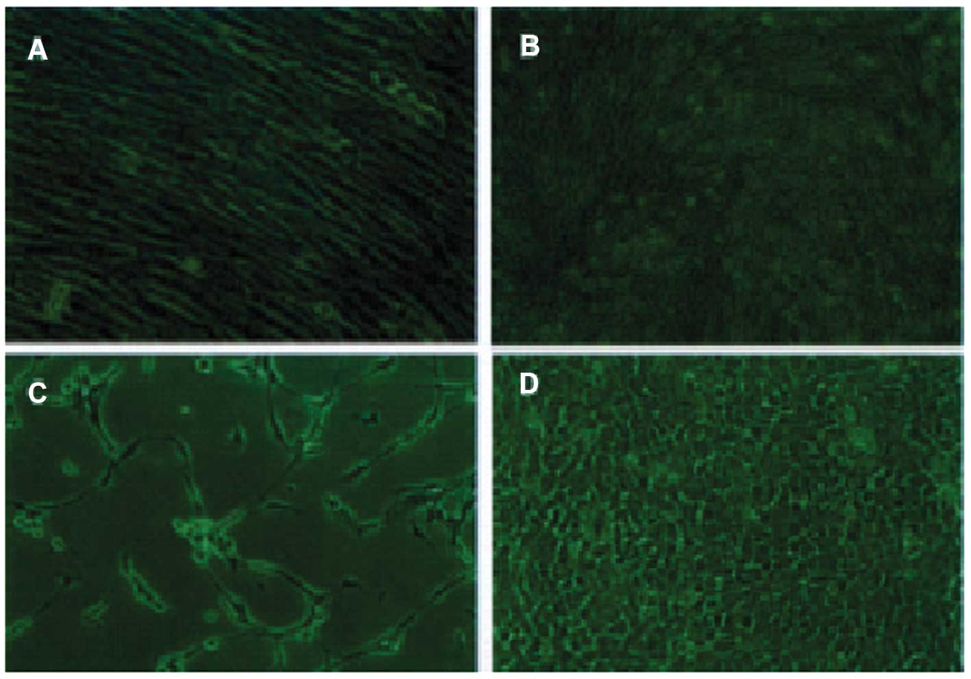
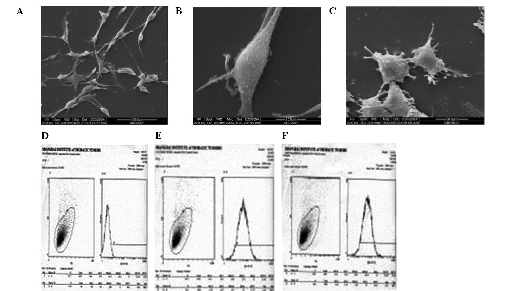
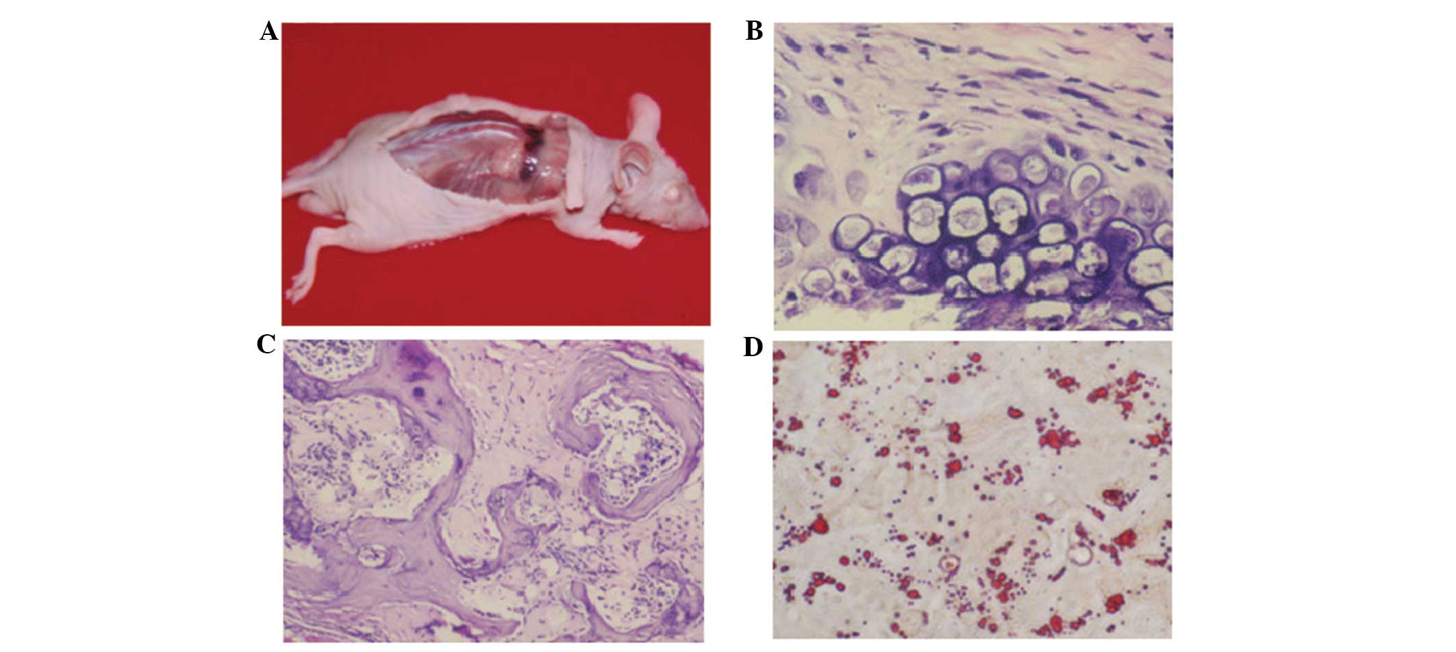
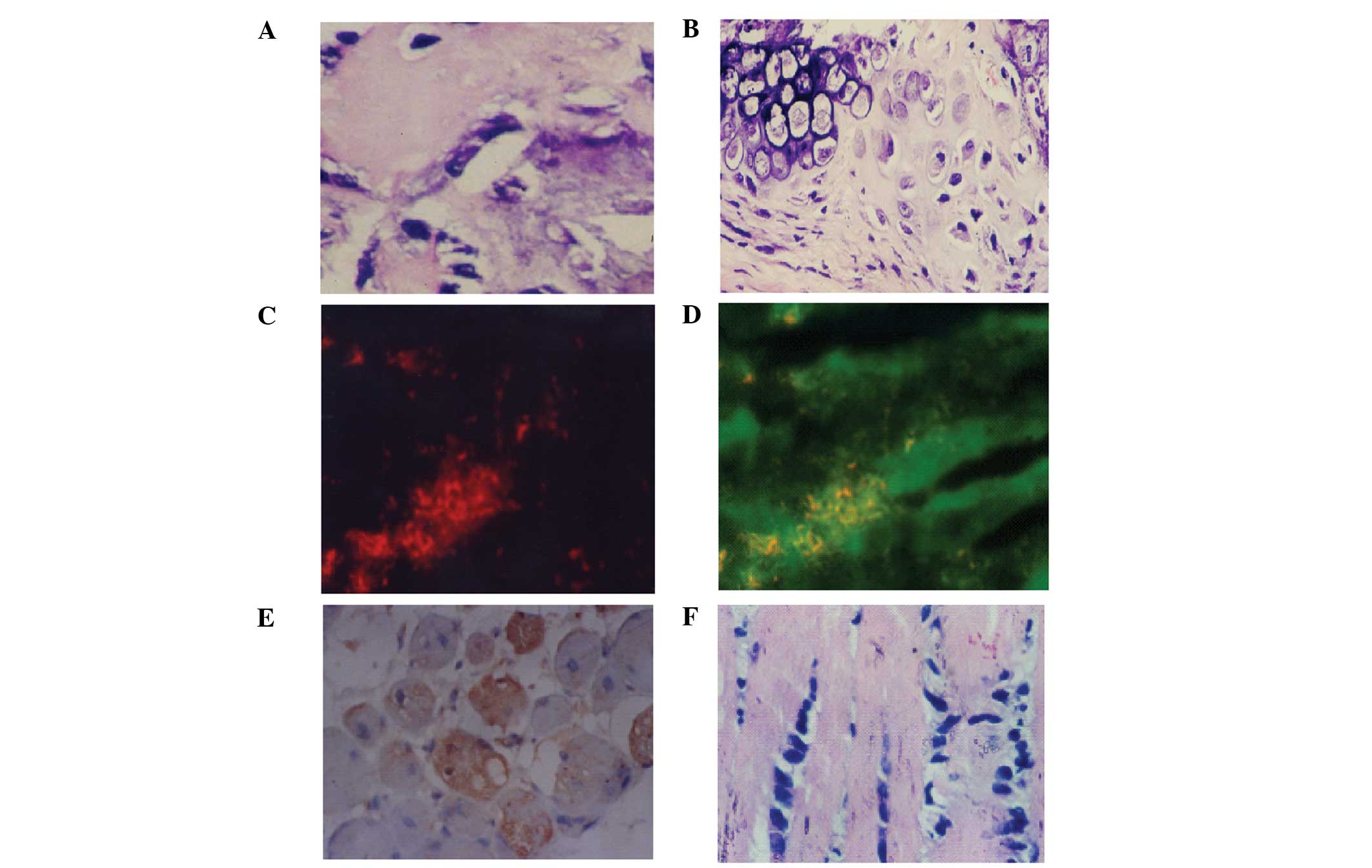
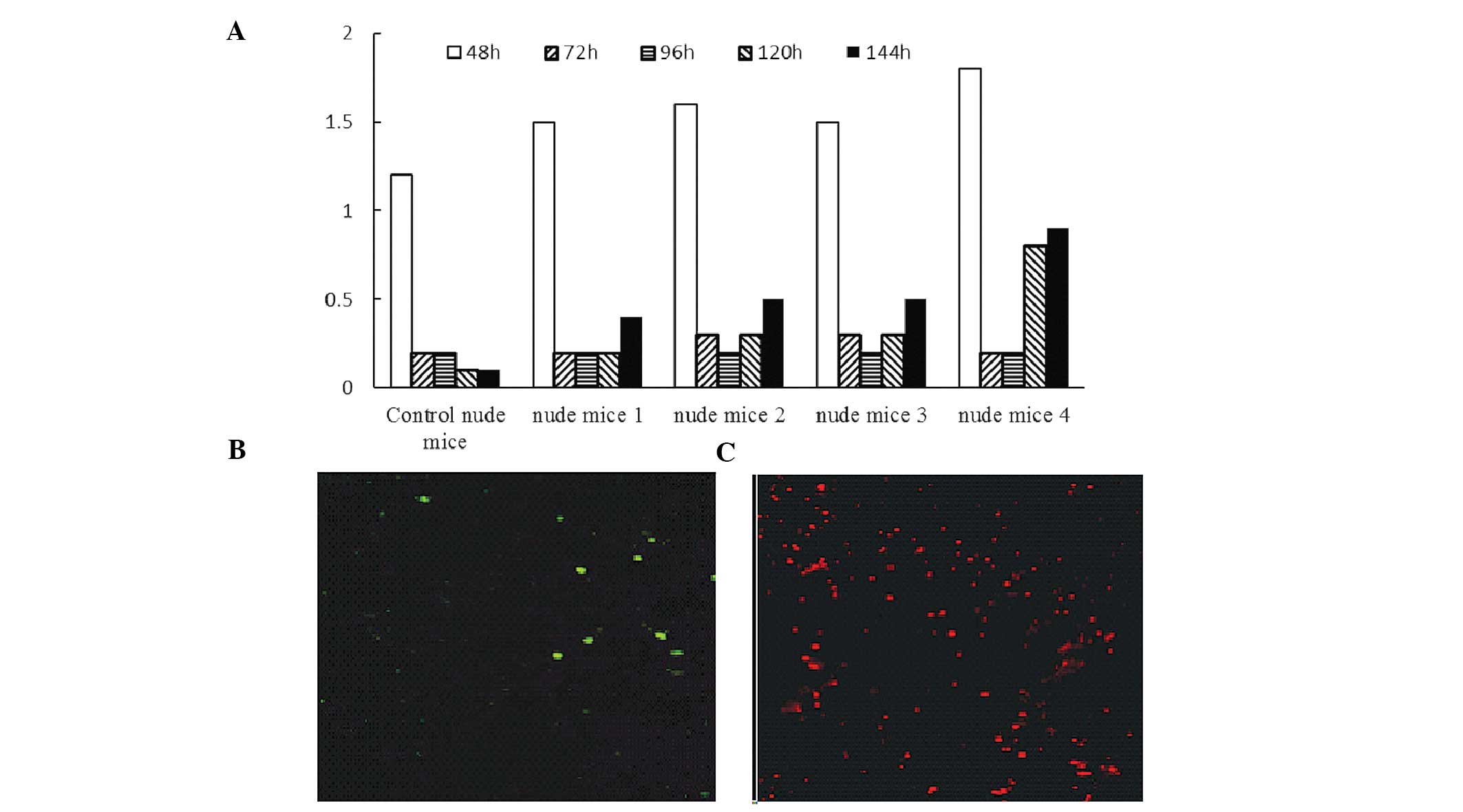
The aim of the present study was to investigate the morphological characteristics and pluripotent differentiation potential of human bone marrow mesenchymal stem cells (hBMMSCs) in vitro and in vivo. Bone marrow cells were isolated from a rib fragment of an adult surgical patient, hBMMSCs were isolated based on plastic adherence and expanded ex vivo and phenotyping was performed. Pluripotent differentiation assays for adipogenesis, myogenesis and osteogenesis were conducted. Hematopoietic reconstruction of sublethally irradiated nude mice was performed by infusion of hBMMSCs. The gene expression profiles of early and late hBMMSCs were examined. The rate of CD31‑positive cells was 31.1% in passage (P)4 hBMMSCs and 18.6% in P10 hBMMSCs. CD105 and CD106 were expressed in 99 and 95% of P25 hBMMSCs, respectively. Lipid droplets appeared at day 18 post induction. For osteogenesis, palpable masses were grossly observed from day 35 post inoculation of hBMMSCs. Hematoxylin and eosin staining further revealed chondrocytes and bone tissues. For myogenesis, at day six post subcutaneous inoculation, hBMMSCs differentiated into myocytes and were positive for myoglobin and MyoD1. In irradiated nude mice reconstituted by hBMMSCs, the white blood cell count briefly decreased following irradiation; however, it gradually recovered. In the irradiated nude mice reconstituted with hBMMSCs, CD45‑ and CD34‑positive cells were detected 72 h post induction. Gene microarray analysis of P7 and P57 hBMMSCs demonstrated that 20 genes were upregulated >2 fold and 40 genes were downregulated >2 fold in P57 hBMMSCs. In conclusion, the isolated HBMMSCs possessed pluripotent differentiation potential and it was feasible and safe to use hBMMSCs within 30 passages.
View Figures
View References

Related Articles

Journal Cover

March-2015
Volume 11 Issue 3

Print ISSN: 1791-2997
Online ISSN:1791-3004

Sign up for eToc alerts

Recommend to Library

Copy and paste a formatted citation
x
Spandidos Publications style
Huang J, Sha H, Wang G, Bao G, Lu S, Luo Q and Tan Q: Isolation and characterization of ex vivo expanded mesenchymal stem cells obtained from a surgical patient. Mol Med Rep 11: 1777-1783, 2015
APA
Huang, J., Sha, H., Wang, G., Bao, G., Lu, S., Luo, Q., & Tan, Q. (2015). Isolation and characterization of ex vivo expanded mesenchymal stem cells obtained from a surgical patient. Molecular Medicine Reports, 11, 1777-1783. https://doi.org/10.3892/mmr.2014.2892
MLA
Huang, J., Sha, H., Wang, G., Bao, G., Lu, S., Luo, Q., Tan, Q."Isolation and characterization of ex vivo expanded mesenchymal stem cells obtained from a surgical patient". Molecular Medicine Reports 11.3 (2015): 1777-1783.
Chicago
Huang, J., Sha, H., Wang, G., Bao, G., Lu, S., Luo, Q., Tan, Q."Isolation and characterization of ex vivo expanded mesenchymal stem cells obtained from a surgical patient". Molecular Medicine Reports 11, no. 3 (2015): 1777-1783. https://doi.org/10.3892/mmr.2014.2892