Introduction
Pulmonary endarterectomy (PEA) can prevent mortality
due to right ventricle failure (1), which occurs in chronic thromboembolic
pulmonary hypertension (CTEPH), a type-4 pulmonary hypertension
(2). A number of gene expression
studies conducted in postmortem lung tissue samples from patients
with CTEPH have indicated that aberrant processing of the messenger
RNA (mRNA) transcriptome in CTEPH may provide a mechanistic
convergence between the diverse genetic heritability of this
disease and the disruption of fundamental signaling pathways,
resulting in the common CTEPH phenotype. Studies conducted in
pulmonary endothelial cells have identified differentially
expressed genes, which may be involved in the pathogenesis of CTEPH
(3,4). However, gene expression regulation is
a complex process, which involves an interplay between DNA sequence
variation, chromatin and epigenetic modifications, protein
transcription factors and regulatory noncoding RNAs.
A previous study by our group examining the role of
the transcriptome in pulmonary artery endomembrane samples
demonstrated that the abnormal expression of mRNA transcripts may
represent a point of convergence in the otherwise heterogeneous
genomics, underlying the development of CTEPH (5). However, the regulatory RNAs inducing
the aberrant mRNA expression levels observed in CTEPH have not been
concurrently assessed. To the best of our knowledge, only a single
study conducted in the tissues of CTEPH patients has demonstrated
that miRNA-759 may influence the susceptibility to the development
of CTEPH (6). Long noncoding RNAs
(lncRNAs), a novel class of regulatory RNAs, have been shown to be
involved in certain fundamental events in gene regulation. However,
the role of these molecules in the pathogenesis of CTEPH remains
unclear (7).
lncRNAs are noncoding RNA molecules that are longer
than 200 nucleotides. lncRNAs were originally considered to be
‘transcriptional noise’; however, their involvement in important
mechanisms controlling the gene expression regulation has been
demonstrated. These mechanisms include targeting transcription
factors, initiating chromatin remodeling, directing methylation
complexes and blocking proximate transcription (8). Aberrant regulation of lncRNAs has
been shown to be associated with a number of diseases, including
certain forms of cancer (9,10).
Numerous lncRNAs have been identified through large-scale analyses
of full-length cDNA sequences in humans, mice and flies. lncRNA
molecules have been shown to play an important role in the control
of imprinting, cell differentiation, immune response, pathogenesis
of various human diseases, tumorigenesis and other biological
processes (11–15). However, the expression and
biological function of lncRNAs in CTEPH remains to be
elucidated.
The aim of the present study was to determine
whether the dysregulation of the lncRNA expression is involved in
the molecular pathogenesis of CTEPH. The lncRNAs expression
profiles of five CTEPH patients were compared with healthy control
individuals (normal tissues). In addition, an assessment of the
transcriptional differences in all known protein-coding mRNAs was
conducted.
Materials and methods
Patient samples
In total, five patients diagnosed with CTEPH (male,
2; female, 3; mean age, 38.2 years; age range, 17–52 years) who had
been referred to Beijing Chaoyang Hospital (Capital Medical
University, Beijing, China) were recruited to the study. The study
was approved by the relevant ethics committee of Beijing Chaoyang
University. All the patients provided informed written consent
prior to participation in the study. Pulmonary angiography and
right heart catheterization were used in the diagnosis of CTEPH and
determination of cardiopulmonary hemodynamics (16). Mean pulmonary artery pressure
>25 mmHg at rest or >30 mmHg during exercise was considered
to indicate the presence of pulmonary hypertension. Pulmonary
vascular resistance (PVR) and the six-minute walk test (6-MWT) were
hemodynamic variables applied to assess the cardiopulmonary
function and prognosis of the CTEPH patients. At inclusion, all the
patients received oral anticoagulants for a minimum of 6 months and
underwent PEA in accordance with the guidelines of the Beijing
Chao-Yang Hospital (Beijing, China). In addition, healthy control
samples were obtained from five lung transplant donors. The control
subjects and patients were matched according to age and gender.
Written informed consent was obtained from the healthy controls or
their families.
RNA extraction
To prepare the samples for microarray profiling,
total RNA was isolated from the CTEPH patient and normal tissue
samples using TRIzol™ reagent (Invitrogen Life Technologies, Inc.,
Burlington, ON, Canada) and purified using an RNeasy Mini kit
(Qiagen, Hilden, German), including a DNase digestion treatment.
RNA concentrations were determined by measuring the sample
absorbance at 260 nm with a NanoDrop 2000 spectrophotometer (Thermo
Fisher Scientific, Inc., Waltham, MA, USA). A260/A280 ratio values
of 1.8–2.1 were set as the quality control standard.
Microarray profiling
cDNA was generated via reverse transcription of RNA
obtained from pulmonary artery endothelium samples using a reverse
transcription kit (miScript II RT kit; Qiagen). cDNA obtained from
pulmonary artery endothelium samples of the CTEPH patients or
normal controls was hybridized to GeneChip® Human Gene
2.0 ST arrays (Affymetrix, Inc., Santa Clara, CA, USA), according
to the manufacturer’s instructions. Affymetrix Expression Console™
software (version 1.2.1; Affymetrix, Inc.) was used for microarray
analysis. The raw data (CEL files) were normalized at the
transcript level using the robust multiarray average method
(17), followed by median
summarization of the transcript expression levels. Subsequently,
gene-level data were filtered to include only the probe sets
derived from the ‘core’ metaprobe list, representing the reference
sequence (RefSeq) genes.
Significant differential gene
analysis
The random variance model (RVM) t-test was used to
filter differentially expressed genes in the control and CTEPH
groups. This test was selected since it can effectively raise the
degrees of freedom when investigating small samples. Following
significance analysis of microarrays and false discovery rate (FDR)
analysis, the differentially expressed genes were identified
according to the predetermined P-value threshold using
BRB-ArrayTools (version 4.3.0 Beta 1; National Cancer Institute,
Bethesda, MD, USA). P<0.05 was considered to indicate a
statistically significant difference (18–20).
The differentially expressed genes were subjected to unsupervised
hierarchical clustering (Cluster 3.0; Stanford University,
Stanford, CA, USA) and TreeView version 3.0 analysis (Stanford
University).
Coexpression network
Gene coexpression networks were constructed based on
the normalized signal intensity of the specific expression of
genes, in order to identify the interactions among genes (21). For each pair of genes, the Pearson
correlation coefficient was calculated in order to identify pairs
with a significant correlation, thus enabling construction of the
network (22).
When conducting a network analysis, the simplest and
most important measure of gene centrality within a network is
degree centrality. Degree centrality is defined as the number of
links a particular node has to other nodes in the network (23). Furthermore, k-cores were introduced
using graph theory in order to simplify the graph topology analysis
and investigate various network properties. A k-core of a network
consists of a subnetwork where all the nodes are connected to at
least k other genes. A k-core of a protein-protein interaction
network usually contains cohesive groups of proteins with similar
functions (23,24).
Network structure analysis aims to locate core
regulatory factors (genes). Within a network, core regulatory
factors connect the majority of nearby genes and have the highest
degree centralities. When evaluating different networks, core
regulatory factors are determined by the degree differences between
the CTEPH and normal tissue samples (25), since they show the highest degree
differences. The network was constructed using Cytoscape software
version 2.8.3 (Cytoscape Consortium, San Diego, CA, USA).
Gene ontology (GO) analysis
Based on the gene ontology database (http://www.geneontology.org/; accessed on January 15,
2014), the significance level of GO terms for the CTEPH-associated
differentially expressed genes was analyzed by two-side Fisher’s
exact test and χ2 test using the Database for
Annotation, Visualization and Integrated Discovery (DAVID) software
version 6.7 (http://david.abcc.ncifcrf.gov/home.jsp; accessed on
January 11, 2014; National Institute of Allergy and Infectious
Diseases, National Institutes of Health, Bethesda, MD, USA)
(26). Differentially expressed
genes were analyzed independently according to whether they were
upregulated or downregulated. P-values were calculated for the
differentially expressed genes in all GO categories. P<0.01 and
FDR<0.01 were considered to indicate statistically significant
results.
Pathway analysis
Based on the Kyoto Encyclopedia of Genes and Genomes
database (http://www.genome.jp/kegg/; accessed
on January 12, 2014; Kanehisa Laboratories, Kyoto, Japan), the
significance levels of CTEPH-associated differentially expressed
gene pathways were analyzed using Pathway-Express version 1.0
(Intelligent Systems and Bioinformatics Laboratory, Detroit, MI,
USA) (27,28). The occurrence of significant
differences from the expected values was assessed using a two-sided
binomial distribution. The number of differentially expressed genes
corresponding to each pathway category was counted and compared
with the number of genes expected for each pathway category. All
the signaling pathways were analyzed using γ P<0.05, provided by
the impact analysis, as the threshold indicating a statistically
significant difference.
Results
Overview of lncRNA profiles
Based on the lncRNAs expression profiles, a number
of differentially expressed lncRNAs were identified between the
CTEPH and healthy control samples. The expression profiles of
lncRNAs in the paired samples were determined by calculating the
log fold change of CTEPH/control samples. Due to the limited sample
size, FDR and P-values were calculated from normalized expression
levels. Hundreds of differentially expressed human lncRNAs were
identified using the RefSeq (www.ncbi.nlm.nih.gov/refseq), Ensembl (www.ensembl.org), lncRNAdb (www.lncrnadb.org), Broad Institute, Human Body Map
lincRNAs (www.broadinstitute.org/genome_bio/human_lincrnas/) and
transcripts of uncertain coding potential catalog (http://www.broadinstitute.org/genome_bio/human_lincrnas/?q=TUCP_transcripts_catalog)
databases in the CTEPH patients and healthy controls.
Using the microarray data, the expression levels of
lncRNAs in the five CTEPH tissue samples were compared with the
matched normal tissue samples. In total, 185 lncRNAs were
identified in which the expression levels were found to be
significantly different between the two groups. The characteristics
of the five CTEPH patients and five normal controls are shown in
Table I. The procedure used to
obtain a sequence may vary depending on the database used for
lncRNA classification. While tens of thousands lncRNAs were
investigated in normal and diseased tissues, only a few hundreds
lncRNAs were found to be significantly upregulated or
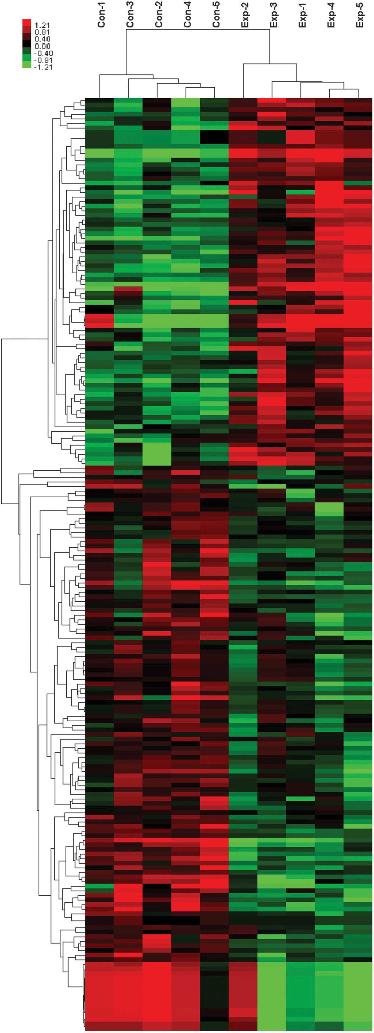
downregulated. Thus, the upregulation or downregulation of lncRNAs
was used to distinguish the CTEPH patient tissues from healthy
tissues (Fig. 1). Compared with
normal tissues, NR_033766 was the most evidently downregulated
lncRNA, whereas TCONS_l2_00010131-XLOC_l2_005462 was upregulated to
the greatest degree (Table II).
Therefore, downregulated lncRNAs were shown to be more prevalent
compared with upregulated lncRNAs in the CTEPH group.
 | Table IClinical characteristics of study
participants. |
Table I
Clinical characteristics of study
participants.
| Group | n | Age, years (mean ±
SD) | Gender, n
(M/F) | Median mPAP
(range), mmHg | PVR, dyn × sec ×
cm−5 | 6WMT, m |
|---|
| Healthy
control | 5 | 35.3±10.6 | 2/3 | - | - | - |
| CTEPH patients | 5 | 38.2±14.7 | 2/3 | 55 (33–78) | 1,075.4 | 454.6667 |
| Table IICollection of the top ten deregulated
lncRNAs detected using microarray analysis in ten CTEPH and control
samples. |
Table II
Collection of the top ten deregulated
lncRNAs detected using microarray analysis in ten CTEPH and control
samples.
| Downregulated in
CTEPH tissues | Upregulated in
CTEPH tissues |
|---|
|
|
|---|
| lncRNA | P-value | Fold changea | lncRNA | P-value | Fold changea |
|---|
| NR_003679 |
5.70×10⁻⁶ | 0.45 |
TCONS_l2_00010131-XLOC_l2_005462 |
3.00×10⁻⁵ | 5.89 |
| NR_033766 |
2.14×10⁻⁵ | 0.16 |
TCONS_l2_00011539-XLOC_l2_005705 |
3.00×10⁻⁵ | 5.89 |
|
TCONS_l2_00004769-XLOC_l2_002469 |
3.52×10⁻⁵ | 0.41 | NR_026544 |
1.16×10⁻⁴ | 2.61 |
|
TCONS_00023959-XLOC_011280 |
4.21×10⁻⁵ | 0.49 |
TCONS_00009277-XLOC_004803 |
1.18×10⁻⁴ | 1.82 |
|
TCONS_00023957-XLOC_011280 |
4.96×10⁻⁵ | 0.52 |
TCONS_l2_00016084-XLOC_l2_008434 |
1.24×10⁻⁴ | 2.02 |
| NR_026799 |
6.78×10⁻⁵ | 0.39 | NR_028406 |
2.36×10⁻⁴ | 2.17 |
|
TCONS_l2_00020176-XLOC_l2_010319 |
9.89×10⁻⁵ | 0.32 |
TCONS_00000337-XLOC_000468 |
4.50×10⁻⁴ | 2.42 |
| NR_026985 |
1.29×10⁻⁴ | 0.46 | NR_002433 |
5.00×10⁻⁴ | 2.46 |
|
TCONS_00000192-XLOC_000173 |
1.42×10⁻⁴ | 0.28 |
TCONS_00028854-XLOC_013966 |
5.18×10⁻⁴ | 1.89 |
| NR_026597 |
1.56×10⁻⁴ | 0.45 | NR_033652 |
5.66×10⁻⁴ | 1.91 |
Overview of mRNA profiles
In total, ≤30,654 coding transcripts were detected
in the ten samples that were examined. Using the RVM t-test method,
880 genes were found to be upregulated and 734 genes were found to
be downregulated in the CTEPH samples, compared with the healthy
controls. The results supported the hypothesis that CTEPH is a
metabolic disease as the mRNA expression level of oxidized
low-density lipoprotein receptor 1 showed the greatest
upregulation, while the mRNA expression level of chordin-like 1
showed the greatest downregulation. Therefore, upregulation of mRNA
expression levels was more prevalent compared with downregulation
in the CTEPH group.
Analysis of nearby lncRNAs and mRNAs
Previous studies have used chromatin-state maps to
identify 3,019 lncRNAs with a clear evolutionary conservation,
which are associated with distinct and diverse biological processes
(such as cell proliferation), RNA binding complexes, immune
surveillance, embryonic stem cell pluripotency, neuronal processes,
morphogenesis, gametogenesis and muscle development (29,30).
Among the 185 differentially expressed lncRNAs identified in the
present study, 74 lncRNAs were shown to have differentially
expressed mRNAs that were overlapping, antisense or nearby. Further
analysis resulted in the identification of nine pairs of
differentially expressed lncRNAs overlapping with mRNAs, nine pairs
of lncRNAs and antisense mRNAs, 340 lncRNAs located upstream of
mRNAs (distance, <300 kb) and 106 lncRNAs located downstream of
mRNAs (distance, <300 kb) in each comparison between CTEPH and
normal control tissues. Among the 464 lncRNA-mRNA pairs, the
expression regulation of 442 lncRNAs and nearby coding genes was in
the same direction (up or down), whereas the expression of 22 pairs
was regulated in opposite directions.
Construction of the coding-noncoding gene
coexpression network
The correlation analysis among differentially
expressed lncRNAs and mRNAs was used to construct a
coding-noncoding (CNC) gene coexpression network. LncRNAs and mRNAs
having Pearson correlation coefficients ≥0.97 were selected and the
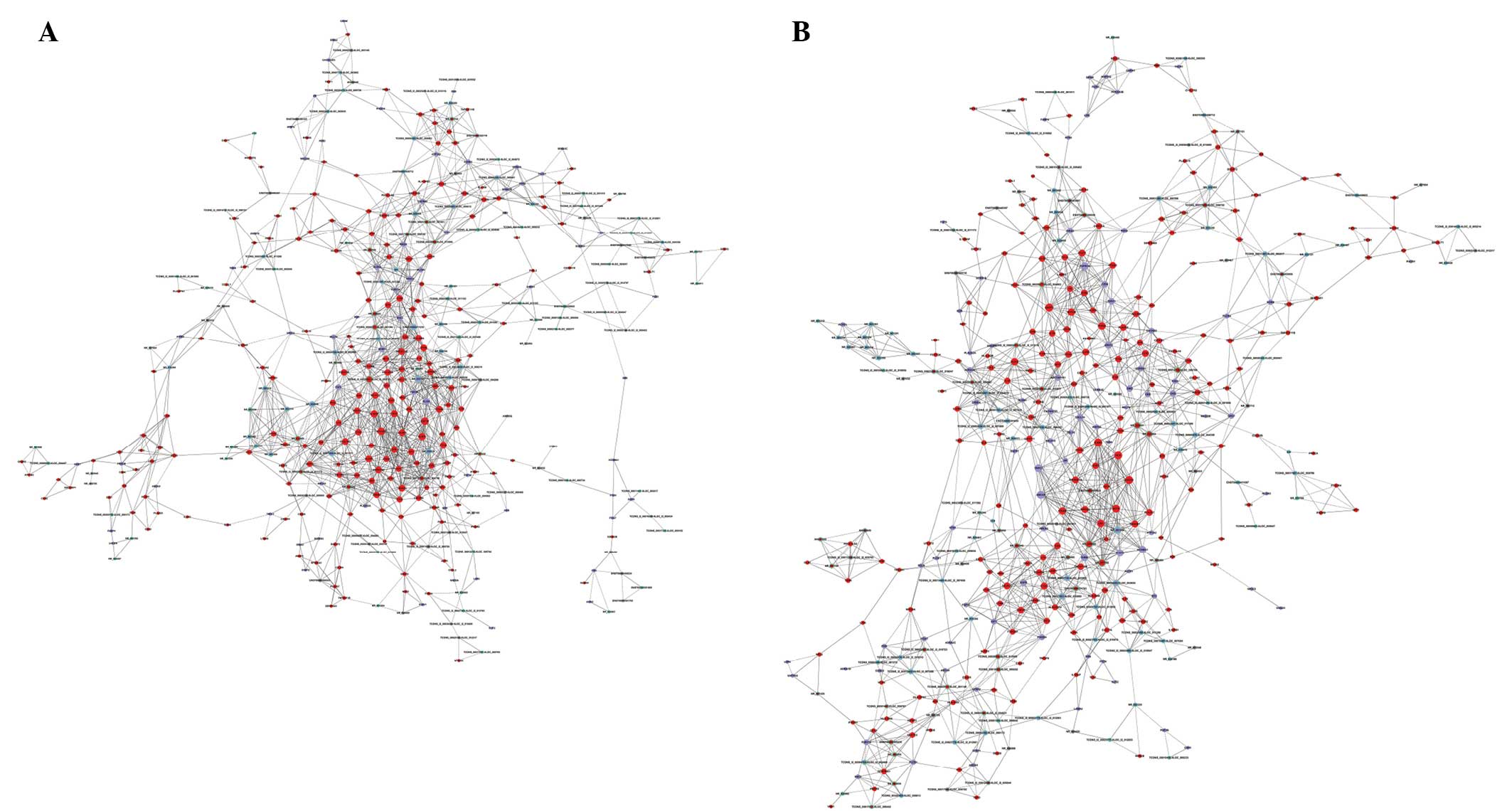
network was constructed using the Cytoscape software. Within this
coexpression network, the CTEPH-CNC network node consisted of 129
lncRNAs and 275 mRNAs, whereas the normal control-CNC network node
consisted of 134 lncRNAs and 294 mRNAs (Fig. 2). A total of 832 network nodes in
the two networks formed 3,239 coexpression pairs of lncRNAs and
mRNAs, with positively correlated expression in the majority of
pairs. Investigation of the CNC network indicated that one mRNA may
correlate with numerous lncRNAs and vice versa.
In order to identify the most significant RNA
molecules in CTEPH, the degree of certain RNAs (k-core) in each
network was normalized and the difference in connectivity (diffK),
representing the differences between the two networks, was
calculated. NR_036693 and NR_027783 were found to be the most
differentially expressed lncRNAs, whilst the expression of the
arginine vasopressin receptor 1A gene showed the greatest
significant difference in the genes examined. The CNC network
presented here, may implicate the inter-regulation of lncRNAs and
mRNAs in the development of CTEPH.
Coexpressed coding gene function
analysis
An important part of research into lncRNAs involves
inferring the possible functions of nearby protein-coding genes
(29,31). In the current study, the
differentially expressed mRNAs of the two CNC networks were
combined. Subsequently, the DAVID functional annotation chart
(32,33) and pathway analysis results were
used to perform functional enrichment analysis of the
differentially regulated protein-coding gene and lncRNA pairs. The
annotation terms showing the greatest significant differences (with
the lowest P-values) were found to be immune response, inflammatory
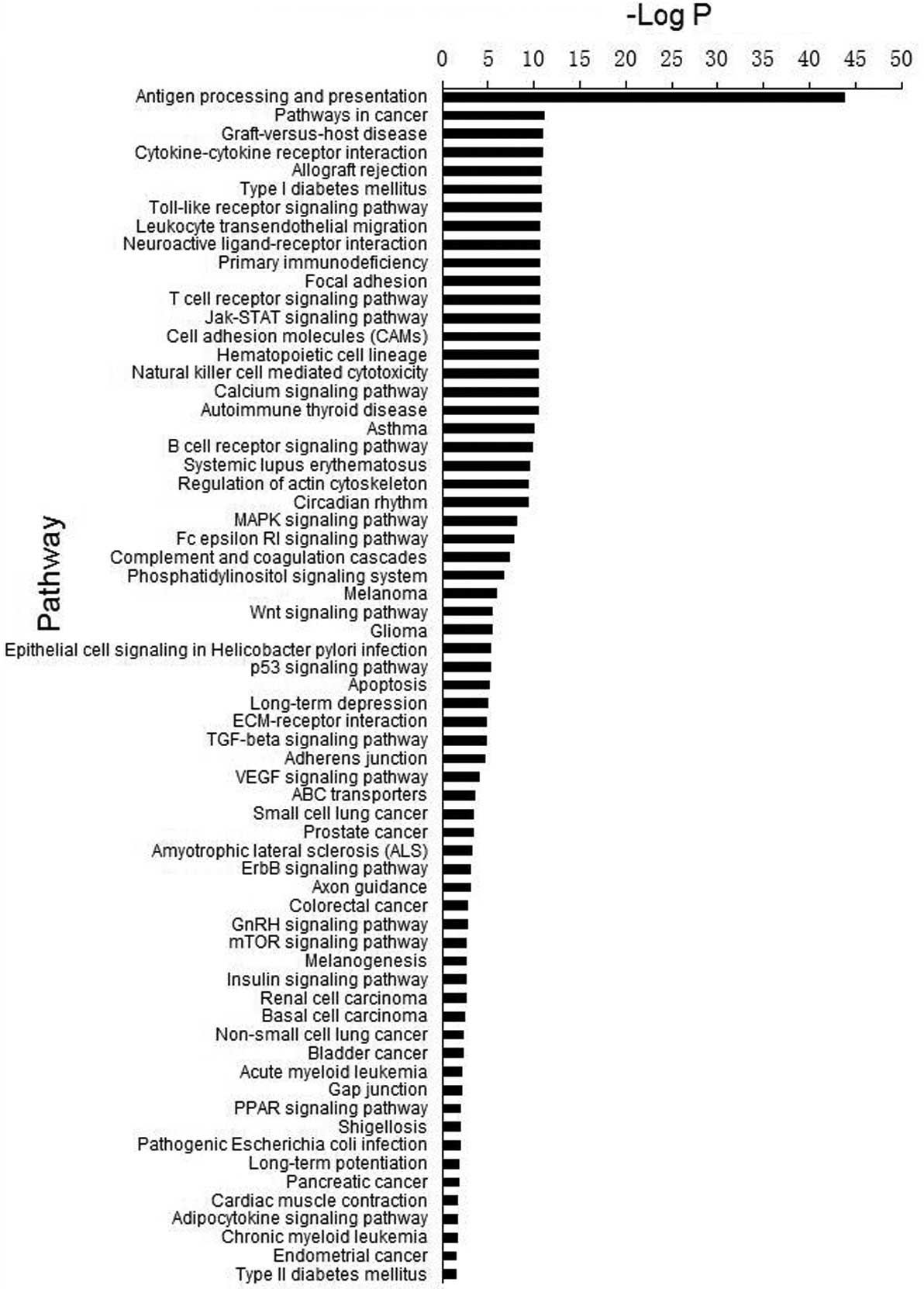
response, defense response and response to wounding (Table III). In addition, the most
important signaling pathways relevant to CTEPH were found to be
antigen processing and presentation, cytokine-cytokine receptor
interaction and leukocyte transendothelial migration (Fig. 3). Therefore, lncRNAs are
hypothesized to modulate the host response through their effect on
nearby protein-coding genes.
| Table IIIGO analysis. |
Table III
GO analysis.
| GO ID | Term | Regulation | P-value | FDR |
|---|
| GO:0006955 | Immune
response | Up |
4.21×10−59 |
7.32×10−56 |
| GO:0006952 | Defense
response | Up |
1.24×10−49 |
2.16×10−46 |
| GO:0006954 | Inflammatory
response | Up |
5.61×10−44 |
9.77×10−41 |
| GO:0009611 | Response to
wounding | Up |
2.88×10−43 |
5.01×10−40 |
| GO:0002684 | Positive regulation
of immune system process | Up |
7.37×10−39 |
1.28×10⁻³⁵ |
| GO:0001775 | Cell
activation | Up |
3.95×10⁻²⁹ |
6.88×10⁻²⁶ |
| GO:0002696 | Positive regulation
of leukocyte activation | Up |
1.01×10⁻²⁷ |
1.76×10⁻²⁴ |
| GO:0050867 | Positive regulation
of cell activation | Up |
4.41×10⁻²⁷ |
7.68×10⁻²⁴ |
| GO:0045321 | Leukocyte
activation | Up |
1.56×10⁻²⁶ |
2.72×10⁻²³ |
| GO:0050865 | Regulation of cell
activation | Up |
9.23×10⁻²⁶ |
1.61×10⁻²² |
| GO:0042110 | T cell
activation | Up |
2.35×10⁻²⁵ |
4.10×10⁻²² |
| GO:0002694 | Regulation of
leukocyte activation | Up |
2.58×10⁻²⁵ |
4.49×10⁻²² |
| GO:0046649 | Lymphocyte
activation | Up |
6.86×10⁻²⁴ |
1.19×10⁻²⁰ |
| GO:0051249 | Regulation of
lymphocyte activation | Up |
5.15×10⁻²² |
8.98×10⁻¹⁹ |
| GO:0051251 | Positive regulation
of lymphocyte activation | Up |
5.97×10⁻²² |
1.04×10⁻¹⁸ |
| GO:0048584 | Positive regulation
of response to stimulus | Up |
1.77×10⁻²¹ |
3.09×10⁻¹⁸ |
| GO:0042330 | Taxis | Up |
4.76×10⁻²¹ |
8.30×10⁻¹⁸ |
| GO:0006935 | Chemotaxis | Up |
4.76×10⁻²¹ |
8.30×10⁻¹⁸ |
| GO:0050870 | Positive regulation
of T cell activation | Up |
4.71×10⁻¹⁹ |
8.21×10⁻¹⁶ |
| GO:0050863 | Regulation of T
cell activation | Up |
1.18×10⁻¹⁸ |
2.06×10⁻¹⁵ |
| GO:0042981 | Regulation of
apoptosis | Up |
5.68×10⁻¹⁸ |
9.89×10⁻¹⁵ |
| GO:0043067 | Regulation of
programmed cell death | Up |
8.67×10⁻¹⁸ |
1.51×10⁻¹⁴ |
| GO:0010941 | Regulation of cell
death | Up |
1.01×10⁻¹⁷ |
1.77×10⁻¹⁴ |
| GO:0001817 | Regulation of
cytokine production | Up |
2.11×10⁻¹⁷ |
3.67×10⁻¹⁴ |
| GO:0050778 | Positive regulation
of immune response | Up |
1.73×10⁻¹⁶ |
3.89×10⁻¹³ |
| GO:0007626 | Locomotory
behavior | Up |
1.26×10⁻¹⁵ |
2.13×10⁻¹² |
| GO:0006928 | Cell motion | Up |
2.59×10⁻¹⁵ |
4.45×10⁻¹² |
| GO:0051240 | Positive regulation
of multicellular organismal process | Up |
4.47×10⁻¹⁵ |
7.74×10⁻¹² |
| GO:0030098 | Lymphocyte
differentiation | Up |
4.87×10⁻¹⁵ |
8.50×10⁻¹² |
| GO:0030217 | T cell
differentiation | Up |
5.86×10⁻¹⁵ |
1.02×10⁻¹¹ |
| GO:0007166 | Cell surface
receptor linked signal transduction | Up |
7.85×10⁻¹⁵ |
1.37×10⁻¹¹ |
| GO:0051094 | Positive regulation
of developmental process | Up |
1.49×10⁻¹⁴ |
2.59×10⁻¹¹ |
| GO:0010033 | Response to organic
substance | Up |
3.12×10⁻¹⁴ |
5.43×10⁻¹¹ |
| GO:0002521 | Leukocyte
differentiation | Up |
4.00×10⁻¹⁴ |
6.96×10⁻¹¹ |
| GO:0002252 | Immune effector
process | Up |
6.25×10⁻¹⁴ |
1.09×10⁻¹⁰ |
| GO:0045619 | Regulation of
lymphocyte differentiation | Up |
7.29×10⁻¹⁴ |
1.27×10⁻¹⁰ |
| GO:0045058 | T cell
selection | Up |
1.03×10⁻¹³ |
1.79×10⁻¹⁰ |
| GO:0042127 | Regulation of cell
proliferation | Up |
1.56×10⁻¹³ |
2.71×10⁻¹⁰ |
| GO:0007610 | Behavior | Up |
3.16×10⁻¹³ |
5.51×10⁻¹⁰ |
| GO:0009617 | Response to
bacterium | Up |
9.56×10⁻¹³ |
1.66×10⁻⁹ |
| GO:0001819 | Positive regulation
of cytokine production | Up |
1.36×10⁻¹² |
2.36×10⁻⁹ |
| GO:0034097 | Response to
cytokine stimulus | Up |
2.55×10⁻¹² |
4.44×10⁻⁹ |
| GO:0002253 | Activation of
immune response | Up |
2.73×10⁻¹² |
4.76×10⁻⁹ |
| GO:0050670 | Regulation of
lymphocyte proliferation | Up |
5.42×10⁻¹² |
9.44×10⁻⁹ |
| GO:0045061 | Thymic T cell
selection | Up |
5.98×10⁻¹² |
1.04×10⁻⁸ |
| GO:0050900 | Leukocyte
migration | Up |
6.39×10⁻¹² |
1.11×10⁻⁸ |
| GO:0070663 | Regulation of
leukocyte proliferation | Up |
6.50×10⁻¹² |
1.13×10⁻⁸ |
| GO:0032944 | Regulation of
mononuclear cell proliferation | Up |
6.50×10⁻¹² |
1.13×10⁻⁸ |
| GO:0030097 | Hemopoiesis | Up |
7.70×10⁻¹² |
1.34×10⁻⁸ |
| GO:0002443 | Leukocyte mediated
immunity | Up |
9.29×10⁻¹² |
1.62×10⁻⁸ |
| GO:0045087 | Innate immune
response | Up |
1.23×10⁻¹¹ |
2.15×10⁻⁸ |
| GO:0007243 | Protein kinase
cascade | Up |
1.71×10⁻¹¹ |
2.99×10⁻⁸ |
| GO:0042102 | Positive regulation
of T cell proliferation | Up |
2.24×10⁻¹¹ |
3.90×10⁻⁸ |
| GO:0002460 | Adaptive immune
response based on somatic recombination of immune receptors built
from immunoglobulin superfamily domains | Up |
2.59×10⁻¹¹ |
4.51×10⁻⁸ |
| GO:0002250 | Adaptive immune
response | Up |
2.59×10⁻¹¹ |
4.51×10⁻⁸ |
| GO:0045597 | Positive regulation
of cell differentiation | Up |
2.99×10⁻¹¹ |
5.20×10⁻⁸ |
| GO:0016477 | Cell migration | Up |
3.00×10⁻¹¹ |
5.22×10⁻⁸ |
| GO:0002757 | Immune
response-activating signal transduction | Up |
3.64×10⁻¹¹ |
6.34×10⁻⁸ |
| GO:0010647 | Positive regulation
of cell communication | Up |
3.66×10⁻¹¹ |
6.38×10⁻⁸ |
| GO:0008284 | Positive regulation
of cell proliferation | Up |
4.73×10⁻¹¹ |
8.23×10⁻⁸ |
| GO:0048534 | Hemopoietic or
lymphoid organ development | Up |
5.60×10⁻¹¹ |
9.76×10⁻⁸ |
| GO:0050671 | Positive regulation
of lymphocyte proliferation | Up |
7.41×10⁻¹¹ |
1.29×10⁻⁷ |
| GO:0070665 | Positive regulation
of leukocyte proliferation | Up |
9.30×10⁻¹¹ |
1.62×10⁻⁷ |
| GO:0032946 | Positive regulation
of mononuclear cell proliferation | Up |
9.30×10⁻¹¹ |
1.62×10⁻⁷ |
| GO:0002764 | Immune
response-regulating signal transduction | Up |
9.30×10⁻¹¹ |
1.62×10⁻⁷ |
| GO:0002449 | Lymphocyte mediated
immunity | Up |
1.02×10⁻¹⁰ |
1.78×10⁻⁷ |
| GO:0007242 | Intracellular
signaling cascade | Up |
1.11×10⁻¹⁰ |
1.94×10⁻⁷ |
| GO:0002237 | Response to
molecule of bacterial origin | Up |
1.23×10⁻¹⁰ |
2.14×10⁻⁷ |
| GO:0045089 | Positive regulation
of innate immune response | Up |
1.61×10⁻¹⁰ |
2.80×10⁻⁷ |
| GO:0045621 | Positive regulation
of lymphocyte differentiation | Up |
1.62×10⁻¹⁰ |
2.82×10⁻⁷ |
| GO:0031349 | Positive regulation
of defense response | Up |
1.78×10⁻¹⁰ |
3.10×10⁻⁷ |
| GO:0002520 | Immune system
development | Up |
1.86×10⁻¹⁰ |
3.25×10⁻⁷ |
| GO:0048870 | Cell motility | Up |
2.74×10⁻¹⁰ |
4.76×10⁻⁷ |
| GO:0051674 | Localization of
cell | Up |
2.74×10⁻¹⁰ |
4.76×10⁻⁷ |
| GO:0009615 | Response to
virus | Up |
3.09×10⁻¹⁰ |
5.39×10⁻⁷ |
| GO:0042129 | Regulation of T
cell proliferation | Up |
3.30×10⁻¹⁰ |
5.74×10⁻⁷ |
| GO:0032496 | Response to
lipopolysaccharide | Up |
3.58×10⁻¹⁰ |
6.24×10⁻⁷ |
| GO:0007159 | Leukocyte
adhesion | Up |
4.19×10⁻¹⁰ |
7.29×10⁻⁷ |
| GO:0045580 | Regulation of T
cell differentiation | Up |
5.34×10⁻¹⁰ |
9.31×10⁻⁷ |
| GO:0043065 | Positive regulation
of apoptosis | Up |
5.62×10⁻¹⁰ |
9.78×10⁻⁷ |
| GO:0043068 | Positive regulation
of programmed cell death | Up |
6.60×10⁻¹⁰ |
1.15×10⁻⁶ |
| GO:0009967 | Positive regulation
of signal transduction | Up |
6.94×10⁻¹⁰ |
1.21×10⁻⁶ |
| GO:0010942 | Positive regulation
of cell death | Up |
7.34×10⁻¹⁰ |
1.28×10⁻⁶ |
| GO:0051092 | Positive regulation
of NF-κB transcription factor activity | Up |
9.07×10⁻¹⁰ |
1.58×10⁻⁶ |
| GO:0019882 | Antigen processing
and presentation | Up |
9.44×10⁻¹⁰ |
1.64×10⁻⁶ |
| GO:0045088 | Regulation of
innate immune response | Up |
1.03×10⁻⁹ |
1.79×10⁻⁶ |
| GO:0019221 | Cytokine-mediated
signaling pathway | Up |
1.44×10⁻⁹ |
2.52×10⁻⁶ |
| GO:0019724 | B cell mediated
immunity | Up |
1.55×10⁻⁹ |
2.71×10⁻⁶ |
| GO:0002504 | Antigen processing
and presentation of peptide or polysaccharide antigen via MHC class
II | Up |
2.18×10⁻⁹ |
3.79×10⁻⁶ |
| GO:0002221 | Pattern recognition
receptor signaling pathway | Up |
5.25×10⁻⁹ |
9.14×10⁻⁶ |
| GO:0032844 | Regulation of
homeostatic process | Up |
5.62×10⁻⁹ |
9.78×10⁻⁶ |
| GO:0008219 | Cell death | Up |
8.22×10⁻⁹ |
1.43×10⁻⁵ |
| GO:0032101 | Regulation of
response to external stimulus | Up |
8.42×10⁻⁹ |
1.47×10⁻⁵ |
| GO:0033077 | T cell
differentiation in the thymus | Up |
8.44×10⁻⁹ |
1.47×10⁻⁵ |
| GO:0006874 | Cellular calcium
ion homeostasis | Up |
9.61×10⁻⁹ |
1.67×10⁻⁵ |
| GO:0016265 | Death | Up |
9.82×10⁻⁹ |
1.71×10⁻⁵ |
| GO:0002683 | Negative regulation
of immune system process | Up |
1.09×10⁻⁸ |
1.90×10⁻⁵ |
| GO:0060326 | Cell
chemotaxis | Up |
1.10×10⁻⁸ |
1.91×10⁻⁵ |
| GO:0002758 | Innate immune
response-activating signal transduction | Up |
1.32×10⁻⁸ |
2.30×10⁻⁵ |
| GO:0002218 | Activation of
innate immune response | Up |
1.32×10⁻⁸ |
2.30×10⁻⁵ |
| GO:0051090 | Regulation of
transcription factor activity | Up |
1.43×10⁻⁸ |
2.49×10⁻⁵ |
| GO:0055074 | Calcium ion
homeostasis | Up |
1.44×10⁻⁸ |
2.51×10⁻⁵ |
| GO:0008283 | Cell
proliferation | Up |
1.52×10⁻⁸ |
2.65×10⁻⁵ |
| GO:0016064 | Immunoglobulin
mediated immune response | Up |
1.60×10⁻⁸ |
2.79×10⁻⁵ |
| GO:0006915 | Apoptosis | Up |
1.80×10⁻⁸ |
3.14×10⁻⁵ |
| GO:0048545 | Response to steroid
hormone stimulus | Up |
1.98×10⁻⁸ |
3.44×10⁻⁵ |
| GO:0012501 | Programmed cell
death | Up |
2.53×10⁻⁸ |
4.41×10⁻⁵ |
| GO:0006875 | Cellular metal ion
homeostasis | Up |
2.69×10⁻⁸ |
4.68×10⁻⁵ |
| GO:0042035 | Regulation of
cytokine biosynthetic process | Up |
3.36×10⁻⁸ |
5.85×10⁻⁵ |
| GO:0045582 | Positive regulation
of T cell differentiation | Up |
3.73×10⁻⁸ |
6.49×10⁻⁵ |
| GO:0051091 | Positive regulation
of transcription factor activity | Up |
4.64×10⁻⁸ |
8.07×10⁻⁵ |
| GO:0001816 | Cytokine
production | Up |
5.16×10⁻⁸ |
8.98×10⁻⁵ |
| GO:0055065 | Metal ion
homeostasis | Up |
5.22×10⁻⁸ |
9.09×10⁻⁵ |
| GO:0042325 | Regulation of
phosphorylation | Up |
5.84×10⁻⁸ |
1.02×10⁻⁴ |
| GO:0045060 | Negative thymic T
cell selection | Up |
6.40×10⁻⁸ |
1.11×10⁻⁴ |
| GO:0040017 | Positive regulation
of locomotion | Up |
7.34×10⁻⁸ |
1.28×10⁻⁴ |
| GO:0007249 | I-κB kinase/NF-κB
cascade | Up |
7.52×10⁻⁸ |
1.31×10⁻⁴ |
| GO:0042108 | Positive regulation
of cytokine biosynthetic process | Up |
7.63×10⁻⁸ |
1.33×10⁻⁴ |
| GO:0006917 | Induction of
apoptosis | Up |
8.00×10⁻⁸ |
1.39×10⁻⁴ |
| GO:0012502 | Induction of
programmed cell death | Up |
8.50×10⁻⁸ |
1.48×10⁻⁴ |
| GO:0051101 | Regulation of DNA
binding | Up |
1.01×10⁻⁷ |
1.75×10⁻⁴ |
| GO:0002697 | Regulation of
immune effector process | Up |
1.03×10⁻⁷ |
1.80×10⁻⁴ |
| GO:0030595 | Leukocyte
chemotaxis | Up |
1.27×10⁻⁷ |
2.22×10⁻⁴ |
| GO:0044093 | Positive regulation
of molecular function | Up |
1.28×10⁻⁷ |
2.22×10⁻⁴ |
| GO:0051174 | Regulation of
phosphorus metabolic process | Up |
1.29×10⁻⁷ |
2.24×10⁻⁴ |
| GO:0019220 | Regulation of
phosphate metabolic process | Up |
1.29×10⁻⁷ |
2.24×10⁻⁴ |
| GO:0050864 | Regulation of B
cell activation | Up |
1.33×10⁻⁷ |
2.31×10⁻⁴ |
| GO:0043383 | Negative T cell
selection | Up |
1.42×10⁻⁷ |
2.47×10⁻⁴ |
| GO:0050730 | Regulation of
peptidyl-tyrosine phosphorylation | Up |
1.59×10⁻⁷ |
2.77×10⁻⁴ |
| GO:0007155 | Cell adhesion | Up |
1.65×10⁻⁷ |
2.87×10⁻⁴ |
| GO:0022610 | Biological
adhesion | Up |
1.70×10⁻⁷ |
2.97×10⁻⁴ |
| GO:0043388 | Positive regulation
of DNA binding | Up |
2.11×10⁻⁷ |
3.68×10⁻⁴ |
| GO:0030005 | Cellular di-,
tri-valent inorganic cation homeostasis | Up |
2.29×10⁻⁷ |
3.99×10⁻⁴ |
| GO:0002822 | Regulation of
adaptive immune response based on somatic recombination of immune
receptors built from immunoglobulin superfamily domains | Up |
2.62×10⁻⁷ |
4.55×10⁻⁴ |
| GO:0007204 | Elevation of
cytosolic calcium ion concentration | Up |
2.67×10⁻⁷ |
4.65×10⁻⁴ |
| GO:0002819 | Regulation of
adaptive immune response | Up |
3.07×10⁻⁷ |
5.35×10⁻⁴ |
| GO:0006916 | Anti-apoptosis | Up |
3.21×10⁻⁷ |
5.58×10⁻⁴ |
| GO:0055066 | Di-, tri-valent
inorganic cation homeostasis | Up |
4.78×10⁻⁷ |
8.33×10⁻⁴ |
| GO:0051480 | Cytosolic calcium
ion homeostasis | Up |
5.76×10⁻⁷ |
1.00×10⁻³ |
| GO:0006468 | Protein amino acid
phosphorylation | Up |
5.91×10⁻⁷ |
1.03×10⁻³ |
| GO:0051099 | Positive regulation
of binding | Up |
5.97×10⁻⁷ |
1.04×10⁻³ |
| GO:0042592 | Homeostatic
process | Up |
7.89×10⁻⁷ |
1.37×10⁻³ |
| GO:0050871 | Positive regulation
of B cell activation | Up |
9.20×10⁻⁷ |
1.60×10⁻³ |
| GO:0051050 | Positive regulation
of transport | Up |
9.32×10⁻⁷ |
1.62×10⁻³ |
| GO:0001932 | Regulation of
protein amino acid phosphorylation | Up |
1.09×10⁻⁶ |
1.90×10⁻³ |
| GO:0030003 | Cellular cation
homeostasis | Up |
1.11×10⁻⁶ |
1.94×10⁻³ |
| GO:0045086 | Positive regulation
of interleukin-2 biosynthetic process | Up |
1.37×10⁻⁶ |
2.39×10⁻³ |
| GO:0051098 | Regulation of
binding | Up |
1.54×10⁻⁶ |
2.68×10⁻³ |
| GO:0051047 | Positive regulation
of secretion | Up |
1.86×10⁻⁶ |
3.23×10⁻³ |
| GO:0002224 | Toll-like receptor
signaling pathway | Up |
2.11×10⁻⁶ |
3.68×10⁻³ |
| GO:0045637 | Regulation of
myeloid cell differentiation | Up |
2.16×10⁻⁶ |
3.76×10⁻³ |
| GO:0045059 | Positive thymic T
cell selection | Up |
2.46×10⁻⁶ |
4.29×10⁻³ |
| GO:0002429 | Immune
response-activating cell surface receptor signaling pathway | Up |
3.05×10⁻⁶ |
5.30×10⁻³ |
| GO:0009725 | Response to hormone
stimulus | Up |
3.08×10⁻⁶ |
5.37×10⁻³ |
| GO:0051241 | Negative regulation
of multicellular organismal process | Up |
3.36×10⁻⁶ |
5.85×10⁻³ |
| GO:0040012 | Regulation of
locomotion | Up |
3.76×10⁻⁶ |
6.55×10⁻³ |
| GO:0070482 | Response to oxygen
levels | Up |
3.85×10⁻⁶ |
6.71×10⁻³ |
| GO:0051270 | Regulation of cell
motion | Up |
4.00×10⁻⁶ |
6.96×10⁻³ |
| GO:0010740 | Positive regulation
of protein kinase cascade | Up |
4.10×10⁻⁶ |
7.15×10⁻³ |
| GO:0006793 | Phosphorus
metabolic process | Up |
4.14×10⁻⁶ |
7.20×10⁻³ |
| GO:0006796 | Phosphate metabolic
process | Up |
4.14×10⁻⁶ |
7.20×10⁻³ |
| GO:0045577 | Regulation of B
cell differentiation | Up |
4.48×10⁻⁶ |
7.80×10⁻³ |
| GO:0002495 | Antigen processing
and presentation of peptide antigen via MHC class II | Up |
4.86×10⁻⁶ |
8.47×10⁻³ |
| GO:0019886 | Antigen processing
and presentation of exogenous peptide antigen via MHC class II | Up |
4.86×10⁻⁶ |
8.47×10⁻³ |
| GO:0051272 | Positive regulation
of cell motion | Up |
4.98×10⁻⁶ |
8.67×10⁻³ |
| GO:0002768 | Immune
response-regulating cell surface receptor signaling pathway | Up |
5.12×10⁻⁶ |
8.92×10⁻³ |
| GO:0055080 | Cation
homeostasis | Up |
5.58×10⁻⁶ |
9.72×10⁻³ |
| GO:0044057 | Regulation of
system process | Down |
1.01×10⁻¹³ |
1.67×10⁻¹⁰ |
| GO:0009719 | Response to
endogenous stimulus | Down |
1.25×10⁻¹² |
2.07×10⁻⁹ |
| GO:0009725 | Response to hormone
stimulus | Down |
2.19×10⁻¹¹ |
3.62×10⁻⁸ |
| GO:0007267 | Cell-cell
signaling | Down |
2.88×10⁻¹¹ |
4.77×10⁻⁸ |
| GO:0010033 | Response to organic
substance | Down |
1.52×10⁻¹⁰ |
2.52×10⁻⁷ |
| GO:0007166 | Cell surface
receptor linked signal transduction | Down |
3.97×10⁻¹⁰ |
6.57×10⁻⁷ |
| GO:0048878 | Chemical
homeostasis | Down |
6.67×10⁻¹⁰ |
1.10×10⁻⁶ |
| GO:0050678 | Regulation of
epithelial cell proliferation | Down |
8.15×10⁻¹⁰ |
1.35×10⁻⁶ |
| GO:0042127 | Regulation of cell
proliferation | Down |
8.51×10⁻¹⁰ |
1.41×10⁻⁶ |
| GO:0050679 | Positive regulation
of epithelial cell proliferation | Down |
7.87×10⁻⁹ |
1.30×10⁻⁵ |
| GO:0007610 | Behavior | Down |
8.57×10⁻⁹ |
1.42×10⁻⁵ |
| GO:0042592 | Homeostatic
process | Down |
1.18×10⁻⁸ |
1.95×10⁻⁵ |
| GO:0050801 | Ion
homeostasis | Down |
6.17×10⁻⁸ |
1.02×10⁻⁴ |
| GO:0055065 | Metal ion
homeostasis | Down |
9.62×10⁻⁸ |
1.59×10⁻⁴ |
| GO:0006873 | Cellular ion
homeostasis | Down |
1.39×10⁻⁷ |
2.30×10⁻⁴ |
| GO:0055082 | Cellular chemical
homeostasis | Down |
1.69×10⁻⁷ |
2.80×10⁻⁴ |
| GO:0032870 | Cellular response
to hormone stimulus | Down |
2.14×10⁻⁷ |
3.54×10⁻⁴ |
| GO:0007169 | Transmembrane
receptor protein tyrosine kinase signaling pathway | Down |
2.36×10⁻⁷ |
3.91×10⁻⁴ |
| GO:0019725 | Cellular
homeostasis | Down |
3.35×10⁻⁷ |
5.53×10⁻⁴ |
| GO:0007167 | Enzyme linked
receptor protein signaling pathway | Down |
3.42×10⁻⁷ |
5.65×10⁻⁴ |
| GO:0008284 | Positive regulation
of cell proliferation | Down |
4.78×10⁻⁷ |
7.91×10⁻⁴ |
| GO:0040012 | Regulation of
locomotion | Down |
5.05×10⁻⁷ |
8.35×10⁻⁴ |
| GO:0006875 | Cellular metal ion
homeostasis | Down |
6.10×10⁻⁷ |
1.01×10⁻³ |
| GO:0008016 | Regulation of heart
contraction | Down |
6.95×10⁻⁷ |
1.15×10⁻³ |
| GO:0048511 | Rhythmic
process | Down |
1.92×10⁻⁶ |
3.17×10⁻³ |
| GO:0055080 | Cation
homeostasis | Down |
2.63×10⁻⁶ |
4.36×10⁻³ |
| GO:0019932 |
Second-messenger-mediated signaling | Down |
3.16×10⁻⁶ |
5.23×10⁻³ |
| GO:0040017 | Positive regulation
of locomotion | Down |
3.58×10⁻⁶ |
5.92×10⁻³ |
| GO:0055074 | Calcium ion
homeostasis | Down |
3.90×10⁻⁶ |
6.45×10⁻³ |
| GO:0010863 | Positive regulation
of phospholipase C activity | Down |
4.14×10⁻⁶ |
6.84×10⁻³ |
| GO:0007202 | Activation of
phospholipase C activity | Down |
4.14×10⁻⁶ |
6.84×10⁻³ |
| GO:0007242 | Intracellular
signaling cascade | Down |
4.31×10⁻⁶ |
7.14×10⁻³ |
| GO:0051969 | Regulation of
transmission of nerve impulse | Down |
5.40×10⁻⁶ |
8.93×10⁻³ |
| GO:0010518 | Positive regulation
of phospholipase activity | Down |
5.88×10⁻⁶ |
9.73×10⁻³ |
Discussion
To the best of our knowledge, no previous studies
have investigated the lncRNA expression profiles in CTEPH or the
association of lncRNA expression with clinical characteristics and
outcomes in CTEPH patients. CTEPH is a polygenic disorder,
resulting from genetic alterations. The description of thousands of
genomic sequences, along with technological developments enabling
the identification of gene expression profiles on a large scale,
have improved the understanding of the pathogenesis of a number of
diseases, including cancer (34).
These advances may also facilitate the development of novel
therapeutic targets, as well as diagnostic and prognostic markers.
Thus, a concerted effort to genetically characterize CTEPH may
provide an improved understanding of the pathogenesis and
development of the disease, as well as help identify novel
personalized treatments. A number of studies have demonstrated that
the expression levels of lncRNAs are dysregulated in numerous human
diseases, including lung cancer and hepatocellular carcinoma
(35–37,38).
In addition, lncRNAs, such as HOTAIR, are involved in the
development and progression of tumors, such as breast cancer
(39).
In the current study, differentially expressed
lncRNAs and nearby coding gene pairs were described. The silencing
or reduced expression of particular lncRNAs has been previously
demonstrated to result in a concomitant reduction in the expression
of nearby protein-coding genes, including numerous proteins which
are known to govern the regulation of cellular differentiation. In
addition, the lncRNAs and nearby coding genes may share upstream
regulation or local transcriptional effects (29,31,40–42).
Certain lncRNAs have been reported to increase gene expression. For
instance, Evf-2 ncRNA forms a complex with the
homeodomain-containing protein Dlx2, which leads to transcriptional
enhancement (43). In addition,
heat-shock RNA-1 ncRNA binding to heat-shock transcription factor 1
has been found to result in the induction of heat-shock proteins
(44). Furthermore, an isoform of
the ncRNA steroid receptor RNA activator is known to be associated
with steroid receptor responsiveness (45). Finally, Ørom et al (46) recently identified that noncoding
RNA-activators 1–7 can enhance the expression of nearby genes.
Thus, analyzing the genes nearby to lncRNAs may assist in
understanding the involvement of lncRNAs in CTEPH. The majority of
lncRNAs have a distinct spatial and temporal specificity in the
process of organismal differentiation and development (47). A previous study, which investigated
1,300 lncRNAs in mice, demonstrated that in particular areas of the
brain, there are different expression patterns of lncRNAs (48). These lncRNA expression signatures
have been detected in prostate carcinoma and hepatic tumors
(49). Thus, differential
expression patterns of lncRNAs may be present in the pulmonary
artery tissues of CTEPH patients, and lncRNAs that are
differentially expressed may result in alterations in cellular
function, which may be associated with the pathogenesis of CTEPH.
In the present study, 464 pairs of differentially expressed
enhancer-like lncRNAs and mRNAs, of which 95.3% (442/464) were
regulated in the same direction (up or down). Therefore, the
present study hypothesized that a number of these lncRNAs enhance
the activation of nearby genes.
Molecular networks are useful in the investigation
of biological processes and can be constructed using results
obtained from co-immunoprecipitation experiments (50) or from algorithmic predictions based
on gene function correlation and expression profiles (51). Network models based on algorithmic
predictions from high throughput gene expression tests may be used
to construct images of the networks regulating gene expression and
metabolic pathways in the groups analyzed. The networks intrinsic
to the CTEPH phenotype are hypothesized to be involved in the
normal functioning of the pulmonary artery endothelium. Based on
the information obtained regarding the expression of lncRNAs and
mRNAs, Pearson correlation coefficients were calculated. Pairs
found to have a significant correlation were selected and used to
construct a network. These results demonstrated an association
between lncRNAs and mRNAs, indicating that lncRNAs may regulate
specific mRNAs, or vice versa. mRNAs are likely to be directly
involved in the pathogenesis of CTEPH, while lncRNAs function
through the epigenetic modification of mRNAs.
Based on previous studies and the results of the
computer analysis conducted in the present study, four lncRNAs with
the largest diffK were further investigated. lncRNA NR_036693 is a
5,255 bp transcript variant 6 of Homo sapiens C-type lectin
domain family 2, member D. This gene encodes a member of the
natural killer cell receptor C-type lectin family. The natural
killer cell has been found to be involved in vascular remodeling,
which may lead to pulmonary arterial hypertension (52,53).
NR_027783 is a 1,199 bp transcript variant 2 from Homo
sapiens spermidine/spermine N1-acetyltransferase 1 (SAT1). The
protein encoded by this gene is a member of the acetyltransferase
family and a rate-limiting enzyme in polyamine metabolism. Numerous
studies have demonstrated that the polyamine regulatory pathway is
a pharmacological target in pulmonary arterial hypertension
(54,55). NR_033766 is a 6,384 bp transcript
variant 7 from Homo sapiens forkhead box P2 (FOXP2). The
FOXP2 gene is involved in the normal development of the areas of
the brain controlling speech and language during embryogenesis. In
addition, FOXP2 may be associated with a number of biological
pathways and cascades, which also influence the development of
language. Mutations in this gene result in speech-language disorder
1 (SPCH1), also termed as autosomal dominant speech and language
disorder with orofacial dyspraxia. NR_001284 is a 2,783 bp
pseudogene, Homo sapiens tenascin XA, the biological
function of which remains unclear.
Identification of the putative functions of genes
associated with lncRNAs may improve the understanding of the
functional role of these molecules (30,32).
Peng et al (56) performed
a functional enrichment analysis on protein-coding genes nearby to
differentially expressed lncRNAs in SARS-CoV infected mice. The
authors identified that the most significant functional group
consisted of annotation terms associated with gene expression,
including transcription regulation, nuclear and DNA-binding
transcription factor activity, as well as the regulation of RNA
metabolism. In the present study, GO functional enrichment analysis
of differentially expressed mRNAs with their coexpressed
differentially expressed lncRNA partners, demonstrated that these
genes were functionally associated with immune response,
inflammatory response, response of wounding and response to
endogenous stimulus. Furthermore, pathway analysis revealed that
antigen processing and presentation, cytokine-cytokine receptor
interaction and leukocyte transendothelial migration may be
involved in the development of CTEPH. Although the function of
lncRNAs in CTEPH still requires further investigation, the present
study hypothesized that the formation of CTEPH may be caused by
certain lncRNAs.
To the best of our knowledge, this is the first
study describing the expression profiles of human lncRNAs in CTEPH
by microarray. The expression levels of a number of lncRNAs were
found to be aberrant in tissue samples from CTEPH patients,
compared with the healthy control tissues. These deregulated
lncRNAs may function as activators or suppressors of genes involved
in the development and progression of the disease. Further
investigation is required to determine whether these lncRNAs may
serve as novel therapeutic targets and diagnostic biomarkers in
CTEPH.
Acknowledgements
This study was supported by a grant from the
High-Level Technic Personnel Training Plan of Beijing Health System
(no. 2011-3-017).
References
|
1
|
Dartevelle P, Fadel E, Mussot S, et al:
Chronic thromboembolic pulmonary hypertension. Eur Respir J.
23:637–648. 2004. View Article : Google Scholar : PubMed/NCBI
|
|
2
|
Simonneau G, Robbins IM, Beghetti M, et
al: Updated clinical classification of pulmonary hypertension. J Am
Coll Cardiol. 54(1 Suppl): S43–S54. 2009. View Article : Google Scholar : PubMed/NCBI
|
|
3
|
Wynants M, Quarck R, Ronisz A, et al:
Effects of C-reactive protein on human pulmonary vascular cells in
chronic thromboembolic pulmonary hypertension. Eur Respir J.
40:886–894. 2012. View Article : Google Scholar : PubMed/NCBI
|
|
4
|
Quarck R, Wynants M, Ronisz A, et al:
Characterization of proximal pulmonary arterial cells from chronic
thromboembolic pulmonary hypertension patients. Respir Res.
13:272012. View Article : Google Scholar : PubMed/NCBI
|
|
5
|
Gu S, Su P, Yan J, et al: Comparison of
gene expression profiles and related pathways in chronic
thromboembolic pulmonary hypertension. Int J Mol Med. 33:277–300.
2014.
|
|
6
|
Chen Z, Nakajima T, Tanabe N, et al:
Susceptibility to chronic thromboembolic pulmonary hypertension may
be conferred by miR-759 via its targeted interaction with
polymorphic fibrinogen alpha gene. Hum Genet. 128:443–452. 2010.
View Article : Google Scholar : PubMed/NCBI
|
|
7
|
Spizzo R, Almeida MI, Colombatti A and
Calin GA: Long non-coding RNAs and cancer: a new frontier of
translational research? Oncogene. 31:4577–4587. 2012. View Article : Google Scholar : PubMed/NCBI
|
|
8
|
Ponting CP, Oliver PL and Reik W:
Evolution and functions of long noncoding RNAs. Cell. 136:629–641.
2009. View Article : Google Scholar : PubMed/NCBI
|
|
9
|
Wapinski O and Chang HY: Long noncoding
RNAs and human disease. Trends Cell Biol. 21:354–361. 2011.
View Article : Google Scholar : PubMed/NCBI
|
|
10
|
Ziats MN and Rennert OM: Aberrant
expression of long noncoding RNAs in autistic brain. J Mol
Neurosci. 49:589–593. 2013. View Article : Google Scholar :
|
|
11
|
Ota T, Suzuki Y, Nishikawa T, et al:
Complete sequencing and characterization of 21,243 full-length
human cDNAs. Nat Genet. 36:40–45. 2004. View Article : Google Scholar : PubMed/NCBI
|
|
12
|
Okazaki Y, Furuno M, Kasukawa T, et al:
FANTOM Consortium and RIKEN Genome Exploration Research Group Phase
I & II Team: Analysis of the mouse transcriptome based on
functional annotation of 60,770 full-length cDNAs. Nature.
420:563–573. 2002. View Article : Google Scholar : PubMed/NCBI
|
|
13
|
Tupy JL, Bailey AM, Dailey G, et al:
Identification of putative noncoding polyadenylated transcripts in
Drosophila melanogaster. Proc Natl Acad Sci USA. 102:5495–5500.
2005. View Article : Google Scholar : PubMed/NCBI
|
|
14
|
Taft RJ, Pang KC, Mercer TR, Dinger M and
Mattick JS: Non-coding RNAs: regulators of disease. J Pathol.
220:126–139. 2010. View Article : Google Scholar
|
|
15
|
Wilusz JE, Sunwoo H and Spector DL: Long
noncoding RNAs: functional surprises from the RNA world. Genes Dev.
23:1494–1504. 2009. View Article : Google Scholar : PubMed/NCBI
|
|
16
|
Auger WR, Fedullo PF, Moser KM, Buchbinder
M and Peterson KL: Chronic major-vessel thromboembolic pulmonary
artery obstruction: appearance at angiography. Radiology.
182:393–398. 1992. View Article : Google Scholar : PubMed/NCBI
|
|
17
|
Kim Y, Doan BQ, Duggal P and Bailey-Wilson
JE: Normalization of microarray expression data using
within-pedigree pool and its effect on linkage analysis. BMC Proc.
1(Suppl 1): S1522007. View Article : Google Scholar
|
|
18
|
Wright GW and Simon RM: A random variance
model for detection of differential gene expression in small
microarray experiments. Bioinformatics. 19:2448–2455. 2003.
View Article : Google Scholar : PubMed/NCBI
|
|
19
|
Yang H, Crawford N, Lukes L, et al:
Metastasis predictive signature profiles pre-exist in normal
tissues. Clin Exp Metastasis. 22:593–603. 2005. View Article : Google Scholar
|
|
20
|
Clarke R, Ressom HW, Wang A, et al: The
properties of high-dimensional data spaces: implications for
exploring gene and protein expression data. Nat Rev Cancer.
8:37–49. 2008. View
Article : Google Scholar :
|
|
21
|
Pujana MA, Han JD, Starita LM, et al:
Network modeling links breast cancer susceptibility and centrosome
dysfunction. Nat Genet. 39:1338–1349. 2007. View Article : Google Scholar : PubMed/NCBI
|
|
22
|
Prieto C, Risueño A, Fontanillo C and De
las Rivas J: Human gene coexpression landscape: confident network
derived from tissue transcriptomic profiles. PLoS One. 3:e39112008.
View Article : Google Scholar : PubMed/NCBI
|
|
23
|
Barabási AL and Oltvai ZN: Network
biology: understanding the cell’s functional organization. Nat Rev
Genet. 5:101–113. 2004. View
Article : Google Scholar
|
|
24
|
Ravasz E, Somera AL, Mongru DA, Oltvai ZN
and Barabási AL: Hierarchical organization of modularity in
metabolic networks. Science. 297:1551–1555. 2002. View Article : Google Scholar : PubMed/NCBI
|
|
25
|
Carlson MR, Zhang B, Fang Z, et al: Gene
connectivity, function, and sequence conservation: predictions from
modular yeast co-expression networks. BMC Genomics. 7:402006.
View Article : Google Scholar : PubMed/NCBI
|
|
26
|
Dennis G Jr, Sherman BT, Hosack DA, et al:
DAVID: Database for annotation, visualization, and integrated
discovery. Genome Biol. 4:32003. View Article : Google Scholar
|
|
27
|
Khatri P, Draghici S, Ostermeier GC and
Krawetz SA: Profiling gene expression using onto-express. Genomics.
79:266–270. 2002. View Article : Google Scholar : PubMed/NCBI
|
|
28
|
Draghici S, Khatri P, Martins RP,
Ostermeier GC and Krawetz SA: Global functional profiling of gene
expression. Genomics. 81:98–104. 2003.PubMed/NCBI
|
|
29
|
Guttman M, Amit I, Garber M, et al:
Chromatin signature reveals over a thousand highly conserved large
non-coding RNAs in mammals. Nature. 458:223–227. 2009. View Article : Google Scholar : PubMed/NCBI
|
|
30
|
Khalil AM, Guttman M, Huarte M, et al:
Many human large intergenic noncoding RNAs associate with
chromatin-modifying complexes and affect gene expression. Proc Natl
Acad Sci USA. 106:11667–11672. 2009. View Article : Google Scholar : PubMed/NCBI
|
|
31
|
Ponjavic J, Oliver PL, Lunter G and
Ponting CP: Genomic and transcriptional co-localization of
protein-coding and long non-coding RNA pairs in the developing
brain. PLos Genet. 5:e10006172009. View Article : Google Scholar : PubMed/NCBI
|
|
32
|
Huang da W, Sherman BT and Lempicki RA:
Systematic and integrative analysis of large gene lists using DAVID
bioinformatics resources. Nat Protoc. 4:44–57. 2009. View Article : Google Scholar : PubMed/NCBI
|
|
33
|
Huang da W, Sherman BT and Lempicki RA:
Bioinformatics enrichment tools: paths toward the comprehensive
functional analysis of large gene lists. Nucleic Acids Res.
37:1–13. 2009. View Article : Google Scholar :
|
|
34
|
Dall’Oglio MF, Coelho RF, Leite KR, et al:
Gene expression profile of renal cell carcinoma clear cell type.
Int Braz J Urol. 36:410–419. 2010. View Article : Google Scholar
|
|
35
|
Tang Y, Wang Y and Teng X:
Sequence-dependent effect of gemcitabine and cisplatin on A549
non-small-cell lung cancer cells. Mol Med Rep. 8:221–226.
2013.PubMed/NCBI
|
|
36
|
Huang JF, Guo YJ, Zhao CX, et al:
Hepatitis B virus X protein (HBx)-related long noncoding RNA
(lncRNA) down-regulated expression by HBx (Dreh) inhibits
hepatocellular carcinoma metastasis by targeting the intermediate
filament protein vimentin. Hepatology. 57:1882–1892. 2013.
View Article : Google Scholar
|
|
37
|
Yang Y, Li H, Hou S, et al: The noncoding
RNA expression profile and the effect of lncRNA AK126698 on
cisplatin resistance in non-small-cell lung cancer Cell. PLos One.
8:e653092013. View Article : Google Scholar : PubMed/NCBI
|
|
38
|
Yu G, Yao W, Wang J, et al: LncRNAs
expression signatures of renal clear cell carcinoma revealed by
microarray. PLoS One. 7:e423772012. View Article : Google Scholar : PubMed/NCBI
|
|
39
|
Gupta RA, Shah N, Wang KC, et al: Long
non-coding RNA HOTAIR reprograms chromatin state to promote cancer
metastasis. Nature. 464:1071–1076. 2010. View Article : Google Scholar : PubMed/NCBI
|
|
40
|
Huarte M, Guttman M, Feldser D, et al: A
large intergenic noncoding RNA induced by p53 mediates global gene
repression in the p53 response. Cell. 142:409–419. 2010. View Article : Google Scholar : PubMed/NCBI
|
|
41
|
Ebisuya M, Yamamoto T, Nakajima M and
Nishida E: Ripples from neighbouring transcription. Nat Cell Biol.
10:1106–1113. 2008. View
Article : Google Scholar
|
|
42
|
Sproul D, Gilbert N and Bickmore WA: The
role of chromatin structure in regulating the expression of
clustered genes. Nat Rev Genet. 6:775–781. 2005. View Article : Google Scholar : PubMed/NCBI
|
|
43
|
Feng J, Bi C, Clark BS, et al: The Evf-2
noncoding RNA is transcribed from the Dlx-5/6 ultraconserved region
and functions as a Dlx-2 transcriptional coactivator. Genes Dev.
20:1470–1484. 2006. View Article : Google Scholar : PubMed/NCBI
|
|
44
|
Shamovsky I, Ivannikov M, Kandel ES,
Gershon D and Nudler E: RNA-mediated response to heat shock in
mammalian cells. Nature. 440:556–560. 2006. View Article : Google Scholar : PubMed/NCBI
|
|
45
|
Lanz RB, McKenna NJ, Onate SA, et al: A
steroid receptor coactivator, SRA, functions as an RNA and is
present in an SRC-1 complex. Cell. 97:17–27. 1999. View Article : Google Scholar : PubMed/NCBI
|
|
46
|
Ørom UA, Derrien T, Beringer M, et al:
Long noncoding RNAs with enhancer-like function in human cells.
Cell. 143:46–58. 2010. View Article : Google Scholar : PubMed/NCBI
|
|
47
|
Yin Z, Guan D, Fan Q, et al: lncRNA
expression signatures in response to enterovirus 71 infection.
Biochem Biophys Res Commun. 430:629–633. 2013. View Article : Google Scholar
|
|
48
|
Mercer TR, Dinger ME, Sunkin SM, Mehler MF
and Mattick JS: Specific expression of long noncoding RNAs in the
mouse brain. Proc Natl Acad Sci USA. 105:716–721. 2008. View Article : Google Scholar : PubMed/NCBI
|
|
49
|
Ren S, Peng Z, Mao JH, et al: RNA-seq
analysis of prostate cancer in the Chinese population identifies
recurrent gene fusions, cancer-associated long noncoding RNAs and
aberrant alternative splicings. Cell Res. 22:806–821. 2012.
View Article : Google Scholar : PubMed/NCBI
|
|
50
|
Smidtas S, Yartseva A, Schächter V and
Képès F: Model of interactions in biology and application to
heterogeneous network in yeast. C R Biol. 329:945–952. 2006.
View Article : Google Scholar : PubMed/NCBI
|
|
51
|
Nikiforova VJ and Willmitzer L: Network
visualization and network analysis. EXS. 97:245–275.
2007.PubMed/NCBI
|
|
52
|
Perros F, Cohen-Kaminsky S, Gambaryan N,
et al: Cytotoxic cells and granulysin in pulmonary arterial
hypertension and pulmonary veno-occlusive disease. Am J Respir Crit
Care Med. 187:189–196. 2013. View Article : Google Scholar
|
|
53
|
Ormiston ML, Chang C, Long LL, et al:
Impaired natural killer cell phenotype and function in idiopathic
and heritable pulmonary arterial hypertension. Circulation.
126:1099–1109. 2012. View Article : Google Scholar : PubMed/NCBI
|
|
54
|
Gillespie MN and Olson JW: Polyamine
regulatory pathways as pharmacologic targets in pulmonary arterial
hypertension. Adv Exp Med Biol. 661:375–389. 2010. View Article : Google Scholar : PubMed/NCBI
|
|
55
|
Babal P, Ruchko M, Ault-Ziel K, et al:
Regulation of ornithine decarboxylase and polyamine import by
hypoxia in pulmonary artery endothelial cells. Am J Physiol Lung
Cell Mol Physiol. 282:L840–L846. 2002.PubMed/NCBI
|
|
56
|
Peng X, Gralinski L, Armour CD, et al:
Unique signatures of long noncoding RNA expression in response to
virus infection and altered innate immune signaling. MBio.
1:e00206–e00210. 2010. View Article : Google Scholar : PubMed/NCBI
|